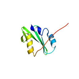
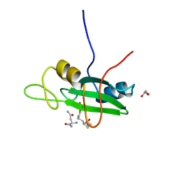
4QSY
 
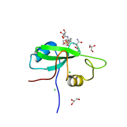
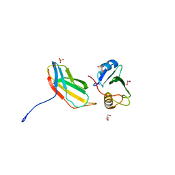
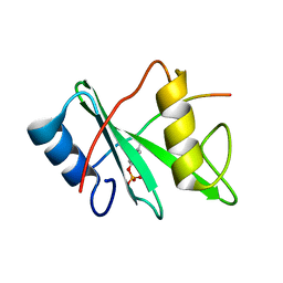 | | SHP2 SH2 domain in complex with GAB1 peptide | | 分子名称: | GRB2-associated-binding protein 1, Tyrosine-protein phosphatase non-receptor type 11 | | 著者 | Gogl, G, Remenyi, A. | | 登録日 | 2014-07-06 | | 公開日 | 2015-07-08 | | 最終更新日 | 2023-12-06 | | 実験手法 | X-RAY DIFFRACTION (2.1 Å) | | 主引用文献 | Selective targeting of GAB adapter protein SHP2 tyrosine phosphatase interaction attenuates ERK signaling
To be Published
|
|
1Z3K
 | |
4P9Z
 | |
3KFJ
 | |
4K44
 | |
4K45
 | |
8X2P
 | | The Crystal Structure of LCK from Biortus. | | 分子名称: | 1,2-ETHANEDIOL, CHLORIDE ION, TETRAETHYLENE GLYCOL, ... | | 著者 | Wang, F, Cheng, W, Lv, Z, Meng, Q, Lu, Y. | | 登録日 | 2023-11-10 | | 公開日 | 2023-11-22 | | 実験手法 | X-RAY DIFFRACTION (1.4 Å) | | 主引用文献 | The Crystal Structure of LCK from Biortus.
To Be Published
|
|
3PQZ
 | | Grb7 SH2 with peptide | | 分子名称: | Growth factor receptor-bound protein 7, cyclic peptide | | 著者 | Wilce, J.A. | | 登録日 | 2010-11-29 | | 公開日 | 2011-07-20 | | 最終更新日 | 2011-09-21 | | 実験手法 | X-RAY DIFFRACTION (2.413 Å) | | 主引用文献 | Structural basis of binding by cyclic nonphosphorylated Peptide antagonists of grb7 implicated in breast cancer progression
J.Mol.Biol., 412, 2011
|
|
3PSJ
 | |
3S8N
 | |
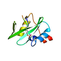
3S9K
 | | Crystal structure of the Itk SH2 domain. | | 分子名称: | CITRIC ACID, Tyrosine-protein kinase ITK/TSK | | 著者 | Joseph, R.E, Ginder, N.D, Hoy, J.A, Nix, J.C, Fulton, B.D, Honzatko, R.B, Andreotti, A.H. | | 登録日 | 2011-06-01 | | 公開日 | 2012-02-08 | | 最終更新日 | 2024-02-28 | | 実験手法 | X-RAY DIFFRACTION (2.354 Å) | | 主引用文献 | Structure of the interleukin-2 tyrosine kinase Src homology 2 domain; comparison between X-ray and NMR-derived structures.
Acta Crystallogr.,Sect.F, 68, 2012
|
|
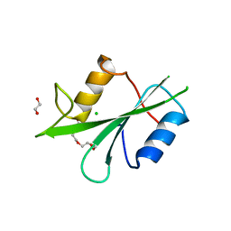
5GJI
 | | PI3K p85 N-terminal SH2 domain/CD28-derived peptide complex | | 分子名称: | GLYCEROL, Phosphatidylinositol 3-kinase regulatory subunit alpha, SULFATE ION, ... | | 著者 | Inaba, S, Numoto, N, Morii, H, Ogawa, S, Ikura, T, Abe, R, Ito, N, Oda, M. | | 登録日 | 2016-06-30 | | 公開日 | 2016-12-14 | | 最終更新日 | 2017-05-10 | | 実験手法 | X-RAY DIFFRACTION (0.9 Å) | | 主引用文献 | Crystal Structures and Thermodynamic Analysis Reveal Distinct Mechanisms of CD28 Phosphopeptide Binding to the Src Homology 2 (SH2) Domains of Three Adaptor Proteins
J. Biol. Chem., 292, 2017
|
|
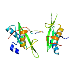
5GJH
 | | Gads SH2 domain/CD28-derived peptide complex | | 分子名称: | GRB2-related adapter protein 2, T-cell-specific surface glycoprotein CD28 | | 著者 | Inaba, S, Numoto, N, Morii, H, Ogawa, S, Ikura, T, Abe, R, Ito, N, Oda, M. | | 登録日 | 2016-06-30 | | 公開日 | 2016-12-14 | | 最終更新日 | 2023-11-15 | | 実験手法 | X-RAY DIFFRACTION (1.2 Å) | | 主引用文献 | Crystal Structures and Thermodynamic Analysis Reveal Distinct Mechanisms of CD28 Phosphopeptide Binding to the Src Homology 2 (SH2) Domains of Three Adaptor Proteins
J. Biol. Chem., 292, 2017
|
|
3S8L
 | |
3S8O
 | |
5EEQ
 | | Grb7 SH2 with the G7-B1 bicyclic peptide inhibitor | | 分子名称: | Bicyclic Peptide Inhibitor, Growth factor receptor-bound protein 7, PHOSPHATE ION | | 著者 | Ambaye, N.D, Watson, G.M, Wilce, M.C.J, Wilce, G.M. | | 登録日 | 2015-10-23 | | 公開日 | 2016-06-15 | | 最終更新日 | 2024-04-17 | | 実験手法 | X-RAY DIFFRACTION (1.6 Å) | | 主引用文献 | Unexpected involvement of staple leads to redesign of selective bicyclic peptide inhibitor of Grb7.
Sci Rep, 6, 2016
|
|
5EEL
 | | Grb7 SH2 with bicyclic peptide inhibitor | | 分子名称: | Bicyclic Peptide Inhibitor, FORMIC ACID, Growth factor receptor-bound protein 7, ... | | 著者 | Watson, G.M, Gunzburg, M.J, Wilce, M.C.J, Wilce, J.A. | | 登録日 | 2015-10-23 | | 公開日 | 2016-06-15 | | 最終更新日 | 2018-12-26 | | 実験手法 | X-RAY DIFFRACTION (2.47 Å) | | 主引用文献 | Unexpected involvement of staple leads to redesign of selective bicyclic peptide inhibitor of Grb7.
Sci Rep, 6, 2016
|
|
3PSK
 | |
3OVE
 | |
3T04
 | | Crystal structure of monobody 7c12/abl1 sh2 domain complex | | 分子名称: | GLYCEROL, MONOBODY 7C12, SULFATE ION, ... | | 著者 | Wojcik, J.B, Wyrzucki, A.M, Koide, S. | | 登録日 | 2011-07-19 | | 公開日 | 2011-11-23 | | 最終更新日 | 2023-09-13 | | 実験手法 | X-RAY DIFFRACTION (2.1 Å) | | 主引用文献 | Targeting the SH2-Kinase Interface in Bcr-Abl Inhibits Leukemogenesis.
Cell(Cambridge,Mass.), 147, 2011
|
|
3WA4
 | | Grb2 SH2 domain/CD28-derived peptide complex | | 分子名称: | ACETIC ACID, CADMIUM ION, Growth factor receptor-bound protein 2, ... | | 著者 | Higo, K, Oda, M, Ito, N. | | 登録日 | 2013-04-23 | | 公開日 | 2014-02-26 | | 最終更新日 | 2023-12-06 | | 実験手法 | X-RAY DIFFRACTION (1.35 Å) | | 主引用文献 | High Resolution Crystal Structure of the Grb2 SH2 Domain with a Phosphopeptide Derived from CD28
Plos One, 8, 2013
|
|
3UYO
 | |
3US4
 | |
1SPS
 | |
1SKJ
 | | COCRYSTAL STRUCTURE OF UREA-SUBSTITUTED PHOSPHOPEPTIDE COMPLEX | | 分子名称: | 4-[3-CARBOXYMETHYL-3-(4-PHOSPHONOOXY-BENZYL)-UREIDO]-4-[(3-CYCLOHEXYL-PROPYL)-METHYL-CARBAMOYL]BUTYRIC ACID, PP60 V-SRC TYROSINE KINASE TRANSFORMING PROTEIN | | 著者 | Holland, D.R, Rubin, J.R. | | 登録日 | 1997-09-18 | | 公開日 | 1998-02-25 | | 最終更新日 | 2023-08-09 | | 実験手法 | X-RAY DIFFRACTION (2 Å) | | 主引用文献 | Design, synthesis, and cocrystal structure of a nonpeptide Src SH2 domain ligand.
J.Med.Chem., 40, 1997
|
|


