Loading
PDBj
MenuPDBj@FacebookPDBj@TwitterPDBj@YouTubewwPDB FoundationwwPDB
RCSB PDBPDBeBMRBAdv. SearchSearch help

3I61
DownloadVisualize
BU of 3i61 by Molmil
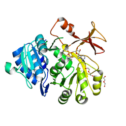
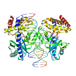
Structure of Mss116p bound to ssRNA and ADP-Beryllium Fluoride
Descriptor: 5'-R(*UP*UP*UP*UP*UP*UP*UP*UP*UP*U)-3', ADENOSINE-5'-DIPHOSPHATE, ATP-dependent RNA helicase MSS116, ...
Authors:Del Campo, M, Lambowitz, A.M.
Deposit date:2009-07-06
Release date:2009-09-08
Last modified:2023-09-06
Method:X-RAY DIFFRACTION (2.1 Å)
Cite:Structure of the Yeast DEAD box protein Mss116p reveals two wedges that crimp RNA
Mol.Cell, 35, 2009
3FJP
DownloadVisualize
BU of 3fjp by Molmil
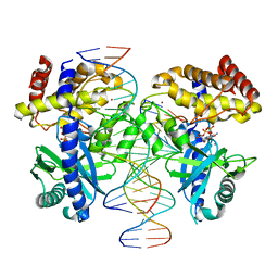
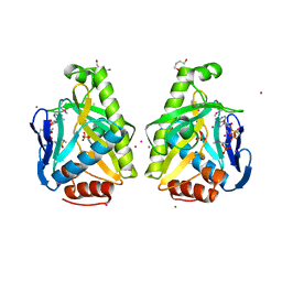
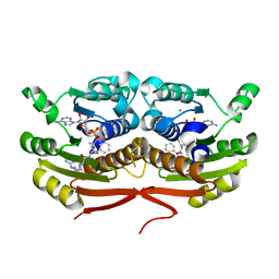
Apo structure of Biotin protein ligase from Aquifex aeolicus
Descriptor: Biotin [acetyl-CoA-carboxylase] ligase, SULFATE ION
Authors:McNae, I.W, Tron, C.M, Baxter, R.L, Walkinshaw, M.D, Campopiano, D.J.
Deposit date:2008-12-15
Release date:2009-06-09
Last modified:2023-11-01
Method:X-RAY DIFFRACTION (2.3 Å)
Cite:Structural and functional studies of the biotin protein ligase from Aquifex aeolicus reveal a critical role for a conserved residue in target specificity.
J.Mol.Biol., 387, 2009
8UZN
DownloadVisualize
BU of 8uzn by Molmil
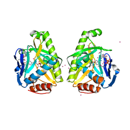
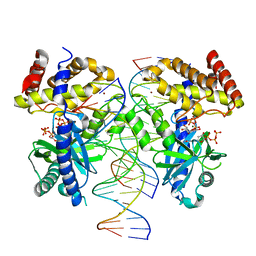
Crystal Structure of Betaine aldehyde dehydrogenase (BetB) from Klebsiella aerogenes (AMP bound)
Descriptor: ADENOSINE MONOPHOSPHATE, Betaine aldehyde dehydrogenase, SODIUM ION
Authors:Seattle Structural Genomics Center for Infectious Disease, Seattle Structural Genomics Center for Infectious Disease (SSGCID)
Deposit date:2023-11-15
Release date:2023-12-06
Method:X-RAY DIFFRACTION (2.15 Å)
Cite:Crystal Structure of Betaine aldehyde dehydrogenase (BetB) from Klebsiella aerogenes (AMP bound)
To be published
6NO7
DownloadVisualize
BU of 6no7 by Molmil
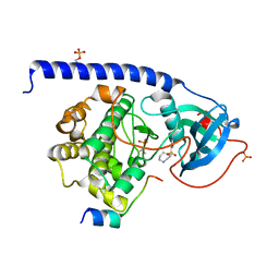
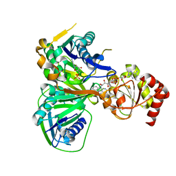
Crystal Structure of the full-length wild-type PKA RIa Holoenzyme
Descriptor: ADENOSINE-5'-TRIPHOSPHATE, MAGNESIUM ION, cAMP-dependent protein kinase catalytic subunit alpha, ...
Authors:Lu, T, Wu, J, Taylor, S.S.
Deposit date:2019-01-15
Release date:2019-07-24
Last modified:2023-10-11
Method:X-RAY DIFFRACTION (3.55 Å)
Cite:Two PKA RI alpha holoenzyme states define ATP as an isoform-specific orthosteric inhibitor that competes with the allosteric activator, cAMP.
Proc.Natl.Acad.Sci.USA, 116, 2019
5T8S
DownloadVisualize
BU of 5t8s by Molmil
Crystal Structure of a S-adenosylmethionine Synthase from Neisseria gonorrhoeae with bound S-adenosylmethionine, AMP, Pyrophosphate, Phosphate, and Magnesium
Descriptor: ADENOSINE MONOPHOSPHATE, MAGNESIUM ION, PHOSPHATE ION, ...
Authors:Seattle Structural Genomics Center for Infectious Disease, Seattle Structural Genomics Center for Infectious Disease (SSGCID)
Deposit date:2016-09-08
Release date:2016-09-28
Last modified:2023-10-04
Method:X-RAY DIFFRACTION (1.7 Å)
Cite:Crystal Structure of a S-adenosylmethionine Synthase from Neisseria gonorrhoeae with bound S-adenosylmethionine, AMP, Pyrophosphate, Phosphate, and Magnesium
to be published
5T8T
DownloadVisualize
BU of 5t8t by Molmil
Crystal Structure of a S-adenosylmethionine Synthase from Neisseria gonorrhoeae with bound AMP and Magnesium
Descriptor: ADENOSINE MONOPHOSPHATE, MAGNESIUM ION, S-adenosylmethionine synthase
Authors:Seattle Structural Genomics Center for Infectious Disease, Seattle Structural Genomics Center for Infectious Disease (SSGCID)
Deposit date:2016-09-08
Release date:2016-09-28
Last modified:2023-10-04
Method:X-RAY DIFFRACTION (2.1 Å)
Cite:Crystal Structure of a S-adenosylmethionine Synthase from Neisseria gonorrhoeae with bound AMP and Magnesium
to be published
3I59
DownloadVisualize
BU of 3i59 by Molmil
Crystal structure of MtbCRP in complex with N6-cAMP
Descriptor: (2R)-N6-(1-Methyl-2-phenylethyl)adenosine-3',5'-cyclic monophosphate, (2S)-N6-(1-Methyl-2-phenylethyl)adenosine-3',5'-cyclic monophosphate, CHLORIDE ION, ...
Authors:Reddy, M.C, Palaninathan, S.K, Bruning, J.B, Thurman, C, Smith, D, Sacchettini, J.C, TB Structural Genomics Consortium (TBSGC)
Deposit date:2009-07-03
Release date:2009-09-08
Last modified:2023-11-22
Method:X-RAY DIFFRACTION (2.29 Å)
Cite:Structural Insights into the Mechanism of the Allosteric Transitions of Mycobacterium tuberculosis cAMP Receptor Protein.
J.Biol.Chem., 284, 2009
6QZR
DownloadVisualize
BU of 6qzr by Molmil
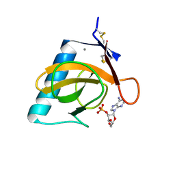
14-3-3 sigma in complex with FOXO1 pT24 peptide
Descriptor: 14-3-3 protein sigma, 2-[3-(2-HYDROXY-1,1-DIHYDROXYMETHYL-ETHYLAMINO)-PROPYLAMINO]-2-HYDROXYMETHYL-PROPANE-1,3-DIOL, Forkhead box protein O1, ...
Authors:Ottmann, C, Wolter, M, Lau, R.A.
Deposit date:2019-03-12
Release date:2019-07-31
Last modified:2024-01-24
Method:X-RAY DIFFRACTION (2.3 Å)
Cite:AMPK and AKT protein kinases hierarchically phosphorylate the N-terminus of the FOXO1 transcription factor, modulating interactions with 14-3-3 proteins.
J.Biol.Chem., 294, 2019
6QZS
DownloadVisualize
BU of 6qzs by Molmil
14-3-3 sigma in complex with FOXO1 pS256 peptide
Descriptor: 14-3-3 protein sigma, 2-[3-(2-HYDROXY-1,1-DIHYDROXYMETHYL-ETHYLAMINO)-PROPYLAMINO]-2-HYDROXYMETHYL-PROPANE-1,3-DIOL, FOXO1 pS256 site, ...
Authors:Ottmann, C, Wolter, M, Lau, R.A.
Deposit date:2019-03-12
Release date:2019-07-31
Last modified:2024-01-24
Method:X-RAY DIFFRACTION (1.9 Å)
Cite:AMPK and AKT protein kinases hierarchically phosphorylate the N-terminus of the FOXO1 transcription factor, modulating interactions with 14-3-3 proteins.
J.Biol.Chem., 294, 2019
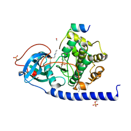
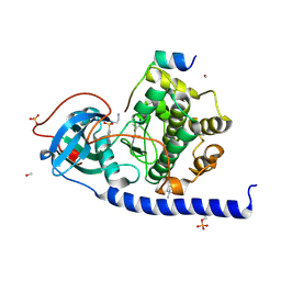
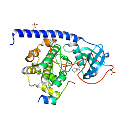
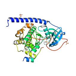
6SQ1
DownloadVisualize
BU of 6sq1 by Molmil
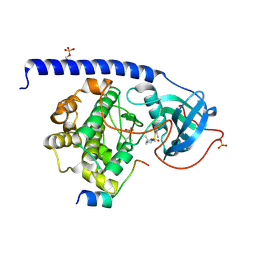
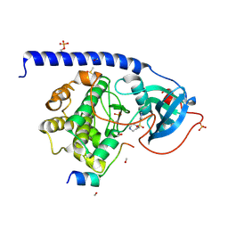
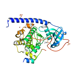
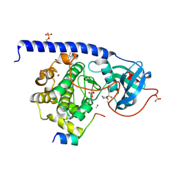
Crystal structure of cAMP-dependent Protein Kinase A (CHO PKA) in complex with Aminofasudil
Descriptor: (4S)-2-METHYL-2,4-PENTANEDIOL, 5-(1,4-diazepan-1-ylsulfonyl)isoquinolin-1-amine, DIMETHYL SULFOXIDE, ...
Authors:Oebbeke, M, Siefker, C, Heine, A, Klebe, G.
Deposit date:2019-09-03
Release date:2020-09-30
Last modified:2024-01-24
Method:X-RAY DIFFRACTION (1.49 Å)
Cite:A crystallographic study of cAMP-dependent Protein Kinase A in complex with different Fasudil-derivatives
To Be Published
7FJ8
DownloadVisualize
BU of 7fj8 by Molmil
KpAckA (PduW) with AMP complex structure
Descriptor: ADENOSINE MONOPHOSPHATE, Probable propionate kinase
Authors:Wu, W, Zhang, Q, Bartlam, M.
Deposit date:2021-08-03
Release date:2022-08-10
Last modified:2023-11-29
Method:X-RAY DIFFRACTION (2.1 Å)
Cite:KpAckA (PduW) with AMP complex structure
To Be Published
4F7Z
DownloadVisualize
BU of 4f7z by Molmil
Conformational dynamics of exchange protein directly activated by cAMP
Descriptor: GLYCEROL, Rap guanine nucleotide exchange factor 4
Authors:White, M.A, Tsalkova, T.N, Mei, F.C, Liu, T, Woods, V.L, Cheng, X.
Deposit date:2012-05-16
Release date:2012-10-03
Last modified:2023-09-13
Method:X-RAY DIFFRACTION (2.6 Å)
Cite:Structural analyses of a constitutively active mutant of exchange protein directly activated by cAMP.
Plos One, 7, 2012
6XB6
DownloadVisualize
BU of 6xb6 by Molmil
Structure of Danaus plexippus poxin cGAMP nuclease
Descriptor: Poxin
Authors:Eaglesham, J.B, McCarty, K.L, Kranzusch, P.J.
Deposit date:2020-06-05
Release date:2020-11-25
Last modified:2024-03-06
Method:X-RAY DIFFRACTION (1.45 Å)
Cite:Structures of diverse poxin cGAMP nucleases reveal a widespread role for cGAS-STING evasion in host-pathogen conflict.
Elife, 9, 2020
4BZP
DownloadVisualize
BU of 4bzp by Molmil
Structure of the Mycobacterium tuberculosis APS kinase CysC in complex with ADP
Descriptor: 1,2-ETHANEDIOL, ADENOSINE-5'-DIPHOSPHATE, BIFUNCTIONAL ENZYME CYSN/CYSC
Authors:Poyraz, O, Lohkamp, B, Schnell, R, Schneider, G.
Deposit date:2013-07-29
Release date:2014-08-06
Last modified:2023-12-20
Method:X-RAY DIFFRACTION (1.47 Å)
Cite:Crystal Structures of the Kinase Domain of the Sulfate-Activating Complex in Mycobacterium Tuberculosis.
Plos One, 10, 2015
1UC3
DownloadVisualize
BU of 1uc3 by Molmil
Crystal Structure of hemoglobin I from river lamprey
Descriptor: Globin, PROTOPORPHYRIN IX CONTAINING FE
Authors:Seki, M, Yao, M, Yazawa, Y, Tanaka, I.
Deposit date:2003-04-08
Release date:2003-04-29
Last modified:2023-10-25
Method:X-RAY DIFFRACTION (2.3 Å)
Cite:Crystal structure analysis of river lamprey hemoglobin I
To be Published
4N96
DownloadVisualize
BU of 4n96 by Molmil
E. coli sliding clamp in complex with 6-nitroindazole
Descriptor: 6-NITROINDAZOLE, CALCIUM ION, CHLORIDE ION, ...
Authors:Yin, Z, Oakley, A.J.
Deposit date:2013-10-19
Release date:2013-11-06
Last modified:2023-09-20
Method:X-RAY DIFFRACTION (1.7 Å)
Cite:Discovery of lead compounds targeting the bacterial sliding clamp using a fragment-based approach.
J.Med.Chem., 57, 2014
4N95
DownloadVisualize
BU of 4n95 by Molmil
E. coli sliding clamp in complex with 5-chloroindoline-2,3-dione
Descriptor: 5-chloro-1H-indole-2,3-dione, CALCIUM ION, CHLORIDE ION, ...
Authors:Yin, Z, Oakley, A.J.
Deposit date:2013-10-19
Release date:2013-11-06
Last modified:2023-09-20
Method:X-RAY DIFFRACTION (1.8 Å)
Cite:Discovery of lead compounds targeting the bacterial sliding clamp using a fragment-based approach.
J.Med.Chem., 57, 2014
4N94
DownloadVisualize
BU of 4n94 by Molmil
E. coli sliding clamp in complex with 3,4-difluorobenzamide
Descriptor: 1,2-ETHANEDIOL, 3,4-difluorobenzamide, CALCIUM ION, ...
Authors:Yin, Z, Oakley, A.J.
Deposit date:2013-10-19
Release date:2013-11-06
Last modified:2023-09-20
Method:X-RAY DIFFRACTION (1.73 Å)
Cite:Discovery of lead compounds targeting the bacterial sliding clamp using a fragment-based approach.
J.Med.Chem., 57, 2014
4NAV
DownloadVisualize
BU of 4nav by Molmil
Crystal structure of hypothetical protein XCC2798 from Xanthomonas campestris, Target EFI-508608
Descriptor: HYPOTHETICAL PROTEIN XCC279
Authors:Kim, J, Toro, R, Bhosle, R, Al Obaidi, N.F, Morisco, L.L, Wasserman, S.R, Sojitra, S, Washington, E, Glenn, A.S, Chowdhury, S, Evans, B, Zhao, S.C, Hillerich, B, Love, J, Seidel, R.D, Imker, H.J, Stead, M, Jacobson, M.P, Gerlt, J.A, Almo, S.C, Enzyme Function Initiative (EFI)
Deposit date:2013-10-22
Release date:2013-11-06
Last modified:2023-09-20
Method:X-RAY DIFFRACTION (2.69 Å)
Cite:Crystal structure of hypothetical protein XCC2798 from Xanthomonas campestris, Target EFI-508608
TO BE PUBLISHED
2E12
DownloadVisualize
BU of 2e12 by Molmil
The crystal structure of XC5848 from Xanthomonas campestris adopting a novel variant of Sm-like motif
Descriptor: Hypothetical protein XCC3642
Authors:Chin, K.-H, Ruan, S.-K, Wang, A.H.-J, Chou, S.-H.
Deposit date:2006-10-17
Release date:2007-10-30
Last modified:2024-03-13
Method:X-RAY DIFFRACTION (1.7 Å)
Cite:XC5848, an ORFan protein from Xanthomonas campestris, adopts a novel variant of Sm-like motif
Proteins, 68, 2007
8GJ1
DownloadVisualize
BU of 8gj1 by Molmil
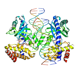
E. coli clamp loader with open clamp on primed template DNA (form 2)
Descriptor: ADENOSINE-5'-DIPHOSPHATE, Beta sliding clamp, DNA polymerase III subunit delta, ...
Authors:Oakley, A.J, Xu, Z.-Q, Dixon, N.E.
Deposit date:2023-03-14
Release date:2024-03-27
Method:ELECTRON MICROSCOPY (3 Å)
Cite:Structural characterisation of the complete cycle of sliding clamp loading in E. coli
To Be Published
8GJ2
DownloadVisualize
BU of 8gj2 by Molmil
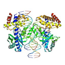
E. coli clamp loader with closed clamp on primed template DNA
Descriptor: ADENOSINE-5'-DIPHOSPHATE, Beta sliding clamp, DNA polymerase III subunit delta, ...
Authors:Oakley, A.J, Xu, Z.-Q, Dixon, N.E.
Deposit date:2023-03-14
Release date:2024-03-27
Method:ELECTRON MICROSCOPY (2.6 Å)
Cite:Structural characterisation of the complete cycle of sliding clamp loading in E. coli
To Be Published
8GJ0
DownloadVisualize
BU of 8gj0 by Molmil
E. coli clamp loader with open clamp on primed template DNA (form 1)
Descriptor: ADENOSINE-5'-DIPHOSPHATE, Beta sliding clamp, DNA polymerase III subunit delta, ...
Authors:Oakley, A.J, Xu, Z.-Q, Dixon, N.E.
Deposit date:2023-03-14
Release date:2024-03-27
Method:ELECTRON MICROSCOPY (2.9 Å)
Cite:Structural characterisation of the complete cycle of sliding clamp loading in E. coli
To Be Published
1KDL
DownloadVisualize
BU of 1kdl by Molmil
Solution structure of the amphipathic domain of YopD from Yersinia
Descriptor: YOPD protein
Authors:Tengel, T, Sethson, I, Francis, M.S.
Deposit date:2001-11-13
Release date:2002-08-21
Last modified:2024-05-01
Method:SOLUTION NMR
Cite:Conformational analysis by CD and NMR spectroscopy of a peptide encompassing the amphipathic domain of YopD from Yersinia.
Eur.J.Biochem., 269, 2002
5I1R
DownloadVisualize
BU of 5i1r by Molmil
Quantitative characterization of configurational space sampled by HIV-1 nucleocapsid using solution NMR and X-ray scattering
Descriptor: Nucleocapsid protein p7, ZINC ION
Authors:Deshmukh, L, Schwieters, C.D, Grishaev, A, Clore, G.M.
Deposit date:2016-02-05
Release date:2016-03-30
Last modified:2024-05-15
Method:SOLUTION NMR, SOLUTION SCATTERING
Cite:Quantitative Characterization of Configurational Space Sampled by HIV-1 Nucleocapsid Using Solution NMR, X-ray Scattering and Protein Engineering.
Chemphyschem, 17, 2016

224201

건을2024-08-28부터공개중

PDB statisticsPDBj update infoContact PDBjnumon