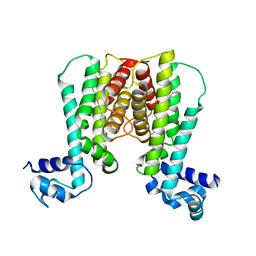
1U9I
 
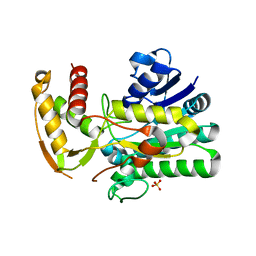
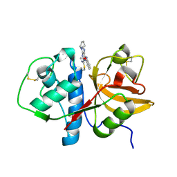
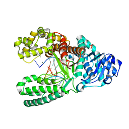
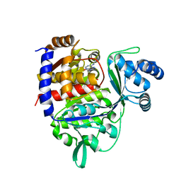
 | | Crystal Structure of Circadian Clock Protein KaiC with Phosphorylation Sites | | 分子名称: | ADENOSINE-5'-TRIPHOSPHATE, KaiC, MAGNESIUM ION | | 著者 | Xu, Y, Mori, T, Pattanayek, R, Pattanayek, S, Egli, M, Johnson, C.H. | | 登録日 | 2004-08-09 | | 公開日 | 2005-04-19 | | 最終更新日 | 2023-08-23 | | 実験手法 | X-RAY DIFFRACTION (2.8 Å) | | 主引用文献 | Identification of key phosphorylation sites in the circadian clock protein KaiC by crystallographic and mutagenetic analyses
PROC.NATL.ACAD.SCI.USA, 101, 2004
|
|
1U9J
 | |
1U9K
 | |
1U9L
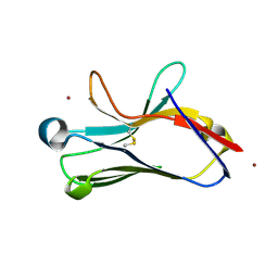
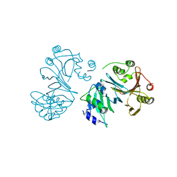
 | | Structural basis for a NusA- protein N interaction | | 分子名称: | GOLD ION, Lambda N, Transcription elongation protein nusA | | 著者 | Bonin, I, Muehlberger, R, Bourenkov, G.P, Huber, R, Bacher, A, Richter, G, Wahl, M.C. | | 登録日 | 2004-08-10 | | 公開日 | 2004-08-31 | | 最終更新日 | 2024-03-13 | | 実験手法 | X-RAY DIFFRACTION (1.9 Å) | | 主引用文献 | Structural basis for the interaction of Escherichia coli NusA with protein N of phage lambda
Proc.Natl.Acad.Sci.Usa, 101, 2004
|
|
1U9M
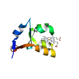
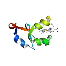 | | Crystal structure of F58W mutant of cytochrome b5 | | 分子名称: | Cytochrome b5, PROTOPORPHYRIN IX CONTAINING FE | | 著者 | Shan, L, Lu, J.-X, Gan, J.-H, Wang, Y.-H, Huang, Z.-X, Xia, Z.-X. | | 登録日 | 2004-08-10 | | 公開日 | 2005-02-01 | | 最終更新日 | 2023-10-25 | | 実験手法 | X-RAY DIFFRACTION (2 Å) | | 主引用文献 | Structure of the F58W mutant of cytochrome b5: the mutation leads to multiple conformations and weakens stacking interactions.
Acta Crystallogr.,Sect.D, 61, 2005
|
|
1U9N
 | | Crystal structure of the transcriptional regulator EthR in a ligand bound conformation opens therapeutic perspectives against tuberculosis and leprosy | | 分子名称: | HEXADECYL OCTANOATE, Transcriptional repressor EthR | | 著者 | Frenois, F, Engohang-Ndong, J, Locht, C, Baulard, A.R, Villeret, V. | | 登録日 | 2004-08-10 | | 公開日 | 2004-11-16 | | 最終更新日 | 2011-07-13 | | 実験手法 | X-RAY DIFFRACTION (2.3 Å) | | 主引用文献 | Structure of EthR in a Ligand Bound Conformation Reveals Therapeutic Perspectives against Tuberculosis
Mol.Cell, 16, 2004
|
|
1U9O
 | | Crystal structure of the transcriptional regulator EthR in a ligand bound conformation | | 分子名称: | HEXADECYL OCTANOATE, Transcriptional repressor EthR | | 著者 | Frenois, F, Engohang-Ndong, J, Locht, C, Baulard, A.R, Villeret, V. | | 登録日 | 2004-08-10 | | 公開日 | 2004-11-16 | | 最終更新日 | 2024-03-13 | | 実験手法 | X-RAY DIFFRACTION (3.3 Å) | | 主引用文献 | Structure of EthR in a Ligand Bound Conformation Reveals Therapeutic Perspectives against Tuberculosis
Mol.Cell, 16, 2004
|
|
1U9P
 | | Permuted single-chain Arc | | 分子名称: | pArc | | 著者 | Tabtiang, R.K, Cezairliyan, B.O, Grant, R.A, Cochrane, J.C, Sauer, R.T. | | 登録日 | 2004-08-10 | | 公開日 | 2005-02-15 | | 最終更新日 | 2024-02-14 | | 実験手法 | X-RAY DIFFRACTION (1.9 Å) | | 主引用文献 | Consolidating critical binding determinants by noncyclic rearrangement of protein secondary structure
Proc.Natl.Acad.Sci.Usa, 102, 2005
|
|
1U9Q
 | | Crystal structure of cruzain bound to an alpha-ketoester | | 分子名称: | [1-(1-METHYL-4,5-DIOXO-PENT-2-ENYLCARBAMOYL)-2-PHENYL-ETHYL]-CARBAMIC ACID BENZYL ESTER, cruzipain | | 著者 | Lange, M, Weston, S.G, Cheng, H, Culliane, M, Fiorey, M.M, Grisostomi, C, Hardy, L.W, Hartstough, D.S, Pallai, P.V, Tilton, R.F, Baldino, C.M, Brinen, L.S, Engel, J.C, Choe, Y, Price, M.S, Craik, C.S. | | 登録日 | 2004-08-10 | | 公開日 | 2005-03-29 | | 最終更新日 | 2023-08-23 | | 実験手法 | X-RAY DIFFRACTION (2.3 Å) | | 主引用文献 | Development of alpha-keto-based inhibitors of cruzain, a cysteine protease implicated in Chagas disease
Bioorg.Med.Chem., 13, 2005
|
|
1U9R
 | |
1U9S
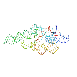 | | Crystal structure of the specificity domain of Ribonuclease P of the A-type | | 分子名称: | BARIUM ION, RIBONUCLEASE P | | 著者 | Krasilnikov, A.S, Xiao, Y, Pan, T, Mondragon, A. | | 登録日 | 2004-08-10 | | 公開日 | 2004-10-26 | | 最終更新日 | 2024-02-14 | | 実験手法 | X-RAY DIFFRACTION (2.9 Å) | | 主引用文献 | Basis for structural diversity in homologous RNAs.
Science, 306, 2004
|
|
1U9T
 | |
1U9U
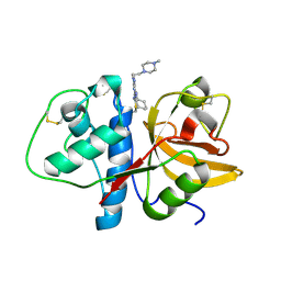
 | | Crystal structure of F58Y mutant of cytochrome b5 | | 分子名称: | Cytochrome b5, PROTOPORPHYRIN IX CONTAINING FE | | 著者 | Shan, L, Lu, J.-X, Gan, J.-H, Wang, Y.-H, Huang, Z.-X, Xia, Z.-X. | | 登録日 | 2004-08-11 | | 公開日 | 2005-02-01 | | 最終更新日 | 2023-10-25 | | 実験手法 | X-RAY DIFFRACTION (1.86 Å) | | 主引用文献 | Structure of the F58W mutant of cytochrome b5: the mutation leads to multiple conformations and weakens stacking interactions.
Acta Crystallogr.,Sect.D, 61, 2005
|
|
1U9V
 | |
1U9W
 | |
1U9X
 | |
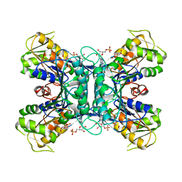
1U9Y
 | | Crystal Structure of Phosphoribosyl Diphosphate Synthase from Methanocaldococcus jannaschii | | 分子名称: | Ribose-phosphate pyrophosphokinase | | 著者 | Kadziola, A, Johansson, E, Jepsen, C.H, McGuire, J, Larsen, S, Hove-Jensen, B. | | 登録日 | 2004-08-11 | | 公開日 | 2005-08-23 | | 最終更新日 | 2024-04-03 | | 実験手法 | X-RAY DIFFRACTION (2.65 Å) | | 主引用文献 | Novel class III phosphoribosyl diphosphate synthase: structure and properties of the tetrameric, phosphate-activated, non-allosterically inhibited enzyme from Methanocaldococcus jannaschii
J.Mol.Biol., 354, 2005
|
|
1U9Z
 | | Crystal Structure of Phosphoribosyl Diphosphate Synthase Complexed with AMP and Ribose 5-Phosphate | | 分子名称: | ADENOSINE MONOPHOSPHATE, RIBOSE-5-PHOSPHATE, Ribose-phosphate pyrophosphokinase | | 著者 | Kadziola, A, Johansson, E, Jepsen, C.H, McGuire, J, Larsen, S, Hove-Jensen, B. | | 登録日 | 2004-08-11 | | 公開日 | 2005-08-23 | | 最終更新日 | 2023-08-23 | | 実験手法 | X-RAY DIFFRACTION (2.8 Å) | | 主引用文献 | Novel class III phosphoribosyl diphosphate synthase: structure and properties of the tetrameric, phosphate-activated, non-allosterically inhibited enzyme from Methanocaldococcus jannaschii
J.Mol.Biol., 354, 2005
|
|
1UA0
 | | Aminofluorene DNA adduct at the pre-insertion site of a DNA polymerase | | 分子名称: | 2-AMINOFLUORENE, DNA polymerase I, DNA primer strand, ... | | 著者 | Hsu, G.W, Kiefer, J.R, Becherel, O.J, Fuchs, R.P.P, Beese, L.S. | | 登録日 | 2004-08-11 | | 公開日 | 2004-09-28 | | 最終更新日 | 2023-08-23 | | 実験手法 | X-RAY DIFFRACTION (2.1 Å) | | 主引用文献 | Observing translesion synthesis of an aromatic amine DNA adduct by a high-fidelity DNA polymerase
J.Biol.Chem., 279, 2004
|
|
1UA1
 | | Structure of aminofluorene adduct paired opposite cytosine at the polymerase active site. | | 分子名称: | 2-AMINOFLUORENE, DNA polymerase I, DNA primer strand, ... | | 著者 | Hsu, G.W, Kiefer, J.R, Becherel, O.J, Fuchs, R.P.P, Beese, L.S. | | 登録日 | 2004-08-11 | | 公開日 | 2004-09-28 | | 最終更新日 | 2024-02-14 | | 実験手法 | X-RAY DIFFRACTION (2 Å) | | 主引用文献 | Observing translesion synthesis of an aromatic amine DNA adduct by a high-fidelity DNA polymerase
J.Biol.Chem., 279, 2004
|
|
1UA2
 | | Crystal Structure of Human CDK7 | | 分子名称: | ADENOSINE-5'-TRIPHOSPHATE, Cell division protein kinase 7 | | 著者 | Lolli, G, Lowe, E.D, Brown, N.R, Johnson, L.N. | | 登録日 | 2004-08-11 | | 公開日 | 2004-12-07 | | 最終更新日 | 2024-04-03 | | 実験手法 | X-RAY DIFFRACTION (3.02 Å) | | 主引用文献 | The Crystal Structure of Human CDK7 and Its Protein Recognition Properties
Structure, 12, 2004
|
|
1UA3
 | |
1UA4
 | | Crystal Structure of an ADP-dependent Glucokinase from Pyrococcus furiosus | | 分子名称: | ADENOSINE MONOPHOSPHATE, ADP-dependent glucokinase, alpha-D-glucopyranose, ... | | 著者 | Ito, S, Jeong, J.J, Yoshioka, I, Koga, S, Fushinobu, S, Shoun, H, Wakagi, T. | | 登録日 | 2003-02-27 | | 公開日 | 2004-02-27 | | 最終更新日 | 2023-12-27 | | 実験手法 | X-RAY DIFFRACTION (1.9 Å) | | 主引用文献 | Crystal structure of an ADP-dependent glucokinase from Pyrococcus furiosus: implications for a sugar-induced conformational change in ADP-dependent kinase
J.Mol.Biol., 331, 2003
|
|
1UA5
 | |
1UA6
 | | Crystal structure of HYHEL-10 FV MUTANT SFSF complexed with HEN EGG WHITE LYSOZYME complex | | 分子名称: | Ig VH,anti-lysozyme, Lysozyme C, lysozyme binding Ig kappa chain V23-J2 region | | 著者 | Kumagai, I, Nishimiya, Y, Kondo, H, Tsumoto, K. | | 登録日 | 2003-02-28 | | 公開日 | 2004-03-09 | | 最終更新日 | 2023-12-27 | | 実験手法 | X-RAY DIFFRACTION (1.9 Å) | | 主引用文献 | Structural consequences of target epitope-directed functional alteration of an antibody. The case of anti-hen lysozyme antibody, HyHEL-10
J.Biol.Chem., 278, 2003
|
|


