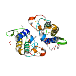
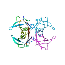
3NG4
 
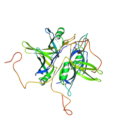
 | | Ternary complex of peptidoglycan recognition protein (PGRP-S) with Maltose and N-Acetylglucosamine at 1.7 A Resolution | | 分子名称: | 2-acetamido-2-deoxy-beta-D-glucopyranose, GLYCEROL, Peptidoglycan recognition protein 1, ... | | 著者 | Sharma, P, Dube, D, Kaur, P, Sharma, S, Singh, T.P. | | 登録日 | 2010-06-10 | | 公開日 | 2010-07-14 | | 最終更新日 | 2023-11-01 | | 実験手法 | X-RAY DIFFRACTION (1.73 Å) | | 主引用文献 | Multiligand specificity of pathogen-associated molecular pattern-binding site in peptidoglycan recognition protein
J.Biol.Chem., 286, 2011
|
|
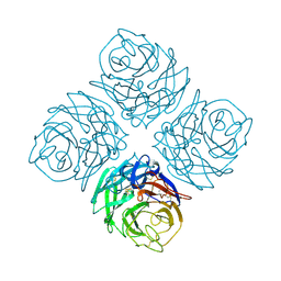
1QVC
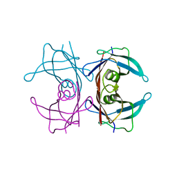
 | | CRYSTAL STRUCTURE ANALYSIS OF SINGLE STRANDED DNA BINDING PROTEIN (SSB) FROM E.COLI | | 分子名称: | SINGLE STRANDED DNA BINDING PROTEIN MONOMER | | 著者 | Matsumoto, T, Morimoto, Y, Shibata, N, Shimamoto, N, Tsukihara, T, Yasuoka, N. | | 登録日 | 1999-07-07 | | 公開日 | 2000-06-05 | | 最終更新日 | 2024-02-14 | | 実験手法 | X-RAY DIFFRACTION (2.2 Å) | | 主引用文献 | Roles of functional loops and the C-terminal segment of a single-stranded DNA binding protein elucidated by X-Ray structure analysis.
J.Biochem.(Tokyo), 127, 2000
|
|
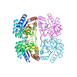
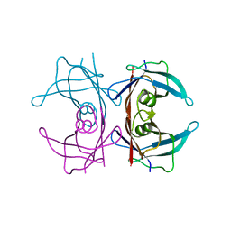
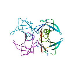
1NNA
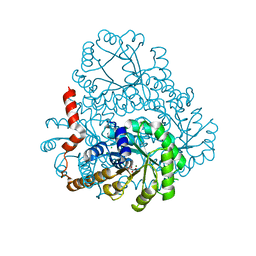
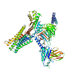
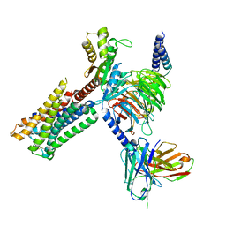
 | | THREE-DIMENSIONAL STRUCTURE OF INFLUENZA A N9 NEURAMINIDASE AND ITS COMPLEX WITH THE INHIBITOR 2-DEOXY 2,3-DEHYDRO-N-ACETYL NEURAMINIC ACID | | 分子名称: | CALCIUM ION, NEURAMINIDASE | | 著者 | Bossart-Whitaker, P, Carson, M, Babu, Y.S, Smith, C.D, Laver, W.G, Air, G.M. | | 登録日 | 1993-03-08 | | 公開日 | 1994-04-30 | | 最終更新日 | 2017-11-29 | | 実験手法 | X-RAY DIFFRACTION (2.5 Å) | | 主引用文献 | Three-dimensional structure of influenza A N9 neuraminidase and its complex with the inhibitor 2-deoxy 2,3-dehydro-N-acetyl neuraminic acid.
J.Mol.Biol., 232, 1993
|
|
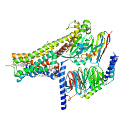
2DRE
 | | Crystal structure of Water-soluble chlorophyll protein from lepidium virginicum at 2.00 angstrom resolution | | 分子名称: | CHLOROPHYLL A, Water-soluble chlorophyll protein | | 著者 | Horigome, D, Satoh, H, Itoh, N, Mitsunaga, K, Oonishi, I, Nakagawa, A, Uchida, A. | | 登録日 | 2006-06-08 | | 公開日 | 2006-12-26 | | 最終更新日 | 2011-07-13 | | 実験手法 | X-RAY DIFFRACTION (2 Å) | | 主引用文献 | Structural mechanism and photoprotective function of water-soluble chlorophyll-binding protein.
J.Biol.Chem., 282, 2007
|
|
2DUA
 | |
3P3S
 | |
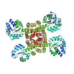
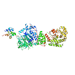
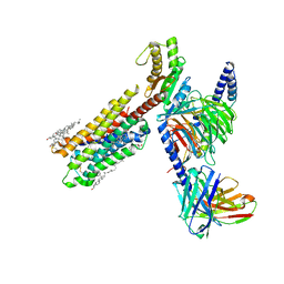
2A2I
 | | Aquifex aeolicus KDO8PS in complex with PEP, A5P, Zn2+ | | 分子名称: | 2-dehydro-3-deoxyphosphooctonate aldolase, ARABINOSE-5-PHOSPHATE, PHOSPHOENOLPYRUVATE, ... | | 著者 | Kona, F, Xu, X, Lu, J, Martin, P, Gatti, D.L. | | 登録日 | 2005-06-22 | | 公開日 | 2006-07-04 | | 最終更新日 | 2024-02-14 | | 実験手法 | X-RAY DIFFRACTION (1.95 Å) | | 主引用文献 | Electronic structure of the metal center in the Cd(2+), Zn(2+), and Cu(2+) substituted forms of KDO8P synthase: implications for catalysis.
Biochemistry, 48, 2009
|
|
3CKY
 | |
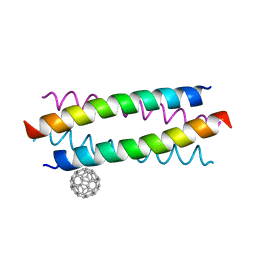
5HKN
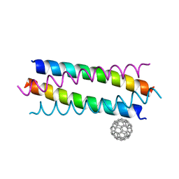 | | Crystal structure de novo designed fullerene organizing protein complex with fullerene | | 分子名称: | (C_{60}-I_{h})[5,6]fullerene, fullerene organizing protein | | 著者 | Paul, J, Acharya, R, Kim, K.-H, Kim, Y.H, Kim, N.H, Grigoryan, G, DeGardo, W.F. | | 登録日 | 2016-01-14 | | 公開日 | 2016-05-04 | | 最終更新日 | 2023-11-08 | | 実験手法 | X-RAY DIFFRACTION (1.761 Å) | | 主引用文献 | Protein-directed self-assembly of a fullerene crystal.
Nat Commun, 7, 2016
|
|
3CYL
 | |
2G4G
 | |
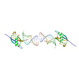
3D71
 | | Crystal structure of E253Q BMRR bound to 22 base pair promoter site | | 分子名称: | BMR promoter DNA, CITRATE ANION, IMIDAZOLE, ... | | 著者 | Newberry, K.J, Huffman, J.L, Brennan, R.G. | | 登録日 | 2008-05-20 | | 公開日 | 2008-08-26 | | 最終更新日 | 2023-08-30 | | 実験手法 | X-RAY DIFFRACTION (2.8 Å) | | 主引用文献 | Structures of BmrR-Drug Complexes Reveal a Rigid Multidrug Binding Pocket and Transcription Activation through Tyrosine Expulsion
J.Biol.Chem., 283, 2008
|
|
5HKR
 | | Crystal structure of de novo designed fullerene organising protein complex with fullerene | | 分子名称: | (C_{60}-I_{h})[5,6]fullerene, fullerene organizing protein | | 著者 | Acharya, R, Kim, Y.H, Grigoryan, G, DeGardo, W.F. | | 登録日 | 2016-01-14 | | 公開日 | 2016-05-04 | | 最終更新日 | 2024-03-20 | | 実験手法 | X-RAY DIFFRACTION (2.35 Å) | | 主引用文献 | Protein-directed self-assembly of a fullerene crystal.
Nat Commun, 7, 2016
|
|
8ID2
 | | Crystal structure of the ubiquitin-like domain in the SF3A1 subunit of human U2 snRNP complexed with the stem-loop 4 of U1 snRNA | | 分子名称: | RNA (5'-R(*GP*GP*GP*GP*AP*CP*UP*GP*CP*GP*UP*UP*CP*GP*CP*GP*CP*UP*UP*UP*CP*CP*CP*C)-3'), Splicing factor 3A subunit 1 | | 著者 | Terawaki, S, Nameki, N, Kuwasako, K. | | 登録日 | 2023-02-12 | | 公開日 | 2023-05-17 | | 最終更新日 | 2024-05-29 | | 実験手法 | X-RAY DIFFRACTION (1.8 Å) | | 主引用文献 | Structural insights into recognition of SL4, the UUCG stem-loop, of human U1 snRNA by the ubiquitin-like domain, including the C-terminal tail in the SF3A1 subunit of U2 snRNP.
J.Biochem., 174, 2023
|
|
8IN9
 | | The structure of the GfsA KSQ-AT didomain in complex with the GfsA ACP domain | | 分子名称: | N-[2-(acetylamino)ethyl]-N~3~-[(2R)-2-hydroxy-3,3-dimethyl-4-(phosphonooxy)butanoyl]-beta-alaninamide, Polyketide synthase | | 著者 | Chisuga, T, Murakami, S, Miyanaga, A, Kudo, F, Eguchi, T. | | 登録日 | 2023-03-09 | | 公開日 | 2023-05-31 | | 最終更新日 | 2023-06-28 | | 実験手法 | X-RAY DIFFRACTION (3.4 Å) | | 主引用文献 | Structure-Based Analysis of Transient Interactions between Ketosynthase-like Decarboxylase and Acyl Carrier Protein in a Loading Module of Modular Polyketide Synthase.
Acs Chem.Biol., 18, 2023
|
|
6XQK
 | | Crystal structure of the D/D domain of PKA from S. cerevisiae | | 分子名称: | CHLORIDE ION, GLYCEROL, cAMP-dependent protein kinase regulatory subunit | | 著者 | Larrieux, N, Gonzalez Bardeci, N, Trajtenberg, F, Buschiazzo, A. | | 登録日 | 2020-07-09 | | 公開日 | 2021-04-14 | | 最終更新日 | 2024-03-06 | | 実験手法 | X-RAY DIFFRACTION (2.56 Å) | | 主引用文献 | The crystal structure of yeast regulatory subunit reveals key evolutionary insights into Protein Kinase A oligomerization.
J.Struct.Biol., 213, 2021
|
|
2G4E
 | |
8IRT
 | | Dopamine Receptor D3R-Gi-Rotigotine complex | | 分子名称: | Guanine nucleotide-binding protein G(I)/G(S)/G(O) subunit gamma-2, Guanine nucleotide-binding protein G(I)/G(S)/G(T) subunit beta-1, Guanine nucleotide-binding protein G(i) subunit alpha-1, ... | | 著者 | Xu, P, Huang, S, Zhuang, Y, Mao, C, Zhang, Y, Wang, Y, Li, H, Jiang, Y, Zhang, Y, Xu, H.E. | | 登録日 | 2023-03-19 | | 公開日 | 2023-06-07 | | 最終更新日 | 2023-11-08 | | 実験手法 | ELECTRON MICROSCOPY (2.7 Å) | | 主引用文献 | Structural genomics of the human dopamine receptor system.
Cell Res., 33, 2023
|
|
8IRS
 | | Dopamine Receptor D2R-Gi-Rotigotine complex | | 分子名称: | Guanine nucleotide-binding protein G(I)/G(S)/G(O) subunit gamma-2, Guanine nucleotide-binding protein G(I)/G(S)/G(T) subunit beta-1, Guanine nucleotide-binding protein G(i) subunit alpha-1, ... | | 著者 | Xu, P, Huang, S, Zhuang, Y, Mao, C, Zhang, Y, Wang, Y, Li, H, Jiang, Y, Zhang, Y, Xu, H.E. | | 登録日 | 2023-03-19 | | 公開日 | 2023-06-07 | | 最終更新日 | 2023-11-08 | | 実験手法 | ELECTRON MICROSCOPY (3 Å) | | 主引用文献 | Structural genomics of the human dopamine receptor system.
Cell Res., 33, 2023
|
|
3D2T
 | |
8IRR
 | | Dopamine Receptor D1R-Gs-Rotigotine complex | | 分子名称: | CHOLESTEROL, D(1A) dopamine receptor, Guanine nucleotide-binding protein G(I)/G(S)/G(O) subunit gamma-2, ... | | 著者 | Xu, P, Huang, S, Zhuang, Y, Mao, C, Zhang, Y, Wang, Y, Li, H, Jiang, Y, Zhang, Y, Xu, H.E. | | 登録日 | 2023-03-19 | | 公開日 | 2023-06-21 | | 最終更新日 | 2023-11-08 | | 実験手法 | ELECTRON MICROSCOPY (3.2 Å) | | 主引用文献 | Structural genomics of the human dopamine receptor system.
Cell Res., 33, 2023
|
|
8IRU
 | | Dopamine Receptor D4R-Gi-Rotigotine complex | | 分子名称: | CHOLESTEROL, D(4) dopamine receptor, Guanine nucleotide-binding protein G(I)/G(S)/G(O) subunit gamma-2, ... | | 著者 | Xu, P, Huang, S, Zhuang, Y, Mao, C, Zhang, Y, Wang, Y, Li, H, Jiang, Y, Zhang, Y, Xu, H.E. | | 登録日 | 2023-03-19 | | 公開日 | 2023-06-21 | | 最終更新日 | 2023-11-08 | | 実験手法 | ELECTRON MICROSCOPY (3.2 Å) | | 主引用文献 | Structural genomics of the human dopamine receptor system.
Cell Res., 33, 2023
|
|
2G3Z
 | |
3CTT
 | |
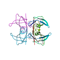
2G9E
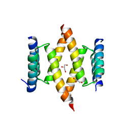
 | | Protonation-mediated structural flexibility in the F conjugation regulatory protein, TRAM | | 分子名称: | Protein traM | | 著者 | Lu, J, Edwards, R.A, Wong, J.J, Manchak, J, Scott, P.G, Frost, L.S, Glover, J.N. | | 登録日 | 2006-03-06 | | 公開日 | 2006-06-13 | | 最終更新日 | 2023-08-30 | | 実験手法 | X-RAY DIFFRACTION (1.8 Å) | | 主引用文献 | Protonation-mediated structural flexibility in the F conjugation regulatory protein, TraM.
Embo J., 25, 2006
|
|


