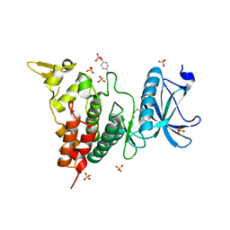
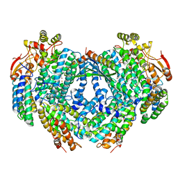
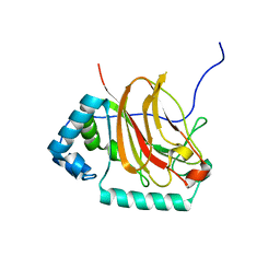
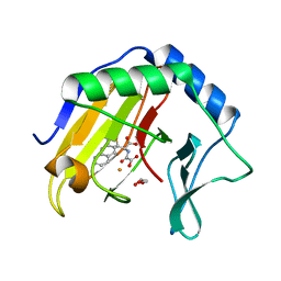
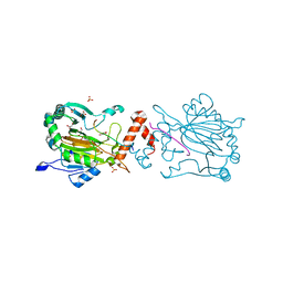
6A1G
 
 | | Crystal structure of human DYRK1A in complex with compound 32 | | 分子名称: | 5,5-dimethyl-8-[1-(piperidin-4-yl)ethenyl]-5,6-dihydrobenzo[h]quinazolin-4-amine, Dual specificity tyrosine-phosphorylation-regulated kinase 1A | | 著者 | Baba, D, Hanzawa, H. | | 登録日 | 2018-06-07 | | 公開日 | 2018-10-03 | | 最終更新日 | 2023-11-22 | | 実験手法 | X-RAY DIFFRACTION (2.15 Å) | | 主引用文献 | Discovery of DS42450411 as a potent orally active hepcidin production inhibitor: Design and optimization of novel 4-aminopyrimidine derivatives.
Bioorg. Med. Chem. Lett., 28, 2018
|
|
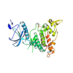
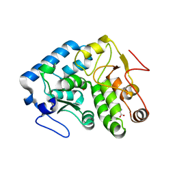
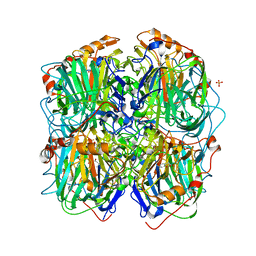
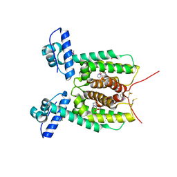
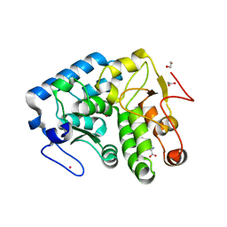
6A1F
 | | Crystal structure of human DYRK1A in complex with compound 14 | | 分子名称: | 8-methoxy-5,5-dimethyl-5,6-dihydrobenzo[h]quinazolin-4-amine, Dual specificity tyrosine-phosphorylation-regulated kinase 1A, SULFATE ION, ... | | 著者 | Baba, D, Hanzawa, H. | | 登録日 | 2018-06-07 | | 公開日 | 2018-10-03 | | 最終更新日 | 2023-11-22 | | 実験手法 | X-RAY DIFFRACTION (1.5 Å) | | 主引用文献 | Discovery of DS42450411 as a potent orally active hepcidin production inhibitor: Design and optimization of novel 4-aminopyrimidine derivatives.
Bioorg. Med. Chem. Lett., 28, 2018
|
|
3TK2
 | |
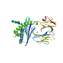
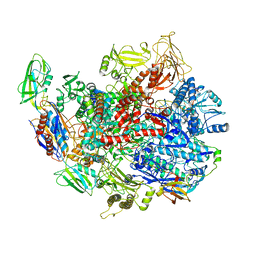
5WJ9
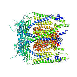 | | Human TRPML1 channel structure in agonist-bound open conformation | | 分子名称: | 2-{2-oxo-2-[(4S)-2,2,4-trimethyl-3,4-dihydroquinolin-1(2H)-yl]ethyl}-1H-isoindole-1,3(2H)-dione, Mucolipin-1 | | 著者 | Schmiege, P, Li, X. | | 登録日 | 2017-07-21 | | 公開日 | 2017-10-18 | | 最終更新日 | 2019-12-18 | | 実験手法 | ELECTRON MICROSCOPY (3.49 Å) | | 主引用文献 | Human TRPML1 channel structures in open and closed conformations.
Nature, 550, 2017
|
|
5WJ5
 | |
4JPX
 | |
1ZHN
 | | Crystal Structure of mouse CD1d bound to the self ligand phosphatidylcholine | | 分子名称: | 2-acetamido-2-deoxy-beta-D-glucopyranose, 7-[(DODECANOYLOXY)METHYL]-4-HYDROXY-N,N,N-TRIMETHYL-9-OXO-3,5,8-TRIOXA-4-PHOSPHADOTRIACONTAN-1-AMINIUM 4-OXIDE, CD1d1 antigen, ... | | 著者 | Giabbai, B, Sidobre, S, Crispin, M.M.D, Sanchez Ruiz, Y, Bachi, A, Kronenberg, M, Wilson, I.A, Degano, M. | | 登録日 | 2005-04-26 | | 公開日 | 2005-07-19 | | 最終更新日 | 2023-08-23 | | 実験手法 | X-RAY DIFFRACTION (2.8 Å) | | 主引用文献 | Crystal structure of mouse CD1d bound to the self ligand phosphatidylcholine: a molecular basis for NKT cell activation
J.Immunol., 175, 2005
|
|
4LZL
 | |
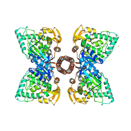
7WFF
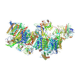 | | Subcomplexes B,M and L in the Cylic electron transfer supercomplex NDH-PSI from Arabidopsis | | 分子名称: | 1,2-DI-O-ACYL-3-O-[6-DEOXY-6-SULFO-ALPHA-D-GLUCOPYRANOSYL]-SN-GLYCEROL, 1,2-DIPALMITOYL-PHOSPHATIDYL-GLYCEROLE, FE2/S2 (INORGANIC) CLUSTER, ... | | 著者 | Pan, X.W, Li, M. | | 登録日 | 2021-12-26 | | 公開日 | 2022-03-16 | | 最終更新日 | 2024-06-26 | | 実験手法 | ELECTRON MICROSCOPY (3.59 Å) | | 主引用文献 | Supramolecular assembly of chloroplast NADH dehydrogenase-like complex with photosystem I from Arabidopsis thaliana.
Mol Plant, 15, 2022
|
|
6D7K
 | | Complex structure of Methane monooxygenase hydroxylase in complex with inhibitory subunit | | 分子名称: | FE (III) ION, FORMIC ACID, HEXANE-1,6-DIOL, ... | | 著者 | Kim, H, Lee, S.J, Cho, U.-S. | | 登録日 | 2018-04-24 | | 公開日 | 2019-06-26 | | 最終更新日 | 2023-10-04 | | 実験手法 | X-RAY DIFFRACTION (2.6 Å) | | 主引用文献 | MMOD-induced structural changes of hydroxylase in soluble methane monooxygenase.
Sci Adv, 5, 2019
|
|
3ZDS
 | | Structure of homogentisate 1,2-dioxygenase in complex with reaction intermediates of homogentisate with oxygen. | | 分子名称: | 2-(3,6-DIHYDROXYPHENYL)ACETIC ACID, 2-(6-oxidanyl-3-oxidanylidene-cyclohexa-1,4-dien-1-yl)ethanoic acid, 2-[(6R)-6-(dioxidanyl)-6-oxidanyl-3-oxidanylidene-cyclohexa-1,4-dien-1-yl]ethanoic acid, ... | | 著者 | Jeoung, J.-H, Bommer, M, Lin, T.-Y, Dobbek, H. | | 登録日 | 2012-11-30 | | 公開日 | 2013-07-24 | | 最終更新日 | 2023-12-20 | | 実験手法 | X-RAY DIFFRACTION (1.7 Å) | | 主引用文献 | Visualizing the Substrate-, Superoxo-, Alkylperoxo- and Product-Bound States at the Non-Heme Fe(II) Site of Homogentisate Dioxygenase
Proc.Natl.Acad.Sci.USA, 110, 2013
|
|
6C6S
 | | CryoEM structure of E.coli RNA polymerase elongation complex bound with RfaH | | 分子名称: | DNA (29-MER), DNA-directed RNA polymerase subunit alpha, DNA-directed RNA polymerase subunit beta, ... | | 著者 | Kang, J.Y, Artsimovitch, I, Landick, R, Darst, S.A. | | 登録日 | 2018-01-19 | | 公開日 | 2018-07-25 | | 最終更新日 | 2024-03-13 | | 実験手法 | ELECTRON MICROSCOPY (3.7 Å) | | 主引用文献 | Structural Basis for Transcript Elongation Control by NusG Family Universal Regulators.
Cell, 173, 2018
|
|
8UPO
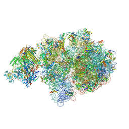 | | Escherichia coli transcription-translation coupled complex class A (TTC-A) containing RfaH bound to ops signal, mRNA with a 21 nt long spacer, and fMet-tRNAs in E-site and P-site of the ribosome | | 分子名称: | 16S rRNA, 23S rRNA, 30S ribosomal protein S1, ... | | 著者 | Molodtsov, V, Wang, C, Ebright, R.H. | | 登録日 | 2023-10-23 | | 公開日 | 2024-05-29 | | 実験手法 | ELECTRON MICROSCOPY (5.5 Å) | | 主引用文献 | Escherichia coli transcription-translation coupled complex class A (TTC-A) containing RfaH bound to ops signal, mRNA with a 21 nt long spacer, and fMet-tRNAs in E-site and P-site of the ribosome
Nat.Struct.Mol.Biol., 2024
|
|
6E0T
 | | C-terminal domain of Fission Yeast OFD1 | | 分子名称: | Prolyl 3,4-dihydroxylase ofd1 | | 著者 | Bianchet, M.A, Amzel, L.M, Espenshade, P.J, Yeh, T. | | 登録日 | 2018-07-06 | | 公開日 | 2019-09-11 | | 最終更新日 | 2024-03-13 | | 実験手法 | X-RAY DIFFRACTION (2.02 Å) | | 主引用文献 | The hypoxic regulator of sterol synthesis nro1 is a nuclear import adaptor.
Structure, 19, 2011
|
|
5X3R
 | | Crystal structure of the SmcR complexed with QStatin | | 分子名称: | 1-(5-bromanylthiophen-2-yl)sulfonylpyrazole, LuxR family transcriptional regulator, SULFATE ION | | 著者 | Jang, S.Y, Hwang, J, Kim, M.H. | | 登録日 | 2017-02-07 | | 公開日 | 2018-01-24 | | 最終更新日 | 2023-11-22 | | 実験手法 | X-RAY DIFFRACTION (2.1 Å) | | 主引用文献 | QStatin, a Selective Inhibitor of Quorum Sensing inVibrioSpecies
MBio, 9, 2018
|
|
3TK4
 | |
3T4H
 | | Crystal Structure of AlkB in complex with Fe(III) and N-Oxalyl-S-(3-nitrobenzyl)-L-cysteine | | 分子名称: | 4-(2-HYDROXYETHYL)-1-PIPERAZINE ETHANESULFONIC ACID, Alpha-ketoglutarate-dependent dioxygenase AlkB, FE (III) ION, ... | | 著者 | Ma, J, Aik, W.S, McDonough, M.A, Schofield, C.J. | | 登録日 | 2011-07-26 | | 公開日 | 2012-03-07 | | 最終更新日 | 2024-02-28 | | 実験手法 | X-RAY DIFFRACTION (1.65 Å) | | 主引用文献 | Dynamic combinatorial mass spectrometry leads to inhibitors of a 2-oxoglutarate-dependent nucleic Acid demethylase.
J.Med.Chem., 55, 2012
|
|
3T4V
 | | Crystal Structure of AlkB in complex with Fe(III) and N-Oxalyl-S-(2-napthalenemethyl)-L-cysteine | | 分子名称: | Alpha-ketoglutarate-dependent dioxygenase AlkB, FE (III) ION, GLYCEROL, ... | | 著者 | Aik, W.S, McDonough, M.A, Schofield, C.J. | | 登録日 | 2011-07-26 | | 公開日 | 2012-03-07 | | 最終更新日 | 2023-09-13 | | 実験手法 | X-RAY DIFFRACTION (1.732 Å) | | 主引用文献 | Dynamic combinatorial mass spectrometry leads to inhibitors of a 2-oxoglutarate-dependent nucleic Acid demethylase.
J.Med.Chem., 55, 2012
|
|
3TCY
 | | Crystallographic structure of phenylalanine hydroxylase from Chromobacterium violaceum (cPAH) bound to phenylalanine in a site distal to the active site | | 分子名称: | 1,2-ETHANEDIOL, COBALT (II) ION, PHENYLALANINE, ... | | 著者 | Ronau, J.A, Abu-Omar, M.M, Das, C. | | 登録日 | 2011-08-09 | | 公開日 | 2012-08-22 | | 最終更新日 | 2023-09-13 | | 実験手法 | X-RAY DIFFRACTION (1.55 Å) | | 主引用文献 | An additional substrate binding site in a bacterial phenylalanine hydroxylase.
Eur.Biophys.J., 42, 2013
|
|
3T3Y
 | |
6K60
 | | Structural and functional basis for HLA-G isoform recognition of immune checkpoint receptor LILRBs | | 分子名称: | Beta-2-microglobulin, HLA class I histocompatibility antigen, alpha chain G, ... | | 著者 | Kuroki, K, Matsubara, H, Kanda, R, Miyashita, N, Shiroishi, M, Fukunaga, Y, Kamishikiryo, J, Fukunaga, A, Hirose, K, Sugita, Y, Kita, S, Ose, T, Maenaka, K. | | 登録日 | 2019-05-31 | | 公開日 | 2019-11-27 | | 最終更新日 | 2023-11-22 | | 実験手法 | X-RAY DIFFRACTION (3.149 Å) | | 主引用文献 | Structural and Functional Basis for LILRB Immune Checkpoint Receptor Recognition of HLA-G Isoforms.
J Immunol., 203, 2019
|
|
1HIG
 | | THREE-DIMENSIONAL STRUCTURE OF RECOMBINANT HUMAN INTERFERON-GAMMA. | | 分子名称: | INTERFERON-GAMMA | | 著者 | Ealick, S.E, Cook, W.J, Vijay-Kumar, S, Carson, M, Nagabhushan, T.L, Trotta, P.P, Bugg, C.E. | | 登録日 | 1991-10-03 | | 公開日 | 1992-04-15 | | 最終更新日 | 2024-02-07 | | 実験手法 | X-RAY DIFFRACTION (3.5 Å) | | 主引用文献 | Three-dimensional structure of recombinant human interferon-gamma.
Science, 252, 1991
|
|
6HL5
 | | Factor Inhibiting HIF (FIH) in complex with zinc, NOG and ASPP1(932-954) | | 分子名称: | Apoptosis-stimulating of p53 protein 1, GLYCEROL, Hypoxia-inducible factor 1-alpha inhibitor, ... | | 著者 | Leissing, T.M, Chowdhury, R, Clifton, I.J, Lu, X, Schofield, C.J. | | 登録日 | 2018-09-10 | | 公開日 | 2019-10-09 | | 最終更新日 | 2024-01-24 | | 実験手法 | X-RAY DIFFRACTION (1.98 Å) | | 主引用文献 | Factor Inhibiting HIF (FIH) in complex with zinc, NOG and ASPP1(932-954)
To Be Published
|
|
5FGJ
 | | Structure of tetrameric rat phenylalanine hydroxylase, residues 1-453 | | 分子名称: | FE (III) ION, MAGNESIUM ION, Phenylalanine-4-hydroxylase | | 著者 | Taylor, A.B, Roberts, K.M, Fitzpatrick, P.F. | | 登録日 | 2015-12-20 | | 公開日 | 2016-05-18 | | 最終更新日 | 2023-09-27 | | 実験手法 | X-RAY DIFFRACTION (3.6 Å) | | 主引用文献 | Domain Movements upon Activation of Phenylalanine Hydroxylase Characterized by Crystallography and Chromatography-Coupled Small-Angle X-ray Scattering.
J.Am.Chem.Soc., 138, 2016
|
|
4QTH
 | | Crystal structure of anti-uPAR Fab 8B12 | | 分子名称: | anti-uPAR antibody, heavy chain, light chain | | 著者 | Zhao, B, Yuan, C, Luo, Z, Huang, M. | | 登録日 | 2014-07-08 | | 公開日 | 2015-02-25 | | 最終更新日 | 2022-08-24 | | 実験手法 | X-RAY DIFFRACTION (2.17 Å) | | 主引用文献 | Stabilizing a flexible interdomain hinge region harboring the SMB binding site drives uPAR into its closed conformation.
J.Mol.Biol., 427, 2015
|
|


