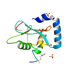
6WQ1
 
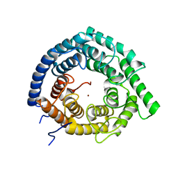 | | Eukaryotic LanCL2 protein | | 分子名称: | LanC-like protein 2, ZINC ION | | 著者 | Nair, S.K, Garg, N. | | 登録日 | 2020-04-28 | | 公開日 | 2021-05-05 | | 最終更新日 | 2023-10-18 | | 実験手法 | X-RAY DIFFRACTION (2.295 Å) | | 主引用文献 | LanCLs add glutathione to dehydroamino acids generated at phosphorylated sites in the proteome.
Cell, 184, 2021
|
|
2RGN
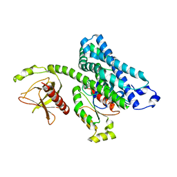
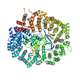
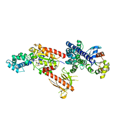 | | Crystal Structure of p63RhoGEF complex with Galpha-q and RhoA | | 分子名称: | GUANOSINE-5'-DIPHOSPHATE, Guanine nucleotide-binding protein G(i) subunit alpha-1,Guanine nucleotide-binding protein G(q) subunit alpha, MAGNESIUM ION, ... | | 著者 | Shankaranarayanan, A, Nance, M.R, Tesmer, J.J.G. | | 登録日 | 2007-10-04 | | 公開日 | 2008-01-15 | | 最終更新日 | 2023-08-30 | | 実験手法 | X-RAY DIFFRACTION (3.5 Å) | | 主引用文献 | Structure of Galphaq-p63RhoGEF-RhoA complex reveals a pathway for the activation of RhoA by GPCRs.
Science, 318, 2007
|
|
6X3A
 | |
6X39
 | |
6X38
 | |
6XJU
 | | Crystal Structure of KPT-8602 bound to CRM1 (E582K, 537-DLTVK-541 to GLCEQ) | | 分子名称: | (2R)-3-{3-[3,5-bis(trifluoromethyl)phenyl]-1H-1,2,4-triazol-1-yl}-2-(pyrimidin-5-yl)propanamide, Exportin-1, GTP-binding nuclear protein Ran, ... | | 著者 | Baumhardt, J.M, Chook, Y.M. | | 登録日 | 2020-06-24 | | 公開日 | 2021-01-27 | | 最終更新日 | 2023-10-18 | | 実験手法 | X-RAY DIFFRACTION (2.193 Å) | | 主引用文献 | Recurrent XPO1 mutations alter pathogenesis of chronic lymphocytic leukemia.
J Hematol Oncol, 14, 2021
|
|
6XVO
 | |
6XX2
 | |
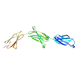
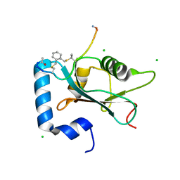
1WJO
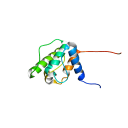 | | Solution structure of the forth CH domain from human plastin 3 T-isoform | | 分子名称: | T-plastin | | 著者 | Tomizawa, T, Kigawa, T, Koshiba, S, Inoue, M, Yokoyama, S, RIKEN Structural Genomics/Proteomics Initiative (RSGI) | | 登録日 | 2004-05-29 | | 公開日 | 2004-11-29 | | 最終更新日 | 2024-05-29 | | 実験手法 | SOLUTION NMR | | 主引用文献 | Solution structure of the forth CH domain from human plastin 3 T-isoform
To be Published
|
|
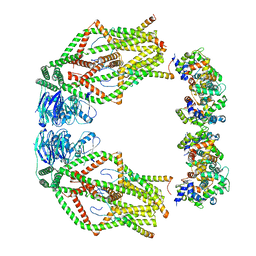
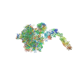
6XU6
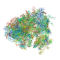 | | Drosophila melanogaster Testis 80S ribosome | | 分子名称: | 18S ribosomal RNA, 28S ribosomal RNA, 2S ribosomal RNA, ... | | 著者 | Hopes, T, Agapiou, M, Norris, K, McCarthy, C.G.P, OConnell, M.J, Fontana, J, Aspden, J.L. | | 登録日 | 2020-01-17 | | 公開日 | 2021-07-28 | | 最終更新日 | 2022-12-21 | | 実験手法 | ELECTRON MICROSCOPY (3.5 Å) | | 主引用文献 | Ribosome heterogeneity in Drosophila melanogaster gonads through paralog-switching.
Nucleic Acids Res., 50, 2022
|
|
3QWX
 | | CED-2 1-174 | | 分子名称: | Cell death abnormality protein 2, SULFATE ION | | 著者 | Kang, Y, Sun, J, Liu, Y. | | 登録日 | 2011-02-28 | | 公開日 | 2011-06-08 | | 最終更新日 | 2024-02-21 | | 実験手法 | X-RAY DIFFRACTION (2.01 Å) | | 主引用文献 | Crystal structure of the cell corpse engulfment protein CED-2 in Caenorhabditis elegans.
Biochem.Biophys.Res.Commun., 410, 2011
|
|
1X86
 | |
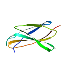
7ZKR
 | | Human GABARAP in complex with stapled peptide Pen3-ortho | | 分子名称: | CHLORIDE ION, Gamma-aminobutyric acid receptor-associated protein, ORTHO-XYLENE, ... | | 著者 | Ueffing, A, Brown, H, Willbold, D, Kritzer, J.A, Weiergraeber, O.H. | | 登録日 | 2022-04-13 | | 公開日 | 2022-08-31 | | 最終更新日 | 2024-01-31 | | 実験手法 | X-RAY DIFFRACTION (1.1 Å) | | 主引用文献 | Structure-Based Design of Stapled Peptides That Bind GABARAP and Inhibit Autophagy.
J.Am.Chem.Soc., 144, 2022
|
|
7ZL7
 | | Human GABARAP in complex with stapled peptide Pen8-ortho | | 分子名称: | CHLORIDE ION, Gamma-aminobutyric acid receptor-associated protein, ORTHO-XYLENE, ... | | 著者 | Ueffing, A, Brown, H, Willbold, D, Kritzer, J.A, Weiergraeber, O.H. | | 登録日 | 2022-04-14 | | 公開日 | 2022-08-31 | | 最終更新日 | 2024-01-31 | | 実験手法 | X-RAY DIFFRACTION (1.55 Å) | | 主引用文献 | Structure-Based Design of Stapled Peptides That Bind GABARAP and Inhibit Autophagy.
J.Am.Chem.Soc., 144, 2022
|
|
2MZN
 | |
6XJS
 | |
6XJV
 | | MCU holocomplex in High-calcium state | | 分子名称: | Calcium uniporter protein, mitochondrial, Calcium uptake protein 1, ... | | 著者 | Wang, Y, Jiang, Y. | | 登録日 | 2020-06-24 | | 公開日 | 2020-09-09 | | 最終更新日 | 2024-03-06 | | 実験手法 | ELECTRON MICROSCOPY (4.17 Å) | | 主引用文献 | Structural insights into the Ca 2+ -dependent gating of the human mitochondrial calcium uniporter.
Elife, 9, 2020
|
|
6YLH
 | |
3S29
 | |
6XX4
 | |
6XJT
 | | Crystal Structure of KPT-8602 bound to CRM1 (537-DLTVK-541 to GLCEQ) | | 分子名称: | (2R)-3-{3-[3,5-bis(trifluoromethyl)phenyl]-1H-1,2,4-triazol-1-yl}-2-(pyrimidin-5-yl)propanamide, Exportin-1, GTP-binding nuclear protein Ran, ... | | 著者 | Baumhardt, J.M, Chook, Y.M. | | 登録日 | 2020-06-24 | | 公開日 | 2021-01-27 | | 最終更新日 | 2023-10-18 | | 実験手法 | X-RAY DIFFRACTION (2.406 Å) | | 主引用文献 | Recurrent XPO1 mutations alter pathogenesis of chronic lymphocytic leukemia.
J Hematol Oncol, 14, 2021
|
|
2W2T
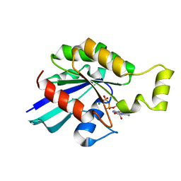 | | Rac2 (G12V) in complex with GDP | | 分子名称: | GUANOSINE-5'-DIPHOSPHATE, MAGNESIUM ION, RAS-RELATED C3 BOTULINUM TOXIN SUBSTRATE 2 | | 著者 | Opaleye, O, Bunney, T.D, Roe, S.M, Pearl, L.H. | | 登録日 | 2008-11-04 | | 公開日 | 2009-05-05 | | 最終更新日 | 2024-05-01 | | 実験手法 | X-RAY DIFFRACTION (1.95 Å) | | 主引用文献 | Structural Insights Into Formation of an Active Signaling Complex between Rac and Phospholipase C Gamma 2.
Mol.Cell, 34, 2009
|
|
2W2V
 | | Rac2 (G12V) in complex with GTPgS | | 分子名称: | GUANOSINE-5'-TRIPHOSPHATE, RAS-RELATED C3 BOTULINUM TOXIN SUBSTRATE 2 | | 著者 | Opaleye, O, Bunney, T.D, Roe, S.M, Pearl, L.H. | | 登録日 | 2008-11-04 | | 公開日 | 2009-05-05 | | 最終更新日 | 2024-05-01 | | 実験手法 | X-RAY DIFFRACTION (2 Å) | | 主引用文献 | Structural Insights Into Formation of an Active Signaling Complex between Rac and Phospholipase C Gamma 2.
Mol.Cell, 34, 2009
|
|
6XVN
 | |
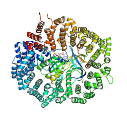
3S28
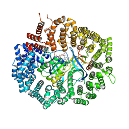
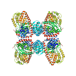
 | | The crystal structure of sucrose synthase-1 in complex with a breakdown product of the UDP-glucose | | 分子名称: | 1,5-anhydro-D-arabino-hex-1-enitol, 1,5-anhydro-D-fructose, MALONIC ACID, ... | | 著者 | Zheng, Y, Garavito, R.M. | | 登録日 | 2011-05-16 | | 公開日 | 2011-08-24 | | 最終更新日 | 2023-09-13 | | 実験手法 | X-RAY DIFFRACTION (2.8 Å) | | 主引用文献 | The Structure of Sucrose Synthase-1 from Arabidopsis thaliana and Its Functional Implications.
J.Biol.Chem., 286, 2011
|
|


