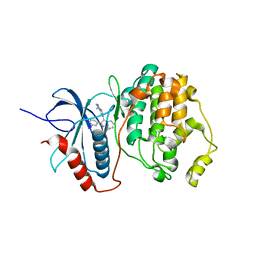
6X93
 
 | | Interleukin-10 signaling complex with IL-10RA and IL-10RB | | 分子名称: | Interleukin-10, Interleukin-10 receptor subunit alpha, Interleukin-10 receptor subunit beta | | 著者 | Saxton, R.A, Tsutsumi, N, Gati, C, Garcia, K.C. | | 登録日 | 2020-06-02 | | 公開日 | 2021-03-17 | | 最終更新日 | 2021-03-31 | | 実験手法 | ELECTRON MICROSCOPY (3.5 Å) | | 主引用文献 | Structure-based decoupling of the pro- and anti-inflammatory functions of interleukin-10.
Science, 371, 2021
|
|
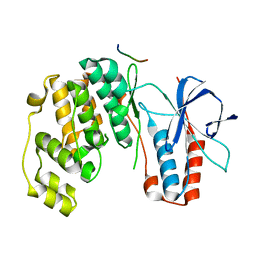
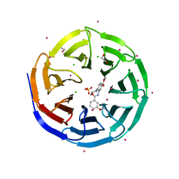
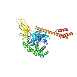
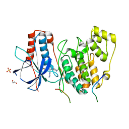
3S7J
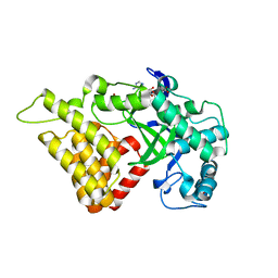 | |
1CQG
 | | HIGH RESOLUTION SOLUTION NMR STRUCTURE OF MIXED DISULFIDE INTERMEDIATE BETWEEN HUMAN THIOREDOXIN (C35A, C62A, C69A, C73A) MUTANT AND A 13 RESIDUE PEPTIDE COMPRISING ITS TARGET SITE IN HUMAN REF-1 (RESIDUES 59-71 OF THE P50 SUBUNIT OF NFKB), NMR, 31 STRUCTURES | | 分子名称: | REF-1 PEPTIDE, THIOREDOXIN | | 著者 | Clore, G.M, Qin, J, Gronenborn, A.M. | | 登録日 | 1996-04-02 | | 公開日 | 1996-08-01 | | 最終更新日 | 2021-11-03 | | 実験手法 | SOLUTION NMR | | 主引用文献 | The solution structure of human thioredoxin complexed with its target from Ref-1 reveals peptide chain reversal.
Structure, 4, 1996
|
|
3DQV
 | | Structural Insights into NEDD8 Activation of Cullin-RING Ligases: Conformational Control of Conjugation | | 分子名称: | Cullin-5, NEDD8, Rbx1, ... | | 著者 | Duda, D.M, Borg, L.A, Scott, D.C, Hunt, H.W, Hammel, M, Schulman, B.A. | | 登録日 | 2008-07-09 | | 公開日 | 2008-09-30 | | 最終更新日 | 2011-07-13 | | 実験手法 | X-RAY DIFFRACTION (3 Å) | | 主引用文献 | Structural insights into NEDD8 activation of cullin-RING ligases: conformational control of conjugation.
Cell(Cambridge,Mass.), 134, 2008
|
|
1TVO
 | | The structure of ERK2 in complex with a small molecule inhibitor | | 分子名称: | 5-(2-PHENYLPYRAZOLO[1,5-A]PYRIDIN-3-YL)-1H-PYRAZOLO[3,4-C]PYRIDAZIN-3-AMINE, Mitogen-activated protein kinase 1 | | 著者 | Kinoshita, T. | | 登録日 | 2004-06-30 | | 公開日 | 2005-09-13 | | 最終更新日 | 2024-03-13 | | 実験手法 | X-RAY DIFFRACTION (2.5 Å) | | 主引用文献 | Identification of a selective ERK inhibitor and structural determination of the inhibitor-ERK2 complex
Biochem.Biophys.Res.Commun., 336, 2005
|
|
4KA3
 | | Structure of MAP kinase in complex with a docking peptide | | 分子名称: | Mitogen-activated protein kinase 14, TGF-beta-activated kinase 1 and MAP3K7-binding protein 1 | | 著者 | Xin, F.J, Wu, J.W. | | 登録日 | 2013-04-22 | | 公開日 | 2013-06-26 | | 最終更新日 | 2023-11-08 | | 実験手法 | X-RAY DIFFRACTION (2.707 Å) | | 主引用文献 | Crystal structure of the p38 alpha MAP kinase in complex with a docking peptide from TAB1
Sci China Life Sci, 56, 2013
|
|
3EMH
 | | Structural basis of WDR5-MLL interaction | | 分子名称: | Mixed-lineage leukemia protein 1, SULFATE ION, WD repeat-containing protein 5 | | 著者 | Song, J.J, Kingston, R.E. | | 登録日 | 2008-09-24 | | 公開日 | 2008-10-07 | | 最終更新日 | 2023-09-06 | | 実験手法 | X-RAY DIFFRACTION (1.37 Å) | | 主引用文献 | WDR5 Interacts with Mixed Lineage Leukemia (MLL) Protein via the Histone H3-binding Pocket.
J.Biol.Chem., 283, 2008
|
|
3UVO
 | | Crystal structure of WDR5 in complex with the WDR5-interacting motif of SET1B | | 分子名称: | Histone-lysine N-methyltransferase SETD1B, WD repeat-containing protein 5 | | 著者 | Zhang, P, Lee, H, Brunzelle, J.S, Couture, J.-F. | | 登録日 | 2011-11-30 | | 公開日 | 2011-12-14 | | 最終更新日 | 2023-09-13 | | 実験手法 | X-RAY DIFFRACTION (2.2 Å) | | 主引用文献 | The plasticity of WDR5 peptide-binding cleft enables the binding of the SET1 family of histone methyltransferases.
Nucleic Acids Res., 40, 2012
|
|
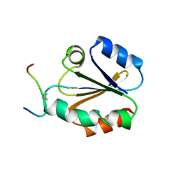
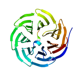
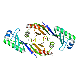
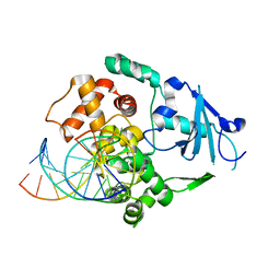
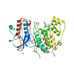
5FQ2
 | |
5EDN
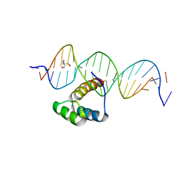 | | Structure of HOXB13-DNA(TCG) complex | | 分子名称: | 2-AMINO-2-HYDROXYMETHYL-PROPANE-1,3-DIOL, DNA (5'-D(P*GP*GP*AP*CP*CP*TP*CP*GP*TP*AP*AP*AP*AP*CP*AP*CP*AP*AP*C)-3'), DNA (5'-D(P*GP*TP*TP*GP*TP*GP*TP*TP*TP*TP*AP*CP*GP*AP*GP*GP*TP*CP*C)-3'), ... | | 著者 | Morgunova, E, Yin, Y, Jolma, A, Popov, A, Taipale, J. | | 登録日 | 2015-10-21 | | 公開日 | 2016-11-09 | | 最終更新日 | 2024-01-10 | | 実験手法 | X-RAY DIFFRACTION (3.2 Å) | | 主引用文献 | Two distinct DNA sequences recognized by transcription factors represent enthalpy and entropy optima.
Elife, 7, 2018
|
|
3UR4
 | | Crystal structure of human WD repeat domain 5 with compound | | 分子名称: | 1,2-ETHANEDIOL, CHLORIDE ION, SULFATE ION, ... | | 著者 | Dong, A, Dombrovski, L, Senisterra, G, Wernimont, A, Wasney, G.A, Allali Hassani, A, Nguyen, K.T, Smil, D, Bolshan, Y, Hajian, T, Poda, G, Chau, I, Al-Awar, R, Bountra, C, Weigelt, J, Edwards, A.M, Arrowsmith, C.H, Brown, P, Schapira, M, Vedadi, M, Wu, H, Structural Genomics Consortium (SGC) | | 登録日 | 2011-11-21 | | 公開日 | 2011-12-14 | | 最終更新日 | 2023-09-13 | | 実験手法 | X-RAY DIFFRACTION (1.8 Å) | | 主引用文献 | Small-molecule inhibition of MLL activity by disruption of its interaction with WDR5.
Biochem. J., 449, 2013
|
|
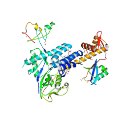
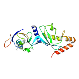
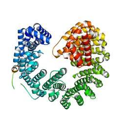
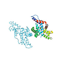
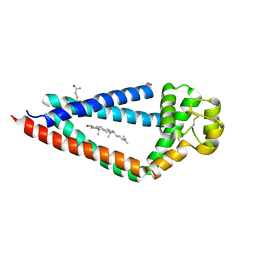
3EIK
 | |
3UVL
 | | Crystal structure of WDR5 in complex with the WDR5-interacting motif of MLL3 | | 分子名称: | Histone-lysine N-methyltransferase MLL3, WD repeat-containing protein 5 | | 著者 | Zhang, P, Lee, H, Brunzelle, J.S, Couture, J.-F. | | 登録日 | 2011-11-30 | | 公開日 | 2011-12-14 | | 最終更新日 | 2023-09-13 | | 実験手法 | X-RAY DIFFRACTION (2.2 Å) | | 主引用文献 | The plasticity of WDR5 peptide-binding cleft enables the binding of the SET1 family of histone methyltransferases.
Nucleic Acids Res., 40, 2012
|
|
5OWU
 | | Kap95:Nup1 complex | | 分子名称: | Importin subunit beta-1, Nucleoporin NUP1 | | 著者 | Stewart, M. | | 登録日 | 2017-09-04 | | 公開日 | 2017-10-25 | | 最終更新日 | 2024-05-08 | | 実験手法 | X-RAY DIFFRACTION (2 Å) | | 主引用文献 | Structural basis for the high-affinity binding of nucleoporin Nup1p to the Saccharomyces cerevisiae importin-beta homologue, Kap95p.
J. Mol. Biol., 349, 2005
|
|
1CQH
 | | HIGH RESOLUTION SOLUTION NMR STRUCTURE OF MIXED DISULFIDE INTERMEDIATE BETWEEN HUMAN THIOREDOXIN (C35A, C62A, C69A, C73A) MUTANT AND A 13 RESIDUE PEPTIDE COMPRISING ITS TARGET SITE IN HUMAN REF-1 (RESIDUES 59-71 OF THE P50 SUBUNIT OF NFKB), NMR, MINIMIZED AVERAGE STRUCTURE | | 分子名称: | REF-1 PEPTIDE, THIOREDOXIN | | 著者 | Clore, G.M, Qin, J, Gronenborn, A.M. | | 登録日 | 1996-04-02 | | 公開日 | 1996-08-01 | | 最終更新日 | 2021-11-03 | | 実験手法 | SOLUTION NMR | | 主引用文献 | The solution structure of human thioredoxin complexed with its target from Ref-1 reveals peptide chain reversal.
Structure, 4, 1996
|
|
3UVN
 | | Crystal structure of WDR5 in complex with the WDR5-interacting motif of SET1A | | 分子名称: | Histone-lysine N-methyltransferase SETD1A, WD repeat-containing protein 5 | | 著者 | Zhang, P, Lee, H, Brunzelle, J.S, Couture, J.-F. | | 登録日 | 2011-11-30 | | 公開日 | 2011-12-14 | | 最終更新日 | 2023-09-13 | | 実験手法 | X-RAY DIFFRACTION (1.792 Å) | | 主引用文献 | The plasticity of WDR5 peptide-binding cleft enables the binding of the SET1 family of histone methyltransferases.
Nucleic Acids Res., 40, 2012
|
|
5E84
 | | ATP-bound state of BiP | | 分子名称: | 78 kDa glucose-regulated protein, ADENOSINE-5'-TRIPHOSPHATE, MAGNESIUM ION, ... | | 著者 | Liu, Q, Yang, J, Nune, M, Zong, Y, Zhou, L. | | 登録日 | 2015-10-13 | | 公開日 | 2016-01-27 | | 最終更新日 | 2023-09-27 | | 実験手法 | X-RAY DIFFRACTION (2.99 Å) | | 主引用文献 | Close and Allosteric Opening of the Polypeptide-Binding Site in a Human Hsp70 Chaperone BiP.
Structure, 23, 2015
|
|
3UVM
 | | Crystal structure of WDR5 in complex with the WDR5-interacting motif of MLL4 | | 分子名称: | Histone-lysine N-methyltransferase MLL4, WD repeat-containing protein 5 | | 著者 | Zhang, P, Lee, H, Brunzelle, J.S, Couture, J.-F. | | 登録日 | 2011-11-30 | | 公開日 | 2011-12-14 | | 最終更新日 | 2023-09-13 | | 実験手法 | X-RAY DIFFRACTION (1.57 Å) | | 主引用文献 | The plasticity of WDR5 peptide-binding cleft enables the binding of the SET1 family of histone methyltransferases.
Nucleic Acids Res., 40, 2012
|
|
1N3C
 | | Structural and biochemical exploration of a critical amino acid in human 8-oxoguanine glycosylase | | 分子名称: | 8-oxoG-containing DNA, CALCIUM ION, DNA complement strand, ... | | 著者 | Norman, D.P, Chung, S.J, Verdine, G.L. | | 登録日 | 2002-10-25 | | 公開日 | 2003-03-04 | | 最終更新日 | 2024-02-14 | | 実験手法 | X-RAY DIFFRACTION (2.7 Å) | | 主引用文献 | Structural and biochemical exploration of a critical amino acid in human 8-oxoguanine glycosylase
Biochemistry, 42, 2003
|
|
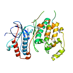
1KXU
 | | CYCLIN H, A POSITIVE REGULATORY SUBUNIT OF CDK ACTIVATING KINASE | | 分子名称: | CYCLIN H | | 著者 | Kim, K.K, Chamberin, H.M, Morgan, D.O, Kim, S.-H. | | 登録日 | 1996-08-08 | | 公開日 | 1997-01-27 | | 最終更新日 | 2024-02-14 | | 実験手法 | X-RAY DIFFRACTION (2.6 Å) | | 主引用文献 | Three-dimensional structure of human cyclin H, a positive regulator of the CDK-activating kinase.
Nat.Struct.Biol., 3, 1996
|
|
4LOO
 | | Structural basis of autoactivation of p38 alpha induced by TAB1 (Monoclinic crystal form) | | 分子名称: | 4-(4-FLUOROPHENYL)-1-(4-PIPERIDINYL)-5-(2-AMINO-4-PYRIMIDINYL)-IMIDAZOLE, Mitogen-activated protein kinase 14, TGF-beta-activated kinase 1 and MAP3K7-binding protein 1 | | 著者 | Chaikuad, A, DeNicola, G.F, Krojer, T, Allerston, C.K, von Delft, F, Arrowsmith, C.H, Edwards, A.M, Bountra, C, Marber, M.S, Knapp, S, Structural Genomics Consortium (SGC) | | 登録日 | 2013-07-13 | | 公開日 | 2013-08-21 | | 最終更新日 | 2023-09-20 | | 実験手法 | X-RAY DIFFRACTION (1.95 Å) | | 主引用文献 | Mechanism and consequence of the autoactivation of p38 alpha mitogen-activated protein kinase promoted by TAB1.
Nat.Struct.Mol.Biol., 20, 2013
|
|
4LOQ
 | | Structural basis of autoactivation of p38 alpha induced by TAB1 (Tetragonal crystal form with bound sulphate) | | 分子名称: | 1,2-ETHANEDIOL, 4-(4-FLUOROPHENYL)-1-(4-PIPERIDINYL)-5-(2-AMINO-4-PYRIMIDINYL)-IMIDAZOLE, Mitogen-activated protein kinase 14, ... | | 著者 | Chaikuad, A, DeNicola, G.F, Yue, W.W, von Delft, F, Arrowsmith, C.H, Edwards, A.M, Bountra, C, Marber, M.S, Knapp, S, Structural Genomics Consortium (SGC) | | 登録日 | 2013-07-13 | | 公開日 | 2013-08-21 | | 最終更新日 | 2023-09-20 | | 実験手法 | X-RAY DIFFRACTION (2.319 Å) | | 主引用文献 | Mechanism and consequence of the autoactivation of p38 alpha mitogen-activated protein kinase promoted by TAB1.
Nat.Struct.Mol.Biol., 20, 2013
|
|
4LOP
 | | Structural basis of autoactivation of p38 alpha induced by TAB1 (Tetragonal crystal form) | | 分子名称: | 1,2-ETHANEDIOL, 4-(4-FLUOROPHENYL)-1-(4-PIPERIDINYL)-5-(2-AMINO-4-PYRIMIDINYL)-IMIDAZOLE, L(+)-TARTARIC ACID, ... | | 著者 | Chaikuad, A, DeNicola, G.F, Krojer, T, Allerston, C.K, von Delft, F, Arrowsmith, C.H, Edwards, A.M, Bountra, C, Marber, M.S, Knapp, S, Structural Genomics Consortium (SGC) | | 登録日 | 2013-07-13 | | 公開日 | 2013-08-21 | | 最終更新日 | 2023-09-20 | | 実験手法 | X-RAY DIFFRACTION (2.049 Å) | | 主引用文献 | Mechanism and consequence of the autoactivation of p38 alpha mitogen-activated protein kinase promoted by TAB1.
Nat.Struct.Mol.Biol., 20, 2013
|
|
5TCX
 | | Crystal structure of human tetraspanin CD81 | | 分子名称: | CD81 antigen, CHOLESTEROL | | 著者 | Zimmerman, B, McMillan, B.J, Seegar, T.C.M, Kruse, A.C, Blacklow, S.C. | | 登録日 | 2016-09-16 | | 公開日 | 2016-11-09 | | 最終更新日 | 2023-10-04 | | 実験手法 | X-RAY DIFFRACTION (2.955 Å) | | 主引用文献 | Crystal Structure of a Full-Length Human Tetraspanin Reveals a Cholesterol-Binding Pocket.
Cell, 167, 2016
|
|
3UVK
 | | Crystal structure of WDR5 in complex with the WDR5-interacting motif of MLL2 | | 分子名称: | 2-[BIS-(2-HYDROXY-ETHYL)-AMINO]-2-HYDROXYMETHYL-PROPANE-1,3-DIOL, Histone-lysine N-methyltransferase MLL2, SULFATE ION, ... | | 著者 | Zhang, P, Lee, H, Brunzelle, J.S, Couture, J.-F. | | 登録日 | 2011-11-30 | | 公開日 | 2011-12-14 | | 最終更新日 | 2023-09-13 | | 実験手法 | X-RAY DIFFRACTION (1.4 Å) | | 主引用文献 | The plasticity of WDR5 peptide-binding cleft enables the binding of the SET1 family of histone methyltransferases.
Nucleic Acids Res., 40, 2012
|
|


