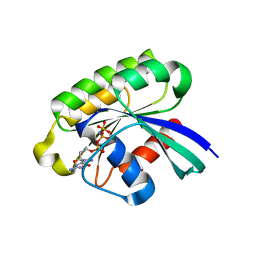
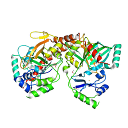
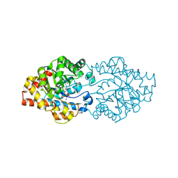
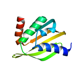
6M1W
 
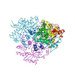 | | Structure of the 2-Aminoisobutyric acid Monooxygenase Hydroxylase | | 分子名称: | Amidohydrolase, CHLORIDE ION, FE (III) ION, ... | | 著者 | Hibi, M, Mikami, B, Ogawa, J. | | 登録日 | 2020-02-26 | | 公開日 | 2021-01-06 | | 最終更新日 | 2024-11-13 | | 実験手法 | X-RAY DIFFRACTION (2.75 Å) | | 主引用文献 | A three-component monooxygenase from Rhodococcus wratislaviensis may expand industrial applications of bacterial enzymes.
Commun Biol, 4, 2021
|
|
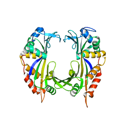
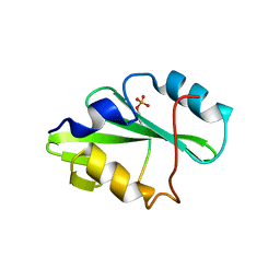
6M2L
 | | Crystal structure of Plasmodium falciparum hexose transporter PfHT1 bound with C3361 | | 分子名称: | (2S,3R,4S,5R,6R)-6-(hydroxymethyl)-4-undec-10-enoxy-oxane-2,3,5-triol, Hexose transporter 1 | | 著者 | Jiang, X, Yuan, Y.Y, Zhang, S, Wang, N, Yan, C.Y, Yan, N. | | 登録日 | 2020-02-27 | | 公開日 | 2020-09-09 | | 最終更新日 | 2024-11-13 | | 実験手法 | X-RAY DIFFRACTION (3.7 Å) | | 主引用文献 | Structural Basis for Blocking Sugar Uptake into the Malaria Parasite Plasmodium falciparum.
Cell, 183, 2020
|
|
3BPQ
 | |
1YZK
 | | GppNHp bound Rab11 GTPase | | 分子名称: | MAGNESIUM ION, PHOSPHOAMINOPHOSPHONIC ACID-GUANYLATE ESTER, Ras-related protein Rab-11A | | 著者 | Eathiraj, S, Pan, X, Ritacco, C, Lambright, D.G. | | 登録日 | 2005-02-28 | | 公開日 | 2005-07-26 | | 最終更新日 | 2024-04-03 | | 実験手法 | X-RAY DIFFRACTION (2 Å) | | 主引用文献 | Structural basis of family-wide Rab GTPase recognition by rabenosyn-5.
Nature, 436, 2005
|
|
1YZU
 | | GppNHp-Bound Rab21 GTPase at 2.50 A Resolution | | 分子名称: | MAGNESIUM ION, PHOSPHOAMINOPHOSPHONIC ACID-GUANYLATE ESTER, Ras-related protein Rab-21 | | 著者 | Eathiraj, S, Pan, X, Ritacco, C, Lambright, D.G. | | 登録日 | 2005-02-28 | | 公開日 | 2005-07-26 | | 最終更新日 | 2024-04-03 | | 実験手法 | X-RAY DIFFRACTION (2.5 Å) | | 主引用文献 | Structural basis of family-wide Rab GTPase recognition by rabenosyn-5.
Nature, 436, 2005
|
|
4D2I
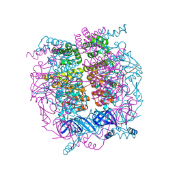 | | Crystal structure of the HerA hexameric DNA translocase from Sulfolobus solfataricus bound to AMP-PNP | | 分子名称: | HERA, MAGNESIUM ION, PHOSPHOAMINOPHOSPHONIC ACID-ADENYLATE ESTER | | 著者 | Rzechorzek, N.J, Blackwood, J.K, Bray, S.M, Maman, J.D, Pellegrini, L, Robinson, N.P. | | 登録日 | 2014-05-09 | | 公開日 | 2014-12-03 | | 最終更新日 | 2024-05-08 | | 実験手法 | X-RAY DIFFRACTION (2.841 Å) | | 主引用文献 | Structure of the Hexameric Hera ATPase Reveals a Mechanism of Translocation-Coupled DNA-End Processing in Archaea
Nat.Commun., 5, 2014
|
|
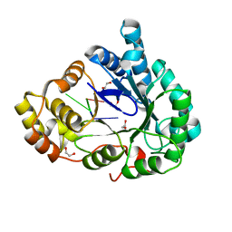
3BV7
 | | Crystal structure of Delta(4)-3-ketosteroid 5-beta-reductase in complex with NADP and glycerol. Resolution: 1.79 A. | | 分子名称: | 3-oxo-5-beta-steroid 4-dehydrogenase, GLYCEROL, NADP NICOTINAMIDE-ADENINE-DINUCLEOTIDE PHOSPHATE | | 著者 | Di Costanzo, L, Drury, J, Penning, T.M, Christianson, D.W. | | 登録日 | 2008-01-04 | | 公開日 | 2008-04-01 | | 最終更新日 | 2023-08-30 | | 実験手法 | X-RAY DIFFRACTION (1.79 Å) | | 主引用文献 | Crystal Structure of Human Liver {Delta}4-3-Ketosteroid 5{beta}-Reductase (AKR1D1) and Implications for Substrate Binding and Catalysis.
J.Biol.Chem., 283, 2008
|
|
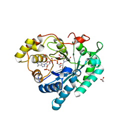
1PJ6
 | | Crystal structure of dimethylglycine oxidase of Arthrobacter globiformis in complex with folic acid | | 分子名称: | FLAVIN-ADENINE DINUCLEOTIDE, FOLIC ACID, N,N-dimethylglycine oxidase, ... | | 著者 | Leys, D, Basran, J, Scrutton, N.S. | | 登録日 | 2003-06-01 | | 公開日 | 2003-10-07 | | 最終更新日 | 2024-10-16 | | 実験手法 | X-RAY DIFFRACTION (1.65 Å) | | 主引用文献 | Channelling and formation of 'active' formaldehyde in dimethylglycine oxidase.
Embo J., 22, 2003
|
|
3BVQ
 | | Crystal Structure of Apo NotI Restriction Endonuclease | | 分子名称: | FE (III) ION, NotI restriction endonuclease, SULFATE ION | | 著者 | Lambert, A.R, Sussman, D, Shen, B, Stoddard, B.L. | | 登録日 | 2008-01-07 | | 公開日 | 2008-01-22 | | 最終更新日 | 2024-10-30 | | 実験手法 | X-RAY DIFFRACTION (2.8 Å) | | 主引用文献 | Structures of the Rare-Cutting Restriction Endonuclease NotI Reveal a Unique Metal Binding Fold Involved in DNA Binding.
Structure, 16, 2008
|
|
6M7K
 | | Structure of mouse RECON (AKR1C13) in complex with cyclic AMP-AMP-GMP (cAAG) | | 分子名称: | 1,2-ETHANEDIOL, Aldo-keto reductase family 1 member C13, cyclic AMP-AMP-GMP | | 著者 | Eaglesham, J.B, Whiteley, A.T, de Oliveira Mann, C.C, Morehouse, B.R, Nieminen, E.A, King, D.S, Lee, A.S.Y, Mekalanos, J.J, Kranzusch, P.J. | | 登録日 | 2018-08-20 | | 公開日 | 2019-02-20 | | 最終更新日 | 2023-10-11 | | 実験手法 | X-RAY DIFFRACTION (1.1 Å) | | 主引用文献 | Bacterial cGAS-like enzymes synthesize diverse nucleotide signals.
Nature, 567, 2019
|
|
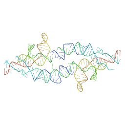
7JKU
 | | Crystal structure of a four-tetrad, parallel, and K+ stabilized Tetrahymena thermophila telomeric G-quadruplex | | 分子名称: | 26-mer DNA, POTASSIUM ION | | 著者 | Yatsunyk, L.A, McCarthy, S.E, Beseiso, D, Gallagher, E.P, Chen, E.V, Miao, J. | | 登録日 | 2020-07-28 | | 公開日 | 2021-07-07 | | 最終更新日 | 2024-04-03 | | 実験手法 | X-RAY DIFFRACTION (1.97 Å) | | 主引用文献 | The first crystal structures of hybrid and parallel four-tetrad intramolecular G-quadruplexes.
Nucleic Acids Res., 50, 2022
|
|
2ID9
 | | 1.85 A Structure of T87I/Y106W Phosphono-CheY | | 分子名称: | Chemotaxis protein cheY | | 著者 | Halkides, C.J, Haas, R.M, McAdams, K.A, Casper, E.S, Santarsiero, B.D, Mesecar, A.D. | | 登録日 | 2006-09-14 | | 公開日 | 2007-09-25 | | 最終更新日 | 2023-08-30 | | 実験手法 | X-RAY DIFFRACTION (1.75 Å) | | 主引用文献 | The structures of T87I phosphono-CheY and T87I/Y106W phosphono-CheY help to explain their binding affinities to the FliM and CheZ peptides.
Arch.Biochem.Biophys., 479, 2008
|
|
3SO7
 | | Organophoshatedegrading enzyme (OpdA)-phosphate complex | | 分子名称: | COBALT (II) ION, PHOSPHATE ION, Phosphotriesterase, ... | | 著者 | Ely, F, Pedroso, M, Gahan, L.R, Ollis, D.L, Guddat, L.W, Schenk, G. | | 登録日 | 2011-06-30 | | 公開日 | 2011-12-07 | | 最終更新日 | 2025-03-26 | | 実験手法 | X-RAY DIFFRACTION (2.2 Å) | | 主引用文献 | Phosphate-bound structure of an organophosphate-degrading enzyme from Agrobacterium radiobacter.
J.Inorg.Biochem., 106, 2011
|
|
4CIJ
 | |
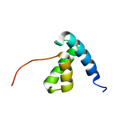
1PAN
 | | A COMPARISON OF NMR SOLUTION STRUCTURES OF THE RECEPTOR BINDING DOMAINS OF PSEUDOMONAS AERUGINOSA PILI STRAINS PAO, KB7, AND PAK: IMPLICATIONS FOR RECEPTOR BINDING AND SYNTHETIC VACCINE DESIGN | | 分子名称: | PAO PILIN, TRANS | | 著者 | Campbell, A.P, Mcinnes, C, Hodges, R.S, Sykes, B.D. | | 登録日 | 1995-10-05 | | 公開日 | 1996-01-29 | | 最終更新日 | 2024-10-30 | | 実験手法 | SOLUTION NMR | | 主引用文献 | Comparison of NMR solution structures of the receptor binding domains of Pseudomonas aeruginosa pili strains PAO, KB7, and PAK: implications for receptor binding and synthetic vaccine design.
Biochemistry, 34, 1995
|
|
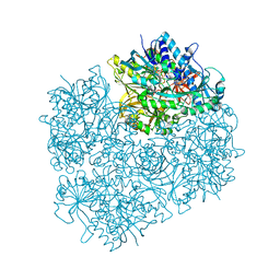
3S8A
 | | Structure of Yeast Ribonucleotide Reductase R293A with dGTP | | 分子名称: | 2'-DEOXYGUANOSINE-5'-TRIPHOSPHATE, MAGNESIUM ION, Ribonucleoside-diphosphate reductase large chain 1 | | 著者 | Ahmad, M.F, Kaushal, P.S, Wan, Q, Wijeratna, S.R, Huang, M, Dealwis, C.D. | | 登録日 | 2011-05-27 | | 公開日 | 2012-04-11 | | 最終更新日 | 2024-02-28 | | 実験手法 | X-RAY DIFFRACTION (2.9 Å) | | 主引用文献 | Structural and biochemical basis of lethal mutant R293A of yeast ribonucleotide reductase
To be Published
|
|
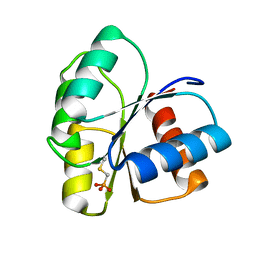
5TPJ
 | | Crystal structure of a de novo designed protein with curved beta-sheet | | 分子名称: | denovo NTF2 | | 著者 | Basanta, B, Oberdorfer, G, Marcos, E, Chidyausiku, T.M, Sankaran, B, Baker, D. | | 登録日 | 2016-10-20 | | 公開日 | 2017-01-25 | | 最終更新日 | 2024-03-06 | | 実験手法 | X-RAY DIFFRACTION (3.101 Å) | | 主引用文献 | Principles for designing proteins with cavities formed by curved beta sheets.
Science, 355, 2017
|
|
5TQS
 | |
3DDC
 | | Crystal Structure of NORE1A in Complex with RAS | | 分子名称: | GTPase HRas, MAGNESIUM ION, PHOSPHOAMINOPHOSPHONIC ACID-GUANYLATE ESTER, ... | | 著者 | Stieglitz, B, Bee, C, Schwarz, D, Yildiz, O, Moshnikova, A, Khokhlatchev, A, Herrmann, C. | | 登録日 | 2008-06-05 | | 公開日 | 2008-07-15 | | 最終更新日 | 2023-11-01 | | 実験手法 | X-RAY DIFFRACTION (1.8 Å) | | 主引用文献 | Novel type of Ras effector interaction established between tumour suppressor NORE1A and Ras switch II
Embo J., 27, 2008
|
|
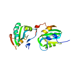
1PDG
 | | CRYSTAL STRUCTURE OF HUMAN PLATELET-DERIVED GROWTH FACTOR BB | | 分子名称: | PLATELET-DERIVED GROWTH FACTOR BB | | 著者 | Oefner, C, Darcy, A.D, Winkler, F.K, Eggimann, B, Hosnag, M. | | 登録日 | 1992-07-14 | | 公開日 | 1994-01-31 | | 最終更新日 | 2024-11-13 | | 実験手法 | X-RAY DIFFRACTION (3 Å) | | 主引用文献 | Crystal structure of human platelet-derived growth factor BB.
EMBO J., 11, 1992
|
|
2IL9
 | |
2KTA
 | | Solution NMR structure of a domain of protein A6KY75 from Bacteroides vulgatus, Northeast Structural Genomics target BvR106A | | 分子名称: | Putative helicase | | 著者 | Mills, J.L, Sukumaran, D.K, Sathyamoorthy, B, Belote, R.L, Ciccosanti, C, Hamilton, K, Acton, T.B, Xiao, R, Swapna, G.V.T, Everett, J.K, Montelione, G.T, Szyperski, T, Northeast Structural Genomics Consortium (NESG) | | 登録日 | 2010-01-26 | | 公開日 | 2010-06-16 | | 最終更新日 | 2024-05-01 | | 実験手法 | SOLUTION NMR | | 主引用文献 | Solution NMR structure of a domain of protein A6KY75 from Bacteroides vulgatus, Northeast Structural Genomics target BvR106A
To be Published
|
|
2ITJ
 | |
4PHJ
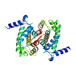 | | The Structural Basis of Differential Inhibition of Human Calpain by Indole and Phenyl alpha-Mercaptoacrylic Acids: Human unliganded protein | | 分子名称: | CALCIUM ION, Calpain small subunit 1 | | 著者 | Adams, S.E, Rizkallah, P.J, Allemann, R.K, Miller, D.J, Hallett, M.B, Robinson, E. | | 登録日 | 2014-05-06 | | 公開日 | 2014-08-13 | | 最終更新日 | 2023-12-20 | | 実験手法 | X-RAY DIFFRACTION (1.6 Å) | | 主引用文献 | The structural basis of differential inhibition of human calpain by indole and phenyl alpha-mercaptoacrylic acids.
J.Struct.Biol., 187, 2014
|
|
3SKT
 | | Crystal structure of the 2'- Deoxyguanosine riboswitch bound to 2'- Deoxyguanosine, manganese Soak | | 分子名称: | 2'-DEOXY-GUANOSINE, MAGNESIUM ION, MANGANESE (II) ION, ... | | 著者 | Pikovskaya, O, Polonskaia, A, Patel, D.J, Serganov, A. | | 登録日 | 2011-06-23 | | 公開日 | 2011-08-17 | | 最終更新日 | 2024-02-28 | | 実験手法 | X-RAY DIFFRACTION (3.1 Å) | | 主引用文献 | Structural principles of nucleoside selectivity in a 2'-deoxyguanosine riboswitch.
Nat.Chem.Biol., 7, 2011
|
|


