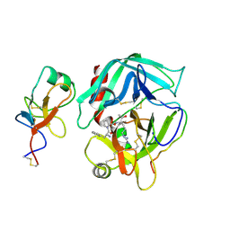
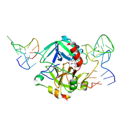
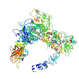
8UF7
 
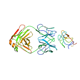 | | Cryo-EM structure of POmAb, a Type-I anti-prothrombin antiphospholipid antibody, bound to kringle-1 of human prothrombin | | 分子名称: | POmAb Heavy Chain, POmAb Light Chain, Prothrombin | | 著者 | Kumar, S, Summers, B, Basore, K, Pozzi, N. | | 登録日 | 2023-10-03 | | 公開日 | 2024-02-14 | | 最終更新日 | 2024-05-22 | | 実験手法 | ELECTRON MICROSCOPY (3.2 Å) | | 主引用文献 | Cryo-EM structure and functional basis of prothrombin recognition by a type I antiprothrombin antiphospholipid antibody.
Blood, 143, 2024
|
|
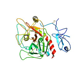
5EDK
 | | Crystal structure of prothrombin deletion mutant residues 146-167 ( Form II ). | | 分子名称: | 2-acetamido-2-deoxy-beta-D-glucopyranose, 2-acetamido-2-deoxy-beta-D-glucopyranose-(1-4)-2-acetamido-2-deoxy-beta-D-glucopyranose, MAGNESIUM ION, ... | | 著者 | Pozzi, N, Chen, Z, Di Cera, E. | | 登録日 | 2015-10-21 | | 公開日 | 2016-01-20 | | 最終更新日 | 2023-11-15 | | 実験手法 | X-RAY DIFFRACTION (3.214 Å) | | 主引用文献 | How the Linker Connecting the Two Kringles Influences Activation and Conformational Plasticity of Prothrombin.
J.Biol.Chem., 291, 2016
|
|
6P9U
 | | Crystal structure of human thrombin mutant W215A | | 分子名称: | 2-acetamido-2-deoxy-beta-D-glucopyranose, Prothrombin, ZINC ION | | 著者 | Pelc, L.A, Koester, S.K, Chen, Z, Di Cera, E. | | 登録日 | 2019-06-10 | | 公開日 | 2019-09-04 | | 最終更新日 | 2024-10-09 | | 実験手法 | X-RAY DIFFRACTION (3.3 Å) | | 主引用文献 | Residues W215, E217 and E192 control the allosteric E*-E equilibrium of thrombin.
Sci Rep, 9, 2019
|
|
2HPP
 | |
2HPQ
 | |
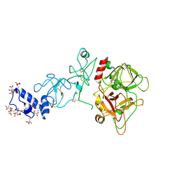
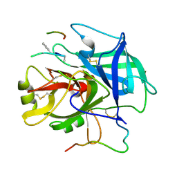
4HZH
 | | Structure of recombinant Gla-domainless prothrombin mutant S525A | | 分子名称: | 2-acetamido-2-deoxy-beta-D-glucopyranose, Prothrombin | | 著者 | Pozzi, N, Niu, W, Gohara, D.W, Chen, Z, Di Cera, E. | | 登録日 | 2012-11-15 | | 公開日 | 2013-06-26 | | 最終更新日 | 2023-09-20 | | 実験手法 | X-RAY DIFFRACTION (3.3 Å) | | 主引用文献 | Crystal structure of prothrombin reveals conformational flexibility and mechanism of activation.
J.Biol.Chem., 288, 2013
|
|
4O03
 | | Crystal structure of Ca2+ bound prothrombin deletion mutant residues 146-167 | | 分子名称: | 2-acetamido-2-deoxy-beta-D-glucopyranose, CALCIUM ION, Prothrombin | | 著者 | Pozzi, N, Chen, Z, Shropshire, D.B, Pelc, L.A, Di Cera, E. | | 登録日 | 2013-12-13 | | 公開日 | 2014-05-21 | | 最終更新日 | 2023-12-06 | | 実験手法 | X-RAY DIFFRACTION (3.38 Å) | | 主引用文献 | The linker connecting the two kringles plays a key role in prothrombin activation.
Proc.Natl.Acad.Sci.USA, 111, 2014
|
|
1NRQ
 | |
5EW2
 | | Human thrombin sandwiched between two DNA aptamers: HD22 and HD1-deltaT12 | | 分子名称: | 2-acetamido-2-deoxy-beta-D-glucopyranose, D-phenylalanyl-N-[(2S,3S)-6-{[amino(iminio)methyl]amino}-1-chloro-2-hydroxyhexan-3-yl]-L-prolinamide, HD1-deltaT12, ... | | 著者 | Pica, A, Russo Krauss, I, Parente, V, Sica, F. | | 登録日 | 2015-11-20 | | 公開日 | 2016-11-30 | | 最終更新日 | 2020-07-29 | | 実験手法 | X-RAY DIFFRACTION (3.59 Å) | | 主引用文献 | Through-bond effects in the ternary complexes of thrombin sandwiched by two DNA aptamers.
Nucleic Acids Res., 45, 2017
|
|
2A45
 | | Crystal structure of the complex between thrombin and the central "E" region of fibrin | | 分子名称: | D-phenylalanyl-N-[(2S,3S)-6-{[amino(iminio)methyl]amino}-1-chloro-2-hydroxyhexan-3-yl]-L-prolinamide, Fibrinogen alpha chain, Fibrinogen beta chain, ... | | 著者 | Pechik, I, Madrazo, J, Gilliland, G.L, Medved, L. | | 登録日 | 2005-06-27 | | 公開日 | 2006-05-02 | | 最終更新日 | 2023-08-23 | | 実験手法 | X-RAY DIFFRACTION (3.65 Å) | | 主引用文献 | Structural basis for sequential cleavage of fibrinopeptides upon fibrin assembly.
Biochemistry, 45, 2006
|
|
7TPP
 | |
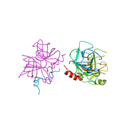
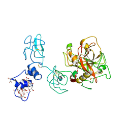
6C2W
 | | Crystal structure of human prothrombin mutant S101C/A470C | | 分子名称: | 2-acetamido-2-deoxy-beta-D-glucopyranose, MAGNESIUM ION, Prothrombin, ... | | 著者 | Chinnaraj, M, Chen, Z, Pelc, L, Grese, Z, Bystranowska, D, Di Cera, E, Pozzi, N. | | 登録日 | 2018-01-09 | | 公開日 | 2018-02-28 | | 最終更新日 | 2023-11-15 | | 実験手法 | X-RAY DIFFRACTION (4.12 Å) | | 主引用文献 | Structure of prothrombin in the closed form reveals new details on the mechanism of activation.
Sci Rep, 8, 2018
|
|
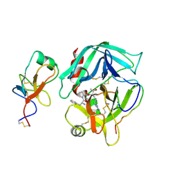
6BJR
 | | Crystal structure of prothrombin mutant S101C/A470C | | 分子名称: | 2-acetamido-2-deoxy-beta-D-glucopyranose, MAGNESIUM ION, Prothrombin, ... | | 著者 | Chinnaraj, M, Chen, Z, Pelc, L, Grese, Z, Bystranowska, D, Di Cera, E, Pozzi, N. | | 登録日 | 2017-11-06 | | 公開日 | 2018-06-27 | | 最終更新日 | 2023-11-15 | | 実験手法 | X-RAY DIFFRACTION (6 Å) | | 主引用文献 | Structure of prothrombin in the closed form reveals new details on the mechanism of activation.
Sci Rep, 8, 2018
|
|
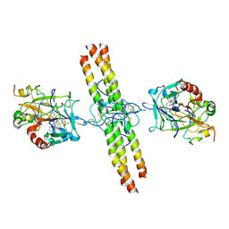
9CTH
 | | Preliminary map of the Prothrombin-prothrombinase complex on nano discs | | 分子名称: | 2-acetamido-2-deoxy-beta-D-glucopyranose, Activated Factor V (FVa) heavy chain, Activated Factor V (FVa) light chain, ... | | 著者 | Stojanovski, B.M, Mohammed, B.M, Di Cera, E. | | 登録日 | 2024-07-25 | | 公開日 | 2024-08-07 | | 最終更新日 | 2024-08-14 | | 実験手法 | ELECTRON MICROSCOPY (6.47 Å) | | 主引用文献 | The Prothrombin-Prothrombinase Interaction.
Subcell Biochem, 104, 2024
|
|
5Z5W
 | |
8BWW
 | |
5Z5X
 | |


