Loading
PDBj
MenuPDBj@FacebookPDBj@TwitterPDBj@YouTubewwPDB FoundationwwPDB
RCSB PDBPDBeBMRBAdv. SearchSearch help
PDB: 142 results

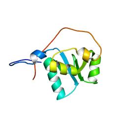
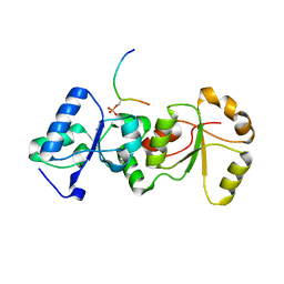
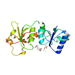
3JVE
DownloadVisualize
BU of 3jve by Molmil
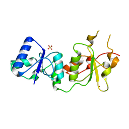
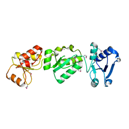
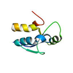
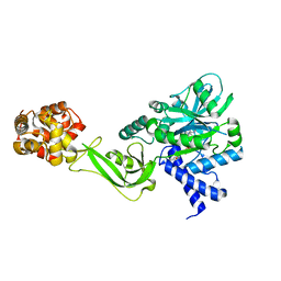
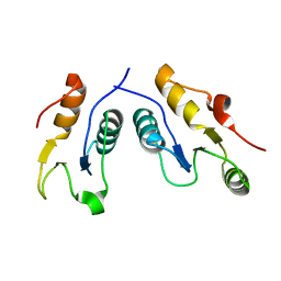
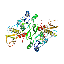
Crystal Structure of the Sixth BRCT Domain of TopBP1
Descriptor: DNA topoisomerase 2-binding protein 1
Authors:Leung, C.C, Kellogg, E, Baker, D, Glover, J.N.M.
Deposit date:2009-09-16
Release date:2010-01-19
Last modified:2024-02-21
Method:X-RAY DIFFRACTION (1.34 Å)
Cite:Insights from the crystal structure of the sixth BRCT domain of topoisomerase IIbeta binding protein 1.
Protein Sci., 19, 2010
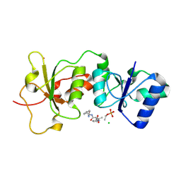
3K16
DownloadVisualize
BU of 3k16 by Molmil
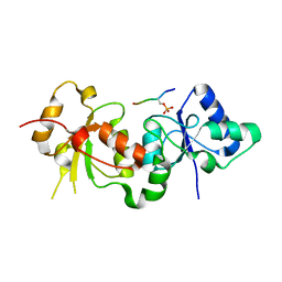
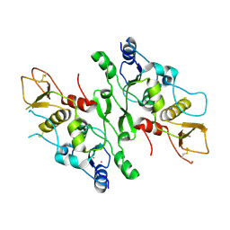
Crystal Structure of BRCA1 BRCT D1840T in complex with a minimal recognition tetrapeptide with a free carboxy C-terminus
Descriptor: Breast cancer type 1 susceptibility protein, CHLORIDE ION, NICKEL (II) ION, ...
Authors:Campbell, S.J, Edwards, R.A, Glover, J.N.
Deposit date:2009-09-25
Release date:2010-03-02
Last modified:2021-10-13
Method:X-RAY DIFFRACTION (3 Å)
Cite:Comparison of the Structures and Peptide Binding Specificities of the BRCT Domains of MDC1 and BRCA1
Structure, 18, 2010
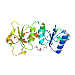
3K0H
DownloadVisualize
BU of 3k0h by Molmil
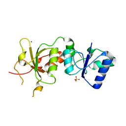
The crystal structure of BRCA1 BRCT in complex with a minimal recognition tetrapeptide with an amidated C-terminus
Descriptor: Breast cancer type 1 susceptibility protein, CHLORIDE ION, NICKEL (II) ION, ...
Authors:Campbell, S.J, Edwards, R.A, Glover, J.N.
Deposit date:2009-09-24
Release date:2010-03-02
Last modified:2017-11-01
Method:X-RAY DIFFRACTION (2.7 Å)
Cite:Comparison of the Structures and Peptide Binding Specificities of the BRCT Domains of MDC1 and BRCA1
Structure, 18, 2010
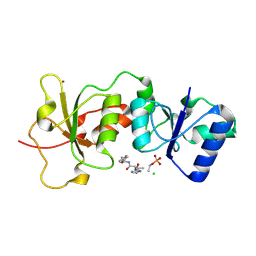
3K15
DownloadVisualize
BU of 3k15 by Molmil
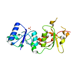
Crystal Structure of BRCA1 BRCT D1840T in complex with a minimal recognition tetrapeptide with an amidated C-terminus
Descriptor: Breast cancer type 1 susceptibility protein, CHLORIDE ION, NICKEL (II) ION, ...
Authors:Campbell, S.J, Edwards, R.A, Glover, J.N.
Deposit date:2009-09-25
Release date:2010-03-02
Last modified:2021-10-13
Method:X-RAY DIFFRACTION (2.8 Å)
Cite:Comparison of the Structures and Peptide Binding Specificities of the BRCT Domains of MDC1 and BRCA1
Structure, 18, 2010
3K0K
DownloadVisualize
BU of 3k0k by Molmil
Crystal Structure of BRCA1 BRCT in complex with a minimal recognition tetrapeptide with a free carboxy C-terminus.
Descriptor: Breast cancer type 1 susceptibility protein, CHLORIDE ION, NICKEL (II) ION, ...
Authors:Campbell, S.J, Edwards, R.A, Glover, J.N.
Deposit date:2009-09-24
Release date:2010-03-02
Last modified:2017-11-01
Method:X-RAY DIFFRACTION (2.7 Å)
Cite:Comparison of the Structures and Peptide Binding Specificities of the BRCT Domains of MDC1 and BRCA1
Structure, 18, 2010
3L46
DownloadVisualize
BU of 3l46 by Molmil
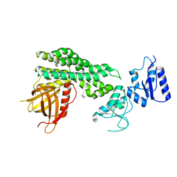
Crystal structure of the second BRCT domain of epithelial cell transforming 2 (ECT2)
Descriptor: Protein ECT2, UNKNOWN ATOM OR ION
Authors:Nedyalkova, L, Tempel, W, Tong, Y, Crombet, L, Wang, H, Guan, X, Arrowsmith, C.H, Edwards, A.M, Bountra, C, Weigelt, J, Bochkarev, A, Park, H, Structural Genomics Consortium (SGC)
Deposit date:2009-12-18
Release date:2010-01-12
Last modified:2023-09-06
Method:X-RAY DIFFRACTION (1.482 Å)
Cite:Crystal structure of the second BRCT domain of epithelial cell transforming 2 (ECT2)
to be published
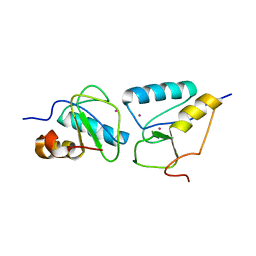
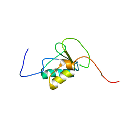
2D8M
DownloadVisualize
BU of 2d8m by Molmil
Solution structure of the first BRCT domain of DNA-repair protein XRCC1
Descriptor: DNA-repair protein XRCC1
Authors:Nagashima, T, Hayashi, F, Yokoyama, S, RIKEN Structural Genomics/Proteomics Initiative (RSGI)
Deposit date:2005-12-06
Release date:2006-06-06
Last modified:2024-05-29
Method:SOLUTION NMR
Cite:Solution structure of the first BRCT domain of DNA-repair protein XRCC1
To be published
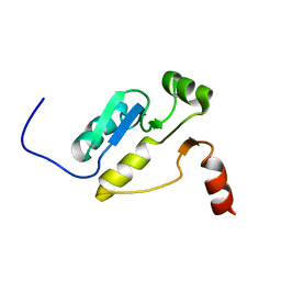
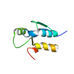
2COU
DownloadVisualize
BU of 2cou by Molmil
Solution structure of the second BRCT domain of epithelial cell transforming 2
Descriptor: ECT2 protein
Authors:Nagashima, T, Hayashi, F, Yokoyama, S, RIKEN Structural Genomics/Proteomics Initiative (RSGI)
Deposit date:2005-05-18
Release date:2005-11-18
Last modified:2024-05-29
Method:SOLUTION NMR
Cite:Solution structure of the second BRCT domain of epithelial cell transforming 2
To be published
2E2W
DownloadVisualize
BU of 2e2w by Molmil
Solution structure of the first BRCT domain of human DNA ligase IV
Descriptor: DNA ligase 4
Authors:Nagashima, T, Hayashi, F, Yokoyama, S, RIKEN Structural Genomics/Proteomics Initiative (RSGI)
Deposit date:2006-11-17
Release date:2006-12-05
Last modified:2024-05-29
Method:SOLUTION NMR
Cite:Solution structure of the first BRCT domain of human DNA ligase IV
TO BE PUBLISHED
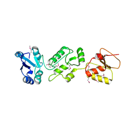
3PXA
DownloadVisualize
BU of 3pxa by Molmil
Impact of BRCA1 BRCT domain missense substitutions on phospho-peptide recognition: G1656D
Descriptor: Breast cancer type 1 susceptibility protein, NICKEL (II) ION, SULFATE ION
Authors:Coquelle, N, Green, R, Glover, J.N.M.
Deposit date:2010-12-09
Release date:2011-04-20
Last modified:2023-09-13
Method:X-RAY DIFFRACTION (2.55 Å)
Cite:Impact of BRCA1 BRCT Domain Missense Substitutions on Phosphopeptide Recognition.
Biochemistry, 50, 2011
3PXD
DownloadVisualize
BU of 3pxd by Molmil
Impact of BRCA1 BRCT domain missense substitutions on phospho-peptide recognition: R1835P
Descriptor: Breast cancer type 1 susceptibility protein, NICKEL (II) ION, SULFATE ION
Authors:Coquelle, N, Green, R, Glover, J.N.M.
Deposit date:2010-12-09
Release date:2011-04-20
Last modified:2023-09-13
Method:X-RAY DIFFRACTION (2.8 Å)
Cite:Impact of BRCA1 BRCT Domain Missense Substitutions on Phosphopeptide Recognition.
Biochemistry, 50, 2011
3PXE
DownloadVisualize
BU of 3pxe by Molmil
Impact of BRCA1 BRCT domain missense substitutions on phospho-peptide recognition: E1836K
Descriptor: Breast cancer type 1 susceptibility protein, phospho peptide
Authors:Coquelle, N, Green, R, Glover, J.N.M.
Deposit date:2010-12-09
Release date:2011-04-20
Last modified:2023-09-13
Method:X-RAY DIFFRACTION (2.85 Å)
Cite:Impact of BRCA1 BRCT Domain Missense Substitutions on Phosphopeptide Recognition.
Biochemistry, 50, 2011
3PXB
DownloadVisualize
BU of 3pxb by Molmil
Impact of BRCA1 BRCT domain missense substitutions on phospho-peptide recognition: T1700A
Descriptor: Breast cancer type 1 susceptibility protein, NICKEL (II) ION, SULFATE ION
Authors:Coquelle, N, Green, R, Glover, J.N.M.
Deposit date:2010-12-09
Release date:2011-04-20
Last modified:2023-09-13
Method:X-RAY DIFFRACTION (2.5 Å)
Cite:Impact of BRCA1 BRCT Domain Missense Substitutions on Phosphopeptide Recognition.
Biochemistry, 50, 2011
3PXC
DownloadVisualize
BU of 3pxc by Molmil
Impact of BRCA1 BRCT domain missense substitutions on phospho-peptide recognition: R1699Q
Descriptor: Breast cancer type 1 susceptibility protein, NICKEL (II) ION, SULFATE ION
Authors:Coquelle, N, Green, R, Glover, J.N.M.
Deposit date:2010-12-09
Release date:2011-04-20
Last modified:2023-09-13
Method:X-RAY DIFFRACTION (2.8 Å)
Cite:Impact of BRCA1 BRCT Domain Missense Substitutions on Phosphopeptide Recognition.
Biochemistry, 50, 2011
3OLC
DownloadVisualize
BU of 3olc by Molmil
Crystal structure of the N-terminal region of TopBP1
Descriptor: DNA topoisomerase 2-binding protein 1
Authors:Huo, Y.G, Bai, L, Xu, M, Jiang, T.
Deposit date:2010-08-25
Release date:2011-03-30
Last modified:2011-07-13
Method:X-RAY DIFFRACTION (2.4 Å)
Cite:Crystal structure of the N-terminal region of human Topoisomerase II beta binding protein 1
Biochem.Biophys.Res.Commun., 401, 2010
3PD7
DownloadVisualize
BU of 3pd7 by Molmil
Crystal Structure of the Sixth BRCT Domain of Human TopBP1
Descriptor: DNA topoisomerase 2-binding protein 1
Authors:Lee, J, Xu, C, Cui, G, Thompson, J.R, Mer, G.
Deposit date:2010-10-22
Release date:2010-12-08
Last modified:2024-02-21
Method:X-RAY DIFFRACTION (1.26 Å)
Cite:Crystal Structure of the Sixth BRCT Domain of Human TopBP1
To be Published
5TT5
DownloadVisualize
BU of 5tt5 by Molmil
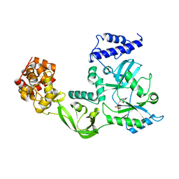
Escherichia coli LigA (K115M) in complex with NAD+
Descriptor: DNA ligase, MAGNESIUM ION, NICOTINAMIDE-ADENINE-DINUCLEOTIDE, ...
Authors:Goldgur, Y, Unciuleac, M.-C, Shuman, S.H.
Deposit date:2016-11-01
Release date:2017-03-08
Last modified:2023-10-04
Method:X-RAY DIFFRACTION (1.552 Å)
Cite:Two-metal versus one-metal mechanisms of lysine adenylylation by ATP-dependent and NAD(+)-dependent polynucleotide ligases.
Proc. Natl. Acad. Sci. U.S.A., 114, 2017
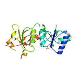
6WH1
DownloadVisualize
BU of 6wh1 by Molmil
Structure of the complex of human DNA ligase III-alpha and XRCC1 BRCT domains
Descriptor: DNA ligase 3 alpha, X-ray repair cross complementing protein 1 variant
Authors:Pourfarjam, Y, Ellenberger, T, Tainer, J.A, Tomkinson, A.E, Kim, I.K.
Deposit date:2020-04-07
Release date:2020-12-02
Last modified:2024-03-06
Method:X-RAY DIFFRACTION (2.4 Å)
Cite:An atypical BRCT-BRCT interaction with the XRCC1 scaffold protein compacts human DNA Ligase III alpha within a flexible DNA repair complex.
Nucleic Acids Res., 49, 2021
2VXC
DownloadVisualize
BU of 2vxc by Molmil
Structure of the Crb2-BRCT2 domain complex with phosphopeptide.
Descriptor: DNA REPAIR PROTEIN RHP9, H2A1 PEPTIDE, PRASEODYMIUM ION
Authors:Kilkenny, M.L, Roe, S.M, Pearl, L.H.
Deposit date:2008-07-03
Release date:2008-08-12
Last modified:2024-05-01
Method:X-RAY DIFFRACTION (3.1 Å)
Cite:Structural and Functional Analysis of the Crb2-Brct2 Domain Reveals Distinct Roles in Checkpoint Signaling and DNA Damage Repair.
Genes Dev., 22, 2008
2VXB
DownloadVisualize
BU of 2vxb by Molmil
Structure of the Crb2-BRCT2 domain
Descriptor: DNA REPAIR PROTEIN RHP9, PRASEODYMIUM ION
Authors:Kilkenny, M.L, Roe, S.M, Pearl, L.H.
Deposit date:2008-07-03
Release date:2008-08-12
Last modified:2019-05-15
Method:X-RAY DIFFRACTION (2.3 Å)
Cite:Structural and Functional Analysis of the Crb2-Brct2 Domain Reveals Distinct Roles in Checkpoint Signaling and DNA Damage Repair.
Genes Dev., 22, 2008
6L30
DownloadVisualize
BU of 6l30 by Molmil
Crystal structure of the epithelial cell transforming 2 (ECT2)
Descriptor: Protein ECT2
Authors:Chen, Z.C, Chen, M.R, Pan, H, Sun, L.F, Shi, P.
Deposit date:2019-10-07
Release date:2020-01-01
Last modified:2023-11-22
Method:X-RAY DIFFRACTION (2.8 Å)
Cite:Structure and regulation of human epithelial cell transforming 2 protein.
Proc.Natl.Acad.Sci.USA, 117, 2020
1DGS
DownloadVisualize
BU of 1dgs by Molmil
CRYSTAL STRUCTURE OF NAD+-DEPENDENT DNA LIGASE FROM T. FILIFORMIS
Descriptor: ADENOSINE MONOPHOSPHATE, DNA LIGASE, ZINC ION
Authors:Lee, J.Y, Chang, C, Song, H.K, Kwon, S.T, Suh, S.W.
Deposit date:1999-11-25
Release date:2000-11-27
Last modified:2011-07-13
Method:X-RAY DIFFRACTION (2.9 Å)
Cite:Crystal structure of NAD(+)-dependent DNA ligase: modular architecture and functional implications.
EMBO J., 19, 2000
4N40
DownloadVisualize
BU of 4n40 by Molmil
Crystal structure of human Epithelial cell-transforming sequence 2 protein
Descriptor: Protein ECT2
Authors:Zou, Y, Shao, Z.H, Li, F.D, Gong, D, Wang, C, Gong, Q, Shi, Y.
Deposit date:2013-10-08
Release date:2014-08-27
Last modified:2017-11-15
Method:X-RAY DIFFRACTION (3.106 Å)
Cite:Crystal structure of triple-BRCT-domain of ECT2 and insights into the binding characteristics to CYK-4
Febs Lett., 588, 2014
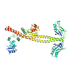
3II6
DownloadVisualize
BU of 3ii6 by Molmil
Structure of human Xrcc4 in complex with the tandem BRCT domains of DNA LigaseIV.
Descriptor: CHLORIDE ION, DNA ligase 4, DNA repair protein XRCC4
Authors:Meesala, S, Junop, M.
Deposit date:2009-07-31
Release date:2009-08-11
Last modified:2024-02-21
Method:X-RAY DIFFRACTION (2.4 Å)
Cite:Structural and functional interaction between the human DNA repair proteins DNA ligase IV and XRCC4
MOL.CELL.BIOL., 11, 2009
3SQD
DownloadVisualize
BU of 3sqd by Molmil
Crystal structure of human PTIP BRCT5/6-gamma H2AX complex
Descriptor: Histone H2A.x, PAX-interacting protein 1
Authors:Yan, W, Shao, Z.H.
Deposit date:2011-07-05
Release date:2011-11-09
Last modified:2023-11-01
Method:X-RAY DIFFRACTION (2.153 Å)
Cite:Crystal structure of human PTIP BRCT5/6-gamma H2AX
To be Published

222926

PDB entries from 2024-07-24

PDB statisticsPDBj update infoContact PDBjnumon