Loading
PDBj
MenuPDBj@FacebookPDBj@TwitterPDBj@YouTubewwPDB FoundationwwPDB
RCSB PDBPDBeBMRBAdv. SearchSearch help
PDB: 260 results

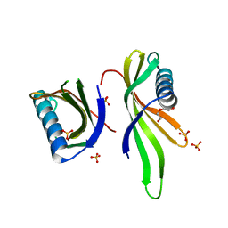
5O7Q
DownloadVisualize
BU of 5o7q by Molmil
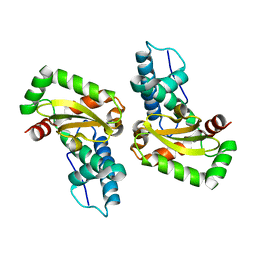
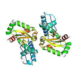
Crystal structure of a single chain monellin mutant (Y65R) pH 5.5
Descriptor: DI(HYDROXYETHYL)ETHER, Monellin chain B,Monellin chain A, SULFATE ION
Authors:Pica, A, Merlino, A.
Deposit date:2017-06-09
Release date:2018-01-10
Last modified:2024-01-17
Method:X-RAY DIFFRACTION (1.72 Å)
Cite:pH driven fibrillar aggregation of the super-sweet protein Y65R-MNEI: A step-by-step structural analysis.
Biochim. Biophys. Acta, 1862, 2017
5N26
DownloadVisualize
BU of 5n26 by Molmil
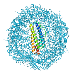
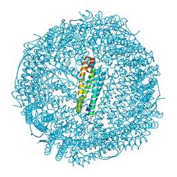
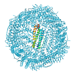
X-ray structure of human heavy chain ferritin in complex with cisplatin
Descriptor: CHLORIDE ION, Cisplatin, Ferritin heavy chain, ...
Authors:Ferraro, G, Merlino, A.
Deposit date:2017-02-07
Release date:2017-08-02
Last modified:2024-01-17
Method:X-RAY DIFFRACTION (2.05 Å)
Cite:Cisplatin Binding Sites in Human H-Chain Ferritin.
Inorg Chem, 56, 2017
4J1B
DownloadVisualize
BU of 4j1b by Molmil
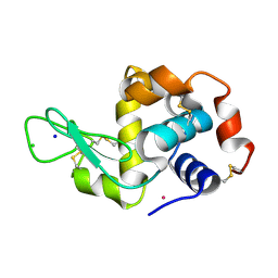
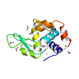
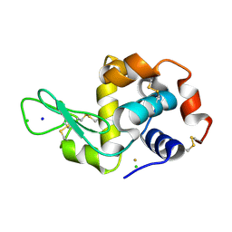
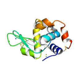
X-ray structure of the adduct between hen egg white lysozyme and AziRu (black crystal)
Descriptor: CHLORIDE ION, Lysozyme C, RUTHENIUM ION, ...
Authors:Vergara, A, Merlino, A.
Deposit date:2013-02-01
Release date:2013-04-03
Last modified:2013-05-08
Method:X-RAY DIFFRACTION (1.66 Å)
Cite:Interaction of Anticancer Ruthenium Compounds with Proteins: High-Resolution X-ray Structures and Raman Microscopy Studies of the Adduct between Hen Egg White Lysozyme and AziRu.
Inorg.Chem., 52, 2013
5ERK
DownloadVisualize
BU of 5erk by Molmil
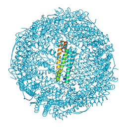
X-ray structure of horse spleen apoferritin (control)
Descriptor: CADMIUM ION, CHLORIDE ION, Ferritin light chain, ...
Authors:Pontillo, N, Merlino, A.
Deposit date:2015-11-14
Release date:2016-03-02
Last modified:2024-05-08
Method:X-RAY DIFFRACTION (2 Å)
Cite:Cisplatin encapsulation within a ferritin nanocage: a high-resolution crystallographic study.
Chem.Commun.(Camb.), 52, 2016
5ERJ
DownloadVisualize
BU of 5erj by Molmil
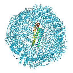
X-ray structure of cisplatin-encapsulated horse spleen apoferritin
Descriptor: CADMIUM ION, CHLORIDE ION, Cisplatin, ...
Authors:Pontillo, N, Merlino, A.
Deposit date:2015-11-14
Release date:2016-03-02
Last modified:2024-05-08
Method:X-RAY DIFFRACTION (1.45 Å)
Cite:Cisplatin encapsulation within a ferritin nanocage: a high-resolution crystallographic study.
Chem.Commun.(Camb.), 52, 2016
4L2C
DownloadVisualize
BU of 4l2c by Molmil
X-ray structure of the C57R mutant of the iron superoxide dismutase from Pseudoalteromonas haloplanktis (crystal form I)
Descriptor: FE (III) ION, Superoxide dismutase [Fe], alpha-D-glucopyranose-(1-1)-alpha-D-glucopyranose
Authors:Russo Krauss, I, Merlino, A, Sica, F.
Deposit date:2013-06-04
Release date:2014-02-26
Last modified:2024-02-28
Method:X-RAY DIFFRACTION (1.66 Å)
Cite:Structural and denaturation studies of two mutants of a cold adapted superoxide dismutase point to the importance of electrostatic interactions in protein stability.
Biochim.Biophys.Acta, 1844, 2014
4L2D
DownloadVisualize
BU of 4l2d by Molmil
X-ray structure of the Fe(II) form of the iron superoxide dismutase from Pseudoalteromonas haloplanktis
Descriptor: FE (II) ION, Superoxide dismutase [Fe], alpha-D-glucopyranose-(1-1)-alpha-D-glucopyranose
Authors:Russo Krauss, I, Merlino, A, Sica, F.
Deposit date:2013-06-04
Release date:2014-02-26
Last modified:2024-02-28
Method:X-RAY DIFFRACTION (2.07 Å)
Cite:Structural and denaturation studies of two mutants of a cold adapted superoxide dismutase point to the importance of electrostatic interactions in protein stability.
Biochim.Biophys.Acta, 1844, 2014
5MIK
DownloadVisualize
BU of 5mik by Molmil
X-ray structure of carboplatin-encapsulated horse spleen apoferritin (rotating anode data)
Descriptor: CADMIUM ION, CHLORIDE ION, Ferritin light chain, ...
Authors:Pontillo, N, Ferraro, G, Helliwell, J.R, Merlino, A.
Deposit date:2016-11-28
Release date:2017-03-15
Last modified:2024-01-17
Method:X-RAY DIFFRACTION (1.96 Å)
Cite:X-ray Structure of the Carboplatin-Loaded Apo-Ferritin Nanocage.
ACS Med Chem Lett, 8, 2017
4I7Y
DownloadVisualize
BU of 4i7y by Molmil
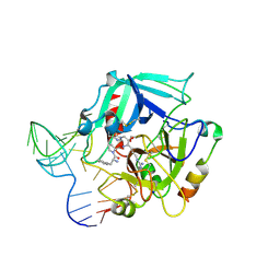
Crystal Structure of Human Alpha Thrombin in Complex with a 27-mer Aptamer Bound to Exosite II
Descriptor: 2-acetamido-2-deoxy-beta-D-glucopyranose, D-phenylalanyl-N-[(2S,3S)-6-{[amino(iminio)methyl]amino}-1-chloro-2-hydroxyhexan-3-yl]-L-prolinamide, DNA (27-MER), ...
Authors:Pica, A, Russo Krauss, I, Merlino, A, Mazzarella, L, Sica, F.
Deposit date:2012-12-01
Release date:2013-10-16
Last modified:2024-10-09
Method:X-RAY DIFFRACTION (2.4 Å)
Cite:Duplex-quadruplex motifs in a peculiar structural organization cooperatively contribute to thrombin binding of a DNA aptamer.
Acta Crystallogr.,Sect.D, 69, 2013
5NJ1
DownloadVisualize
BU of 5nj1 by Molmil
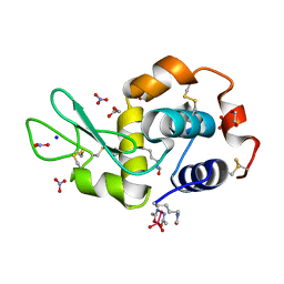
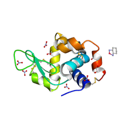
The X-ray structure of the adduct formed in the reaction between hen egg white lysozyme and arsenoplatin-1
Descriptor: 1,2-ETHANEDIOL, Lysozyme C, NITRATE ION, ...
Authors:Ferraro, G, Merlino, A.
Deposit date:2017-03-27
Release date:2018-05-16
Last modified:2024-01-17
Method:X-RAY DIFFRACTION (1.85 Å)
Cite:Arsenoplatin-1 Is a Dual Pharmacophore Anticancer Agent.
J.Am.Chem.Soc., 141, 2019
5NJ7
DownloadVisualize
BU of 5nj7 by Molmil
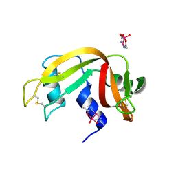
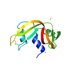
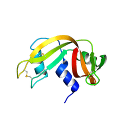
The X-ray structure of the adduct formed in the reaction between bovine pancreatic ribonuclease and arsenoplatin-1
Descriptor: Ribonuclease pancreatic, arsenoplatin-1
Authors:Ferraro, G, Merlino, A.
Deposit date:2017-03-28
Release date:2018-05-16
Last modified:2024-11-06
Method:X-RAY DIFFRACTION (2.15 Å)
Cite:Arsenoplatin-1 Is a Dual Pharmacophore Anticancer Agent.
J.Am.Chem.Soc., 141, 2019
5MIJ
DownloadVisualize
BU of 5mij by Molmil
X-ray structure of carboplatin-encapsulated horse spleen apoferritin
Descriptor: CADMIUM ION, CHLORIDE ION, Ferritin light chain, ...
Authors:Pontillo, N, Ferraro, G, Helliwell, J.R, Merlino, A.
Deposit date:2016-11-28
Release date:2017-03-15
Last modified:2024-05-08
Method:X-RAY DIFFRACTION (1.49 Å)
Cite:X-ray Structure of the Carboplatin-Loaded Apo-Ferritin Nanocage.
ACS Med Chem Lett, 8, 2017
5OLE
DownloadVisualize
BU of 5ole by Molmil
X-ray structure of the adduct formed upon reaction of hen egg white lysozyme with a tetranuclear Pt-thiosemicarbazone compound
Descriptor: 1,2-ETHANEDIOL, DIMETHYL SULFOXIDE, Lysozyme C, ...
Authors:Ferraro, G, Merlino, A.
Deposit date:2017-07-27
Release date:2018-08-29
Last modified:2024-10-16
Method:X-RAY DIFFRACTION (1.78 Å)
Cite:Reactions of a tetranuclear Pt-thiosemicarbazone complex with model proteins.
J. Inorg. Biochem., 181, 2018
5OLD
DownloadVisualize
BU of 5old by Molmil
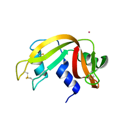
X-ray structure of the adduct formed upon reaction of ribonuclease A with a tetranuclear Pt-thiosemicarbazone compound
Descriptor: DIMETHYL SULFOXIDE, PLATINUM (II) ION, Ribonuclease pancreatic
Authors:Ferraro, G, Merlino, A.
Deposit date:2017-07-27
Release date:2018-08-29
Last modified:2024-01-17
Method:X-RAY DIFFRACTION (1.78 Å)
Cite:Reactions of a tetranuclear Pt-thiosemicarbazone complex with model proteins.
J. Inorg. Biochem., 181, 2018
4PPO
DownloadVisualize
BU of 4ppo by Molmil
First Crystal Structure for an Oxaliplatin-Protein Complex
Descriptor: 1,2-ETHANEDIOL, CYCLOHEXANE-1(R),2(R)-DIAMINE-PLATINUM(II), Lysozyme C, ...
Authors:Messori, L, Merlino, A.
Deposit date:2014-02-27
Release date:2015-01-14
Last modified:2024-10-30
Method:X-RAY DIFFRACTION (1.73 Å)
Cite:The X-ray structure of the complex formed in the reaction between oxaliplatin and lysozyme.
Chem.Commun.(Camb.), 50, 2014
4KXH
DownloadVisualize
BU of 4kxh by Molmil
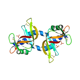
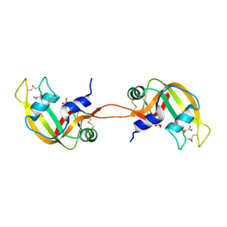
The X-ray crystal structure of a dimeric variant of human pancreatic ribonuclease
Descriptor: CHLORIDE ION, Ribonuclease pancreatic, SODIUM ION, ...
Authors:Pica, A, Merlino, A, Mazzarella, L, Sica, F.
Deposit date:2013-05-26
Release date:2013-10-02
Last modified:2017-11-15
Method:X-RAY DIFFRACTION (2.7 Å)
Cite:Three-dimensional domain swapping and supramolecular protein assembly: insights from the X-ray structure of a dimeric swapped variant of human pancreatic RNase.
Acta Crystallogr.,Sect.D, 69, 2013
4L55
DownloadVisualize
BU of 4l55 by Molmil
X-ray structure of the adduct between bovine pancreatic ribonuclease and AziRu
Descriptor: RUTHENIUM ION, Ribonuclease pancreatic
Authors:Vergara, A, Russo Krauss, I, Merlino, A.
Deposit date:2013-06-10
Release date:2014-01-29
Last modified:2024-10-09
Method:X-RAY DIFFRACTION (1.65 Å)
Cite:Investigating the ruthenium metalation of proteins: X-ray structure and Raman microspectroscopy of the complex between RNase A and AziRu.
Inorg.Chem., 52, 2013
4L0A
DownloadVisualize
BU of 4l0a by Molmil
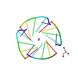
X-ray structure of an all LNA quadruplex
Descriptor: DNA/RNA (5'-R(*(TLN)P*(LCG)P*(LCG)P*(LCG)P*(TLN))-3'), GLYCEROL, POTASSIUM ION
Authors:Russo Krauss, I, Parkinson, G, Merlino, A, Mazzarella, L, Sica, F.
Deposit date:2013-05-31
Release date:2014-03-05
Last modified:2023-09-20
Method:X-RAY DIFFRACTION (1.7 Å)
Cite:A regular thymine tetrad and a peculiar supramolecular assembly in the first crystal structure of an all-LNA G-quadruplex.
Acta Crystallogr.,Sect.D, 70, 2014
4IAU
DownloadVisualize
BU of 4iau by Molmil
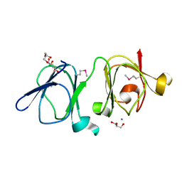
Atomic resolution structure of Geodin, a beta-gamma crystallin from Geodia cydonium
Descriptor: Beta-gamma-crystallin, CALCIUM ION, GLYCEROL
Authors:Vergara, A, Grassi, M, Sica, F, Mazzarella, L, Merlino, A.
Deposit date:2012-12-07
Release date:2013-06-05
Last modified:2024-10-16
Method:X-RAY DIFFRACTION (0.99 Å)
Cite:A novel interdomain interface in crystallins: structural characterization of the [beta][gamma]-crystallin from Geodia cydonium at 0.99 A resolution
Acta Crystallogr.,Sect.D, 69, 2013
4LFX
DownloadVisualize
BU of 4lfx by Molmil
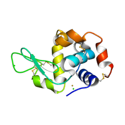
X-ray structure of the adduct between hen egg white lysozyme and Auoxo6, a dinuclear gold(III) complex with -dioxo bridges linking the two metal centers
Descriptor: CHLORIDE ION, GOLD ION, Lysozyme C, ...
Authors:Vergara, A, Merlino, A.
Deposit date:2013-06-27
Release date:2013-10-02
Last modified:2024-10-30
Method:X-RAY DIFFRACTION (2.1 Å)
Cite:The mode of action of anticancer gold-based drugs: a structural perspective.
Chem.Commun.(Camb.), 49, 2013
4LGK
DownloadVisualize
BU of 4lgk by Molmil
X-ray structure of the adduct between hen egg white lysozyme and Au2Phen, a dinuclear gold(III) complex with -dioxo bridges linking the two metal centers
Descriptor: CHLORIDE ION, GOLD ION, Lysozyme C
Authors:Vergara, A, Merlino, A.
Deposit date:2013-06-28
Release date:2013-10-02
Last modified:2024-10-30
Method:X-RAY DIFFRACTION (1.96 Å)
Cite:The mode of action of anticancer gold-based drugs: a structural perspective.
Chem.Commun.(Camb.), 49, 2013
4LFP
DownloadVisualize
BU of 4lfp by Molmil
X-ray structure of the adduct between hen egg white lysozyme and a homoleptic gold(I) complex with the saccharynate ligand
Descriptor: CHLORIDE ION, GOLD ION, Lysozyme C, ...
Authors:Vergara, A, Merlino, A.
Deposit date:2013-06-27
Release date:2013-10-02
Last modified:2024-10-16
Method:X-RAY DIFFRACTION (1.72 Å)
Cite:The mode of action of anticancer gold-based drugs: a structural perspective.
Chem.Commun.(Camb.), 49, 2013
4OT4
DownloadVisualize
BU of 4ot4 by Molmil
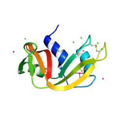
X-ray Structure of the Adduct formed between cisplatin and Ribonuclease A
Descriptor: Cisplatin, Ribonuclease pancreatic
Authors:Messori, L, Merlino, A.
Deposit date:2014-02-13
Release date:2014-07-09
Last modified:2024-11-06
Method:X-RAY DIFFRACTION (1.85 Å)
Cite:Cisplatin binding to proteins: molecular structure of the ribonuclease a adduct.
Inorg.Chem., 53, 2014
5NA9
DownloadVisualize
BU of 5na9 by Molmil
The X-ray structure of bovine pancreatic ribonuclease incubated in the presence of an excess of carboplatin (1:10 ratio)
Descriptor: CESIUM ION, CHLORIDE ION, Ribonuclease pancreatic, ...
Authors:Ferraro, G, Merlino, A.
Deposit date:2017-02-27
Release date:2017-08-16
Last modified:2024-10-16
Method:X-RAY DIFFRACTION (2.07 Å)
Cite:A comparison study on RNase A oligomerization induced by cisplatin, carboplatin and oxaliplatin.
J. Inorg. Biochem., 173, 2017
4N4C
DownloadVisualize
BU of 4n4c by Molmil
Crystal structure of the C-terminal swapped dimer of a Bovine seminal ribonuclease mutant
Descriptor: PHOSPHATE ION, Seminal ribonuclease
Authors:Pica, A, Russo Krauss, I, Merlino, A, Sica, F.
Deposit date:2013-10-08
Release date:2013-11-06
Last modified:2023-09-20
Method:X-RAY DIFFRACTION (2.48 Å)
Cite:The multiple forms of bovine seminal ribonuclease: Structure and stability of a C-terminal swapped dimer.
Febs Lett., 587, 2013

227111

PDB entries from 2024-11-06

PDB statisticsPDBj update infoContact PDBjnumon