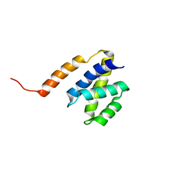
2MSU
 
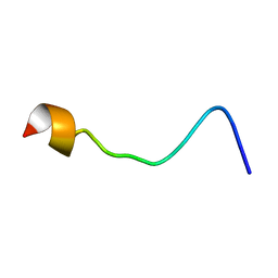 | |
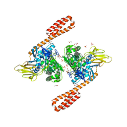
8H1D
 | | Solid-state NMR Structure of Aquaporin Z in its Native Cellular Membranes | | 分子名称: | Aquaporin Z | | 著者 | Xie, H, Zhao, Y, Zhao, W, Chen, Y, Liu, M, Yang, J. | | 登録日 | 2022-10-02 | | 公開日 | 2022-11-09 | | 最終更新日 | 2024-05-15 | | 実験手法 | SOLID-STATE NMR | | 主引用文献 | Solid-state NMR structure determination of a membrane protein in E. coli cellular inner membrane.
Sci Adv, 9, 2023
|
|
5YKJ
 | | Structural basis of the thiol resolving mechanism in yeast mitochondrial 1-Cys peroxiredoxin via glutathione/thioredoxin systems | | 分子名称: | GLYCEROL, Peroxiredoxin PRX1, mitochondrial, ... | | 著者 | Li, C.C, Yang, J, Yang, M.J, Liu, L, Peng, C.T, Li, T, He, L.H, Song, Y.J, Zhu, Y.B, Zhao, N.L, Zhao, C, Bao, R. | | 登録日 | 2017-10-14 | | 公開日 | 2018-10-24 | | 最終更新日 | 2019-11-06 | | 実験手法 | X-RAY DIFFRACTION (1.53 Å) | | 主引用文献 | Structural basis of the thiol resolving mechanism in yeast mitochondrial 1-Cys peroxiredoxin via glutathione/thioredoxin systems
To be published
|
|
2JMX
 | | OSCP-NT (1-120) in complex with N-terminal (1-25) alpha subunit from F1-ATPase | | 分子名称: | ATP synthase O subunit, mitochondrial, ATP synthase subunit alpha heart isoform | | 著者 | Carbajo, R.J, Neuhaus, D, Kellas, F.A, Yang, J, Runswick, M.J, Montgomery, M.G, Walker, J.E. | | 登録日 | 2006-12-12 | | 公開日 | 2007-04-24 | | 最終更新日 | 2023-12-20 | | 実験手法 | SOLUTION NMR | | 主引用文献 | How the N-terminal Domain of the OSCP Subunit of Bovine F(1)F(o)-ATP Synthase Interacts with the N-terminal Region of an Alpha Subunit
J.Mol.Biol., 368, 2007
|
|
2JPS
 | | NAB2 N-terminal domain | | 分子名称: | Nuclear polyadenylated RNA-binding protein NAB2 | | 著者 | Grant, R, Marshall, N.J, Yang, J, Fasken, M, Kelly, S, Harreman, M.T, Neuhaus, D, Corbett, A.H, Stewart, M. | | 登録日 | 2007-05-23 | | 公開日 | 2008-03-18 | | 最終更新日 | 2023-12-20 | | 実験手法 | SOLUTION NMR | | 主引用文献 | Structure of the N-terminal Mlp1-binding domain of the Saccharomyces cerevisiae mRNA-binding protein, Nab2
J.Mol.Biol., 376, 2008
|
|
5YKW
 | | Structural basis of the thiol resolving mechanism in yeast mitochondrial 1-Cys peroxiredoxin via glutathione/thioredoxin systems | | 分子名称: | Thioredoxin-3, mitochondrial, peptide THR-PRO-VAL-CYS-THR-THR-GLU-VAL | | 著者 | Li, C.C, Yang, J, Yang, M.J, Liu, L, Peng, C.T, Li, T, He, L.H, Song, Y.J, Zhu, Y.B, Zhao, N.L, Zhao, C, Bao, R. | | 登録日 | 2017-10-16 | | 公開日 | 2018-10-24 | | 最終更新日 | 2019-11-06 | | 実験手法 | X-RAY DIFFRACTION (2.08 Å) | | 主引用文献 | Structural basis of the thiol resolving mechanism in yeast mitochondrial 1-Cys peroxiredoxin via glutathione/thioredoxin systems
to be published
|
|
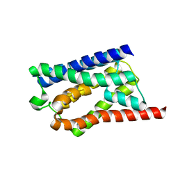
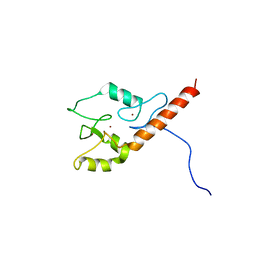
2KQC
 | | Second PBZ domain of human APLF protein | | 分子名称: | Aprataxin and PNK-like factor, ZINC ION | | 著者 | Neuhaus, D, Eustermann, S, Brockmann, C, Yang, J. | | 登録日 | 2009-11-04 | | 公開日 | 2010-01-19 | | 最終更新日 | 2024-05-22 | | 実験手法 | SOLUTION NMR | | 主引用文献 | Solution structures of the two PBZ domains from human APLF and their interaction with poly(ADP-ribose).
Nat.Struct.Mol.Biol., 17, 2010
|
|
2KQB
 | | First PBZ domain of human APLF protein | | 分子名称: | Aprataxin and PNK-like factor, ZINC ION | | 著者 | Neuhaus, D, Eustermann, S, Brockmann, C, Yang, J. | | 登録日 | 2009-11-04 | | 公開日 | 2010-01-19 | | 最終更新日 | 2024-05-08 | | 実験手法 | SOLUTION NMR | | 主引用文献 | Solution structures of the two PBZ domains from human APLF and their interaction with poly(ADP-ribose).
Nat.Struct.Mol.Biol., 17, 2010
|
|
8J4I
 | | Unveiling the Role of Human Prohibitin 2 as a Mitochondrial Calcium Channel in Parkinson's Disease | | 分子名称: | Prohibitin-2 | | 著者 | Li, Y.Y, Fang, Y, Gao, Y.X, Ding, W, Yang, J, Liu, Y, Shen, B, Wang, J.F, Zhou, S. | | 登録日 | 2023-04-20 | | 公開日 | 2024-06-05 | | 実験手法 | SOLUTION NMR | | 主引用文献 | Human Prohibitin 2 is a Mitochondrial Ca2+-Selective Channel
To Be Published
|
|
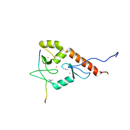
2KQD
 | | First PBZ domain of human APLF protein in complex with ribofuranosyladenosine | | 分子名称: | ADENOSINE, Aprataxin and PNK-like factor, ZINC ION, ... | | 著者 | Neuhaus, D, Eustermann, S, Brockmann, C, Yang, J. | | 登録日 | 2009-11-04 | | 公開日 | 2010-01-19 | | 最終更新日 | 2024-05-01 | | 実験手法 | SOLUTION NMR | | 主引用文献 | Solution structures of the two PBZ domains from human APLF and their interaction with poly(ADP-ribose).
Nat.Struct.Mol.Biol., 17, 2010
|
|
2KQE
 | | Second PBZ domain of human APLF protein in complex with ribofuranosyladenosine | | 分子名称: | ADENOSINE, Aprataxin and PNK-like factor, ZINC ION, ... | | 著者 | Neuhaus, D, Eustermann, S, Brockmann, C, Yang, J. | | 登録日 | 2009-11-04 | | 公開日 | 2010-01-19 | | 最終更新日 | 2024-05-01 | | 実験手法 | SOLUTION NMR | | 主引用文献 | Solution structures of the two PBZ domains from human APLF and their interaction with poly(ADP-ribose).
Nat.Struct.Mol.Biol., 17, 2010
|
|
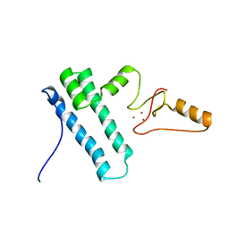
2L31
 | | Human PARP-1 zinc finger 2 | | 分子名称: | Poly [ADP-ribose] polymerase 1, ZINC ION | | 著者 | Neuhaus, D, Eustermann, S, Yang, J, Videler, H. | | 登録日 | 2010-08-30 | | 公開日 | 2011-02-02 | | 最終更新日 | 2024-05-01 | | 実験手法 | SOLUTION NMR | | 主引用文献 | The DNA-binding domain of human PARP-1 interacts with DNA single-strand breaks as a monomer through its second zinc finger.
J.Mol.Biol., 407, 2011
|
|
2L30
 | | Human PARP-1 zinc finger 1 | | 分子名称: | Poly [ADP-ribose] polymerase 1, ZINC ION | | 著者 | Neuhaus, D, Eustermann, S, Yang, J, Videler, H. | | 登録日 | 2010-08-30 | | 公開日 | 2011-02-02 | | 最終更新日 | 2024-05-01 | | 実験手法 | SOLUTION NMR | | 主引用文献 | The DNA-binding domain of human PARP-1 interacts with DNA single-strand breaks as a monomer through its second zinc finger.
J.Mol.Biol., 407, 2011
|
|
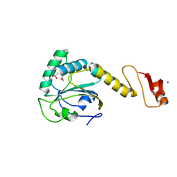
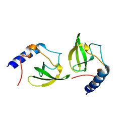
2K0A
 | | 1H, 15N and 13C chemical shift assignments for Rds3 protein | | 分子名称: | Pre-mRNA-splicing factor RDS3, ZINC ION | | 著者 | Loening, N, van Roon, A, Yang, J, Nagai, K, Neuhaus, D. | | 登録日 | 2008-01-31 | | 公開日 | 2008-07-22 | | 最終更新日 | 2024-05-29 | | 実験手法 | SOLUTION NMR | | 主引用文献 | Solution structure of the U2 snRNP protein Rds3p reveals a knotted zinc-finger motif.
Proc.Natl.Acad.Sci.Usa, 105, 2008
|
|
2LD1
 | |
2N8A
 | | 1H, 13C and 15N chemical shift assignments and solution structure for PARP-1 F1F2 domains in complex with a DNA single-strand break | | 分子名称: | DNA (45-MER), Poly [ADP-ribose] polymerase 1, ZINC ION | | 著者 | Neuhaus, D, Eustermann, S, Yang, J, Wu, W. | | 登録日 | 2015-10-08 | | 公開日 | 2015-12-02 | | 最終更新日 | 2024-05-01 | | 実験手法 | SOLUTION NMR | | 主引用文献 | Structural Basis of Detection and Signaling of DNA Single-Strand Breaks by Human PARP-1.
Mol.Cell, 60, 2015
|
|
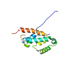
2MY1
 | | Solution structure of Bud31p | | 分子名称: | Pre-mRNA-splicing factor BUD31, ZINC ION | | 著者 | van Roon, A.M, Yang, J, Mathieu, D, Bermel, W, Nagai, K, Neuhaus, D. | | 登録日 | 2015-01-19 | | 公開日 | 2015-03-11 | | 最終更新日 | 2024-05-15 | | 実験手法 | SOLUTION NMR | | 主引用文献 | (113) Cd NMR Experiments Reveal an Unusual Metal Cluster in the Solution Structure of the Yeast Splicing Protein Bud31p.
Angew.Chem.Int.Ed.Engl., 54, 2015
|
|
2HQE
 | | Crystal structure of human P100 Tudor domain: Large fragment | | 分子名称: | P100 Co-activator tudor domain | | 著者 | Shah, N, Zhao, M, Cheng, C, Xu, H, Yang, J, Silvennoinen, O, Liu, Z.J, Wang, B.C, Southeast Collaboratory for Structural Genomics (SECSG) | | 登録日 | 2006-07-18 | | 公開日 | 2007-07-03 | | 最終更新日 | 2023-08-30 | | 実験手法 | X-RAY DIFFRACTION (2 Å) | | 主引用文献 | Crystal Structure of a large fragment of the Human P100 Tudor Domain
To be Published
|
|
2HQX
 | | Crystal structure of human P100 tudor domain conserved region | | 分子名称: | P100 CO-ACTIVATOR TUDOR DOMAIN | | 著者 | Zhao, M, Liu, Z.J, Xu, H, Yang, J, Silvennoinen, O, Wang, B.C, Southeast Collaboratory for Structural Genomics (SECSG) | | 登録日 | 2006-07-19 | | 公開日 | 2006-10-10 | | 最終更新日 | 2023-08-30 | | 実験手法 | X-RAY DIFFRACTION (1.42 Å) | | 主引用文献 | Crystal Structure of Human P100 Tudor Domain Conserved Region
To be Published
|
|
2LBM
 | |
2O4X
 | | Crystal structure of human P100 tudor domain | | 分子名称: | Staphylococcal nuclease domain-containing protein 1 | | 著者 | Shaw, N, Zhao, M, Cheng, C, Xu, H, Yang, J, Silvennoinen, O, Rao, Z, Wang, B.C, Liu, Z.J. | | 登録日 | 2006-12-05 | | 公開日 | 2007-02-13 | | 最終更新日 | 2023-12-27 | | 実験手法 | X-RAY DIFFRACTION (2 Å) | | 主引用文献 | Crystal structure of human P100 tudor domain
To be Published
|
|
6ASY
 | | BiP-ATP2 | | 分子名称: | 78 kDa glucose-regulated protein, ADENOSINE-5'-TRIPHOSPHATE, GLYCEROL, ... | | 著者 | Liu, Q, Yang, J, Zong, Y, Columbus, L, Zhou, L. | | 登録日 | 2017-08-26 | | 公開日 | 2017-12-06 | | 最終更新日 | 2024-03-13 | | 実験手法 | X-RAY DIFFRACTION (1.85 Å) | | 主引用文献 | Conformation transitions of the polypeptide-binding pocket support an active substrate release from Hsp70s.
Nat Commun, 8, 2017
|
|
7D77
 | | Cryo-EM structure of the cortisol-bound adhesion receptor GPR97-Go complex | | 分子名称: | (11alpha,14beta)-11,17,21-trihydroxypregn-4-ene-3,20-dione, Adhesion G protein-coupled receptor G3; GPR97, CHOLESTEROL, ... | | 著者 | Ping, Y, Mao, C, Xiao, P, Zhao, R, Jiang, Y, Yang, Z, An, W, Shen, D, Yang, F, Zhang, H, Qu, C, Shen, Q, Tian, C, Li, Z, Li, S, Wang, G, Tao, X, Wen, X, Zhong, Y, Yang, J, Yi, F, Yu, X, Xu, E, Zhang, Y, Sun, J. | | 登録日 | 2020-10-03 | | 公開日 | 2021-02-03 | | 最終更新日 | 2021-02-10 | | 実験手法 | ELECTRON MICROSCOPY (2.9 Å) | | 主引用文献 | Structures of the glucocorticoid-bound adhesion receptor GPR97-G o complex.
Nature, 589, 2021
|
|
7D76
 | | Cryo-EM structure of the beclomethasone-bound adhesion receptor GPR97-Go complex | | 分子名称: | (8~{S},9~{R},10~{S},11~{S},13~{S},14~{S},16~{S},17~{R})-9-chloranyl-10,13,16-trimethyl-11,17-bis(oxidanyl)-17-(2-oxidanylethanoyl)-6,7,8,11,12,14,15,16-octahydrocyclopenta[a]phenanthren-3-one, Adhesion G protein-coupled receptor G3; GPR97, CHOLESTEROL, ... | | 著者 | Ping, Y, Mao, C, Xiao, P, Zhao, R, Jiang, Y, Yang, Z, An, W, Shen, D, Yang, F, Zhang, H, Qu, C, Shen, Q, Tian, C, Li, Z, Li, S, Wang, G, Tao, X, Wen, X, Zhong, Y, Yang, J, Yi, F, Yu, X, Xu, E, Zhang, Y, Sun, J. | | 登録日 | 2020-10-03 | | 公開日 | 2021-02-03 | | 最終更新日 | 2021-02-10 | | 実験手法 | ELECTRON MICROSCOPY (3.1 Å) | | 主引用文献 | Structures of the glucocorticoid-bound adhesion receptor GPR97-G o complex.
Nature, 589, 2021
|
|
7R5R
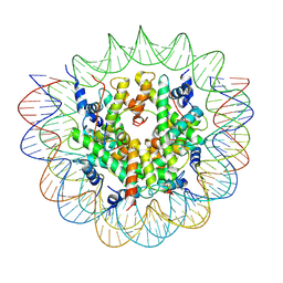 | | Structure of the human CCAN CENP-A alpha-satellite complex | | 分子名称: | Centromere protein C, DNA (171-MER), Histone H2A type 1-C, ... | | 著者 | Yatskevich, S, Muir, K.W, Bellini, D, Zhang, Z, Yang, J, Tischer, T, Predin, M, Dendooven, T, McLaughlin, S.H, Barford, D. | | 登録日 | 2022-02-11 | | 公開日 | 2022-04-27 | | 最終更新日 | 2022-06-01 | | 実験手法 | ELECTRON MICROSCOPY (2.44 Å) | | 主引用文献 | Structure of the human inner kinetochore bound to a centromeric CENP-A nucleosome.
Science, 376, 2022
|
|


