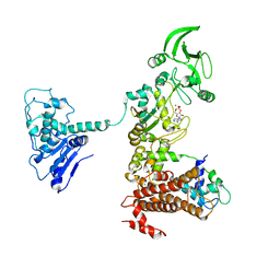
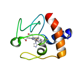
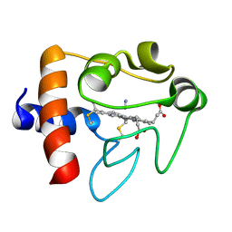
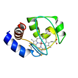
1Y0V
 
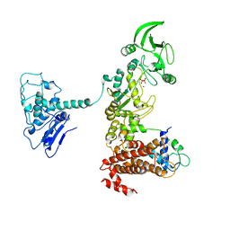 | | Crystal structure of anthrax edema factor (EF) in complex with calmodulin and pyrophosphate | | 分子名称: | CALCIUM ION, Calmodulin, Calmodulin-sensitive adenylate cyclase, ... | | 著者 | Shen, Y, Zhukovskaya, N.L, Guo, Q, Florian, J, Tang, W.-J. | | 登録日 | 2004-11-16 | | 公開日 | 2005-05-03 | | 最終更新日 | 2024-02-14 | | 実験手法 | X-RAY DIFFRACTION (3.6 Å) | | 主引用文献 | Calcium-independent calmodulin binding and two-metal-ion catalytic mechanism of anthrax edema factor
Embo J., 24, 2005
|
|
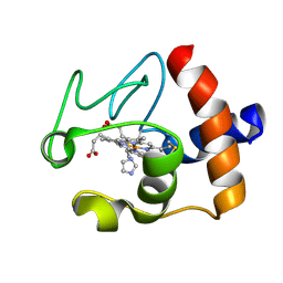
1XFY
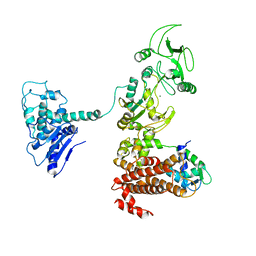 | | Crystal structure of anthrax edema factor (EF) in complex with calmodulin | | 分子名称: | CALCIUM ION, Calmodulin 2, Calmodulin-sensitive adenylate cyclase, ... | | 著者 | Shen, Y, Zhukovskaya, N.L, Guo, Q, Florian, J, Tang, W.J. | | 登録日 | 2004-09-15 | | 公開日 | 2005-05-03 | | 最終更新日 | 2024-02-14 | | 実験手法 | X-RAY DIFFRACTION (3.3 Å) | | 主引用文献 | Calcium-independent calmodulin binding and two-metal-ion catalytic mechanism of anthrax edema factor.
EMBO J., 24, 2005
|
|
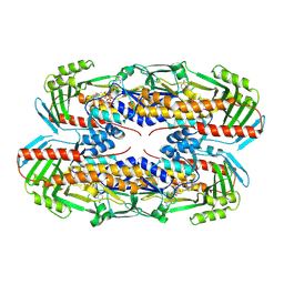
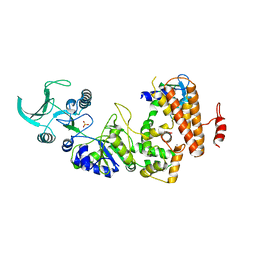
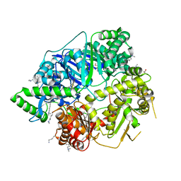
4KOD
 | | Structure of p97 N-D1 R155H mutant in complex with ADP | | 分子名称: | ADENOSINE-5'-DIPHOSPHATE, Transitional endoplasmic reticulum ATPase | | 著者 | Xia, D, Tang, W.K. | | 登録日 | 2013-05-11 | | 公開日 | 2013-11-13 | | 最終更新日 | 2024-02-28 | | 実験手法 | X-RAY DIFFRACTION (2.96 Å) | | 主引用文献 | Altered Intersubunit Communication Is the Molecular Basis for Functional Defects of Pathogenic p97 Mutants.
J.Biol.Chem., 288, 2013
|
|
1XFX
 | | Crystal structure of anthrax edema factor (EF) in complex with calmodulin in the presence of 10 millimolar exogenously added calcium chloride | | 分子名称: | CALCIUM ION, Calmodulin 2, Calmodulin-sensitive adenylate cyclase, ... | | 著者 | Shen, Y, Zhukovskaya, N.L, Guo, Q, Florian, J, Tang, W.J. | | 登録日 | 2004-09-15 | | 公開日 | 2005-05-03 | | 最終更新日 | 2017-12-20 | | 実験手法 | X-RAY DIFFRACTION (3.2 Å) | | 主引用文献 | Calcium-independent calmodulin binding and two-metal-ion catalytic mechanism of anthrax edema factor.
EMBO J., 24, 2005
|
|
1XFW
 | | Crystal structure of anthrax edema factor (EF) in complex with calmodulin and 3'5' cyclic AMP (cAMP) | | 分子名称: | ADENOSINE-3',5'-CYCLIC-MONOPHOSPHATE, CALCIUM ION, Calmodulin 2, ... | | 著者 | Shen, Y, Zhukovskaya, N.L, Guo, Q, Florian, J, Tang, W.J. | | 登録日 | 2004-09-15 | | 公開日 | 2005-05-03 | | 最終更新日 | 2024-02-14 | | 実験手法 | X-RAY DIFFRACTION (3.4 Å) | | 主引用文献 | Calcium-independent calmodulin binding and two-metal-ion catalytic mechanism of anthrax edema factor.
EMBO J., 24, 2005
|
|
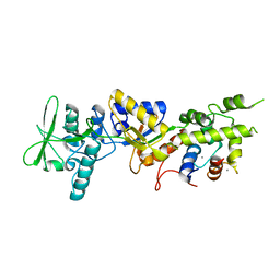
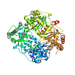
4KLN
 | | Structure of p97 N-D1 A232E mutant in complex with ATPgS | | 分子名称: | MAGNESIUM ION, PHOSPHOTHIOPHOSPHORIC ACID-ADENYLATE ESTER, Transitional endoplasmic reticulum ATPase | | 著者 | Xia, D, Tang, W.K. | | 登録日 | 2013-05-07 | | 公開日 | 2013-11-13 | | 最終更新日 | 2024-02-28 | | 実験手法 | X-RAY DIFFRACTION (2.62 Å) | | 主引用文献 | Altered Intersubunit Communication Is the Molecular Basis for Functional Defects of Pathogenic p97 Mutants.
J.Biol.Chem., 288, 2013
|
|
1XFV
 | | Crystal structure of anthrax edema factor (EF) in complex with calmodulin and 3' deoxy-ATP | | 分子名称: | 3'-DEOXYADENOSINE-5'-TRIPHOSPHATE, CALCIUM ION, Calmodulin 2, ... | | 著者 | Shen, Q, Zhukovskaya, N.L, Guo, Q, Florian, J, Tang, W.J. | | 登録日 | 2004-09-15 | | 公開日 | 2005-05-03 | | 最終更新日 | 2024-02-14 | | 実験手法 | X-RAY DIFFRACTION (3.35 Å) | | 主引用文献 | Calcium-independent calmodulin binding and two-metal-ion catalytic mechanism of anthrax edema factor.
EMBO J., 24, 2005
|
|
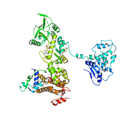
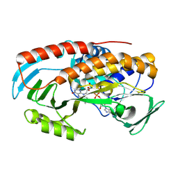
1K8T
 | | Crystal structure of the adenylyl cyclase domain of anthrax edema factor (EF) | | 分子名称: | CALMODULIN-SENSITIVE ADENYLATE CYCLASE, NICKEL (II) ION, SULFATE ION | | 著者 | Drum, C.L, Yan, S.-Z, Bard, J, Shen, Y.-Q, Lu, D, Soelaiman, S, Grabarek, Z, Bohm, A, Tang, W.-J. | | 登録日 | 2001-10-25 | | 公開日 | 2002-01-23 | | 最終更新日 | 2024-04-03 | | 実験手法 | X-RAY DIFFRACTION (2.6 Å) | | 主引用文献 | Structural basis for the activation of anthrax adenylyl cyclase exotoxin by calmodulin
Nature, 415, 2002
|
|
8TWF
 | |
3H44
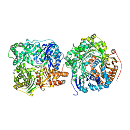 | |
3HGZ
 | | Crystal structure of human insulin-degrading enzyme in complex with amylin | | 分子名称: | Insulin-degrading enzyme, Islet amyloid polypeptide, ZINC ION | | 著者 | Guo, Q, Bian, Y, Tang, W.J. | | 登録日 | 2009-05-14 | | 公開日 | 2009-12-08 | | 最終更新日 | 2021-10-13 | | 実験手法 | X-RAY DIFFRACTION (2.91 Å) | | 主引用文献 | Molecular Basis for the Recognition and Cleavages of IGF-II, TGF-alpha, and Amylin by Human Insulin-Degrading Enzyme.
J.Mol.Biol., 395, 2010
|
|
3TN2
 | | structure analysis of MIP1-beta P8A | | 分子名称: | C-C motif chemokine 4, ZINC ION | | 著者 | Guo, Q, Tang, W.J. | | 登録日 | 2011-09-01 | | 公開日 | 2012-09-05 | | 最終更新日 | 2018-08-22 | | 実験手法 | X-RAY DIFFRACTION (1.6 Å) | | 主引用文献 | Structures of human CCL18, CCL3, and CCL4 reveal molecular determinants for quaternary structures and sensitivity to insulin-degrading enzyme.
J.Mol.Biol., 427, 2015
|
|
1FI9
 | | SOLUTION STRUCTURE OF THE IMIDAZOLE COMPLEX OF CYTOCHROME C | | 分子名称: | CYTOCHROME C, HEME C, IMIDAZOLE | | 著者 | Banci, L, Bertini, I, Liu, G, Lu, J, Reddig, T, Tang, W, Wu, Y, Zhu, D. | | 登録日 | 2000-08-03 | | 公開日 | 2000-08-23 | | 最終更新日 | 2022-02-23 | | 実験手法 | SOLUTION NMR | | 主引用文献 | Effects of extrinsic imidazole ligation on the molecular and electronic structure of cytochrome c
J.Biol.Inorg.Chem., 6, 2001
|
|
8TWG
 | |
4L3T
 | | Crystal Structure of Substrate-free Human Presequence Protease | | 分子名称: | ACETATE ION, GLYCEROL, Presequence protease, ... | | 著者 | King, J.V, Liang, W.G, Tang, W.J. | | 登録日 | 2013-06-06 | | 公開日 | 2013-07-03 | | 最終更新日 | 2014-07-23 | | 実験手法 | X-RAY DIFFRACTION (2.03 Å) | | 主引用文献 | Molecular basis of substrate recognition and degradation by human presequence protease.
Structure, 22, 2014
|
|
1FI7
 | | Solution structure of the imidazole complex of cytochrome C | | 分子名称: | CYTOCHROME C, HEME C, IMIDAZOLE | | 著者 | Banci, L, Bertini, I, Liu, G, Lu, J, Reddig, T, Tang, W, Wu, Y, Zhu, D. | | 登録日 | 2000-08-03 | | 公開日 | 2000-08-23 | | 最終更新日 | 2022-02-23 | | 実験手法 | SOLUTION NMR | | 主引用文献 | Effects of extrinsic imidazole ligation on the molecular and electronic structure of cytochrome c
J.Biol.Inorg.Chem., 6, 2001
|
|
1I5T
 | | SOLUTION STRUCTURE OF CYANOFERRICYTOCHROME C | | 分子名称: | CYANIDE ION, CYTOCHROME C, HEME C | | 著者 | Yao, Y, Qian, C, Ye, K, Wang, J, Tang, W. | | 登録日 | 2001-02-28 | | 公開日 | 2001-03-21 | | 最終更新日 | 2022-02-23 | | 実験手法 | SOLUTION NMR | | 主引用文献 | Solution structure of cyanoferricytochrome c: ligand-controlled conformational flexibility and electronic structure of the heme moiety.
J.Biol.Inorg.Chem., 7, 2002
|
|
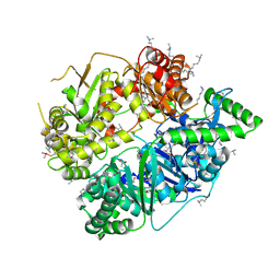
1K93
 | | Crystal structure of the adenylyl cyclase domain of anthrax edema factor (EF) in complex with calmodulin | | 分子名称: | CALCIUM ION, CALMODULIN, CALMODULIN-SENSITIVE ADENYLATE CYCLASE, ... | | 著者 | Drum, C.L, Yan, S.-Z, Bard, J, Shen, Y.-Q, Lu, D, Soelaiman, S, Grabarek, Z, Bohm, A, Tang, W.-J. | | 登録日 | 2001-10-26 | | 公開日 | 2002-01-23 | | 最終更新日 | 2024-04-03 | | 実験手法 | X-RAY DIFFRACTION (2.95 Å) | | 主引用文献 | Structural basis for the activation of anthrax adenylyl cyclase exotoxin by calmodulin.
Nature, 415, 2002
|
|
1YRT
 | | Crystal Structure analysis of the adenylyl cyclaes catalytic domain of adenylyl cyclase toxin of Bordetella pertussis in presence of c-terminal calmodulin | | 分子名称: | Bifunctional hemolysin-adenylate cyclase, CALCIUM ION, Calmodulin | | 著者 | Guo, Q, Shen, Y, Lee, Y.S, Gibbs, C.S, Mrksich, M, Tang, W.J. | | 登録日 | 2005-02-04 | | 公開日 | 2006-01-17 | | 最終更新日 | 2024-02-14 | | 実験手法 | X-RAY DIFFRACTION (2.1 Å) | | 主引用文献 | Structural basis for the interaction of Bordetella pertussis adenylyl cyclase toxin with calmodulin.
Embo J., 24, 2005
|
|
1YRU
 | |
1M60
 | | Solution Structure of Zinc-substituted cytochrome c | | 分子名称: | ZINC SUBSTITUTED HEME C, Zinc-substituted cytochrome c | | 著者 | Qian, C, Yao, Y, Tong, Y, Wang, J, Tang, W. | | 登録日 | 2002-07-11 | | 公開日 | 2002-08-07 | | 最終更新日 | 2022-02-23 | | 実験手法 | SOLUTION NMR | | 主引用文献 | Structural analysis of zinc-substituted cytochrome c.
J.Biol.Inorg.Chem., 8, 2003
|
|
4NGE
 | | Crystal Structure of Human Presequence Protease in Complex with Amyloid-beta (1-40) | | 分子名称: | ACETATE ION, Beta-amyloid protein 40, GLYCEROL, ... | | 著者 | King, J.V, Liang, W.G, Tang, W.J. | | 登録日 | 2013-11-01 | | 公開日 | 2014-05-14 | | 最終更新日 | 2014-07-23 | | 実験手法 | X-RAY DIFFRACTION (2.704 Å) | | 主引用文献 | Molecular basis of substrate recognition and degradation by human presequence protease.
Structure, 22, 2014
|
|
4GSC
 | | Structure analysis of insulin degrading enzyme with compound bdm41559 ((s)-2-[2-(carboxymethyl-phenethyl-amino)-acetylamino]-3-(1h-imidazol-4-yl)-propionic acid methyl ester) | | 分子名称: | Insulin-degrading enzyme, ZINC ION, methyl N-(carboxymethyl)-N-(2-phenylethyl)glycyl-L-histidinate | | 著者 | Guo, Q, Deprez-Poulain, R, Deprez, B, Tang, W.J. | | 登録日 | 2012-08-27 | | 公開日 | 2013-08-28 | | 最終更新日 | 2023-09-13 | | 実験手法 | X-RAY DIFFRACTION (2.81 Å) | | 主引用文献 | Imidazole-derived 2-[N-carbamoylmethyl-alkylamino]acetic acids, substrate-dependent modulators of insulin-degrading enzyme in amyloid-beta hydrolysis.
Eur.J.Med.Chem., 79, 2014
|
|
1ZOT
 | | crystal structure analysis of the CyaA/C-Cam with PMEAPP | | 分子名称: | (ADENIN-9-YL-ETHOXYMETHYL)-HYDROXYPHOSPHINYL-DIPHOSPHATE, CALCIUM ION, Calmodulin, ... | | 著者 | Guo, Q, Tang, W.J. | | 登録日 | 2005-05-13 | | 公開日 | 2005-08-09 | | 最終更新日 | 2024-02-14 | | 実験手法 | X-RAY DIFFRACTION (2.2 Å) | | 主引用文献 | Structural basis for the interaction of Bordetella pertussis adenylyl cyclase toxin with calmodulin.
Embo J., 24, 2005
|
|
4KO8
 | | Structure of p97 N-D1 R155H mutant in complex with ATPgS | | 分子名称: | MAGNESIUM ION, PHOSPHOTHIOPHOSPHORIC ACID-ADENYLATE ESTER, Transitional endoplasmic reticulum ATPase | | 著者 | Xia, D, Tang, W.K. | | 登録日 | 2013-05-11 | | 公開日 | 2013-11-13 | | 最終更新日 | 2024-02-28 | | 実験手法 | X-RAY DIFFRACTION (1.98 Å) | | 主引用文献 | Altered Intersubunit Communication Is the Molecular Basis for Functional Defects of Pathogenic p97 Mutants.
J.Biol.Chem., 288, 2013
|
|


