5J17
 
 | |
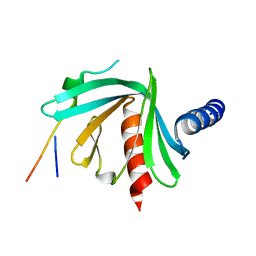
3R83
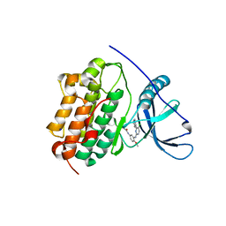
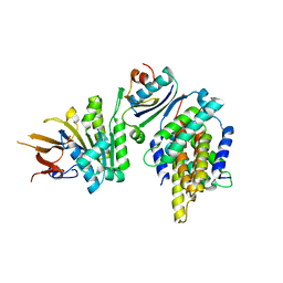
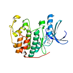 | | CDK2 in complex with inhibitor KVR-2-92 | | 分子名称: | 1,2-ETHANEDIOL, 4-(4-aminopiperidin-1-yl)-2-{[(2-aminopyrimidin-5-yl)methyl]amino}-5-nitrobenzamide, Cyclin-dependent kinase 2 | | 著者 | Betzi, S, Alam, R, Han, H, Becker, A, Schonbrunn, E. | | 登録日 | 2011-03-23 | | 公開日 | 2012-08-08 | | 最終更新日 | 2023-09-13 | | 実験手法 | X-RAY DIFFRACTION (1.75 Å) | | 主引用文献 | Structure-guided optimization of novel CDK2 inhibitors discovered by high-throughput screening
To be Published
|
|
5JS7
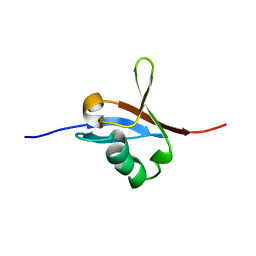
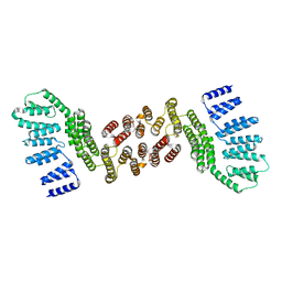
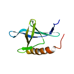
 | | Structural model of a apo G-protein alpha subunit determined with NMR residual dipolar couplings and SAXS | | 分子名称: | Guanine nucleotide-binding protein G(i) subunit alpha-1 | | 著者 | Goricanec, D, Stehle, R, Grigoriu, S, Wagner, G, Hagn, F. | | 登録日 | 2016-05-07 | | 公開日 | 2016-06-29 | | 最終更新日 | 2024-06-19 | | 実験手法 | SOLUTION NMR | | 主引用文献 | Conformational dynamics of a G-protein alpha subunit is tightly regulated by nucleotide binding.
Proc.Natl.Acad.Sci.USA, 113, 2016
|
|
5JS8
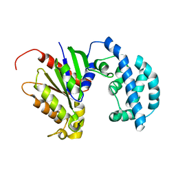
 | | Structural Model of a Protein alpha subunit in complex with GDP obtained with SAXS and NMR residual couplings | | 分子名称: | Guanine nucleotide-binding protein G(i) subunit alpha-1 | | 著者 | Goricanec, D, Stehle, R, Grigoriu, S, Wagner, G, Hagn, F. | | 登録日 | 2016-05-07 | | 公開日 | 2016-06-29 | | 最終更新日 | 2024-06-19 | | 実験手法 | SOLUTION NMR | | 主引用文献 | Conformational dynamics of a G-protein alpha subunit is tightly regulated by nucleotide binding.
Proc.Natl.Acad.Sci.USA, 113, 2016
|
|
7X55
 | |
5JPZ
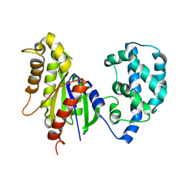
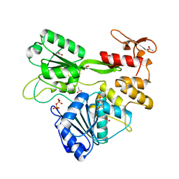
 | | Crystal structure of HAT domain of human Squamous Cell Carcinoma Antigen Recognized By T Cells 3, SART3 (TIP110) | | 分子名称: | Squamous cell carcinoma antigen recognized by T-cells 3 | | 著者 | Grazette, A, Harper, S, Emsley, J, Layfield, R, Dreveny, I. | | 登録日 | 2016-05-04 | | 公開日 | 2016-05-11 | | 最終更新日 | 2024-05-08 | | 実験手法 | X-RAY DIFFRACTION (3.045 Å) | | 主引用文献 | unpublished
To Be Published
|
|
4LI5
 | | EGFR-K IN COMPLEX WITH N-[3-[[5-chloro-4-(1H-indol-3-yl)pyrimidin-2-yl]amino]-4-methoxy-phenyl] Prop-2-enamide | | 分子名称: | Epidermal growth factor receptor, N-(3-{[5-chloro-4-(1H-indol-3-yl)pyrimidin-2-yl]amino}-4-methoxyphenyl)propanamide, SODIUM ION | | 著者 | Debreczeni, J.E, Seiffert, G.B, Kiefersauer, R, Augustin, M, Nagel, S, Ward, R, Anderton, M, Ashton, S, Bethel, P, Box, M, Butterworth, S, Colclough, N, Chroley, C, Chuaqui, C, Cross, D, Eberlein, C, Finlay, R, Hill, G, Grist, M, Klinowska, T, Lane, C, Martin, S, Orme, J, Smith, P, Wang, F, Waring, M. | | 登録日 | 2013-07-02 | | 公開日 | 2013-08-28 | | 最終更新日 | 2024-02-28 | | 実験手法 | X-RAY DIFFRACTION (2.64 Å) | | 主引用文献 | Structure- and Reactivity-Based Development of Covalent Inhibitors of the Activating and Gatekeeper Mutant Forms of the Epidermal Growth Factor Receptor (EGFR).
J.Med.Chem., 56, 2013
|
|
3BA0
 | | Crystal structure of full-length human MMP-12 | | 分子名称: | ACETOHYDROXAMIC ACID, CALCIUM ION, Macrophage metalloelastase, ... | | 著者 | Bertini, I, Calderone, V, Fragai, M, Jaiswal, R, Luchinat, C, Melikian, M, Myonas, E, Svergun, D.I. | | 登録日 | 2007-11-07 | | 公開日 | 2008-07-29 | | 最終更新日 | 2023-11-01 | | 実験手法 | X-RAY DIFFRACTION (3 Å) | | 主引用文献 | Evidence of reciprocal reorientation of the catalytic and hemopexin-like domains of full-length MMP-12.
J.Am.Chem.Soc., 130, 2008
|
|
5JTE
 | | Cryo-EM structure of an ErmBL-stalled ribosome in complex with A-, P-, and E-tRNA | | 分子名称: | 16S ribosomal RNA, 23S ribosomal RNA, 30S ribosomal protein S10, ... | | 著者 | Arenz, S, Bock, L.V, Graf, M, Innis, C.A, Beckmann, R, Grubmueller, H, Vaiana, A.C, Wilson, D.N. | | 登録日 | 2016-05-09 | | 公開日 | 2016-07-20 | | 最終更新日 | 2024-10-16 | | 実験手法 | ELECTRON MICROSCOPY (3.6 Å) | | 主引用文献 | A combined cryo-EM and molecular dynamics approach reveals the mechanism of ErmBL-mediated translation arrest.
Nat Commun, 7, 2016
|
|
5K7L
 | |
1GT3
 | | Complex of Bovine Odorant Binding Protein with dihydromyrcenol | | 分子名称: | (3S)-1-octen-3-ol, 2,6-DIMETHYL-7-OCTEN-2-OL, ODORANT-BINDING PROTEIN | | 著者 | Vincent, F, Ramoni, R, Spinelli, S, Grolli, S, Conti, V, Cambillau, C, Tegoni, M. | | 登録日 | 2002-01-10 | | 公開日 | 2003-10-03 | | 最終更新日 | 2023-12-13 | | 実験手法 | X-RAY DIFFRACTION (1.8 Å) | | 主引用文献 | Crystal Structures of Bovine Odorant-Binding Protein in Complex with Odorant Molecules.
Eur.J.Biochem., 271, 2004
|
|
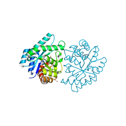
5KA8
 | | Protein Tyrosine Phosphatase 1B L192A mutant, open state | | 分子名称: | 2-AMINO-2-HYDROXYMETHYL-PROPANE-1,3-DIOL, CHLORIDE ION, Tyrosine-protein phosphatase non-receptor type 1 | | 著者 | Choy, M.S, Peti, W, Page, R. | | 登録日 | 2016-06-01 | | 公開日 | 2017-03-01 | | 最終更新日 | 2023-09-27 | | 実験手法 | X-RAY DIFFRACTION (1.971 Å) | | 主引用文献 | Conformational Rigidity and Protein Dynamics at Distinct Timescales Regulate PTP1B Activity and Allostery.
Mol. Cell, 65, 2017
|
|
5JP4
 | |
3JU2
 | | CRYSTAL STRUCTURE OF PROTEIN SMc04130 FROM Sinorhizobium meliloti 1021 | | 分子名称: | GLYCEROL, ZINC ION, uncharacterized protein SMc04130 | | 著者 | Patskovsky, Y, Foti, R, Ramagopal, U, Malashkevich, V, Toro, R, Freeman, J, Miller, S, Sauder, J.M, Raushel, F.M, Burley, S.K, Almo, S.C, New York SGX Research Center for Structural Genomics (NYSGXRC) | | 登録日 | 2009-09-14 | | 公開日 | 2009-09-22 | | 最終更新日 | 2024-02-21 | | 実験手法 | X-RAY DIFFRACTION (1.8 Å) | | 主引用文献 | CRYSTAL STRUCTURE OF PROTEIN SMc04130 FROM Sinorhizobium meliloti
To be Published
|
|
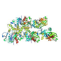
5JRQ
 | | BRAFV600E Kinase Domain In Complex with Chemically Linked Vemurafenib Inhibitor VEM-6-VEM | | 分子名称: | DIMETHYL SULFOXIDE, GLYCEROL, N-{2,4-difluoro-3-[5-(4-methoxyphenyl)-1H-pyrrolo[2,3-b]pyridine-3-carbonyl]phenyl}propane-1-sulfonamide, ... | | 著者 | Grasso, M.J, Marmorstein, R. | | 登録日 | 2016-05-06 | | 公開日 | 2016-09-14 | | 最終更新日 | 2023-10-18 | | 実験手法 | X-RAY DIFFRACTION (2.287 Å) | | 主引用文献 | Chemically Linked Vemurafenib Inhibitors Promote an Inactive BRAF(V600E) Conformation.
Acs Chem.Biol., 11, 2016
|
|
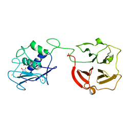
1HAX
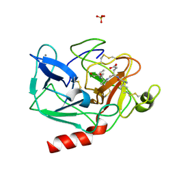 | | Snapshots of serine protease catalysis: (A) acyl-enzyme intermediate between porcine pancreatic elastase and human beta-casomorphin-7 at pH 5 | | 分子名称: | BETA-CASOMORPHIN-7, CALCIUM ION, ELASTASE 1, ... | | 著者 | Wilmouth, R.C, Edman, K, Neutze, R, Wright, P.A, Clifton, I.J, Schneider, T.R, Schofield, C.J, Hajdu, J. | | 登録日 | 2001-04-10 | | 公開日 | 2001-08-02 | | 最終更新日 | 2023-12-13 | | 実験手法 | X-RAY DIFFRACTION (1.6 Å) | | 主引用文献 | X-Ray Snapshots of Serine Protease Catalysis Reveal a Tetrahedral Intermediate
Nat.Struct.Biol., 8, 2001
|
|
5JWR
 | | Crystal structure of foldswitch-stabilized KaiB in complex with the N-terminal CI domain of KaiC and a dimer of KaiA C-terminal domains from Thermosynechococcus elongatus | | 分子名称: | Circadian clock protein KaiA, Circadian clock protein KaiB, Circadian clock protein kinase KaiC, ... | | 著者 | Tseng, R, Goularte, N.F, Chavan, A, Luu, J, Chang, Y.G, Heilser, J, Tripathi, S, LiWang, A, Partch, C.L. | | 登録日 | 2016-05-12 | | 公開日 | 2017-03-29 | | 最終更新日 | 2024-03-06 | | 実験手法 | X-RAY DIFFRACTION (2.61 Å) | | 主引用文献 | Structural basis of the day-night transition in a bacterial circadian clock.
Science, 355, 2017
|
|
1XKE
 | | Solution structure of the second Ran-binding domain from human RanBP2 | | 分子名称: | Ran-binding protein 2 | | 著者 | Geyer, J.P, Doeker, R, Kremer, W, Zhao, X, Kuhlmann, J, Kalbitzer, H.R. | | 登録日 | 2004-09-28 | | 公開日 | 2005-04-19 | | 最終更新日 | 2024-05-29 | | 実験手法 | SOLUTION NMR | | 主引用文献 | Solution structure of the Ran-binding domain 2 of RanBP2 and its interaction with the C terminus of Ran.
J.Mol.Biol., 348, 2005
|
|
4NOF
 | | Crystal structure of the second Ig domain from mouse Polymeric Immunoglobulin receptor [PSI-NYSGRC-006220] | | 分子名称: | 2-acetamido-2-deoxy-beta-D-glucopyranose, GLYCEROL, Polymeric immunoglobulin receptor | | 著者 | Sampathkumar, P, Kumar, P.R, Ahmed, M, Banu, R, Bhosle, R, Calarese, D.A, Celikgil, A, Chamala, S, Chan, M.K, Chowdhury, S, Fiser, A, Garforth, S.J, Glenn, A.S, Hillerich, B, Khafizov, K, Attonito, J, Love, J.D, Patel, H, Patel, R, Seidel, R.D, Smith, B, Stead, M, Casadevall, A, Almo, S.C, New York Structural Genomics Research Consortium (NYSGRC), Atoms-to-Animals: The Immune Function Network (IFN) | | 登録日 | 2013-11-19 | | 公開日 | 2013-12-04 | | 最終更新日 | 2023-09-20 | | 実験手法 | X-RAY DIFFRACTION (1.65 Å) | | 主引用文献 | Crystal structure of the second Ig domain from mouse Polymeric Immunoglobulin receptor
to be published
|
|
5JRZ
 | |
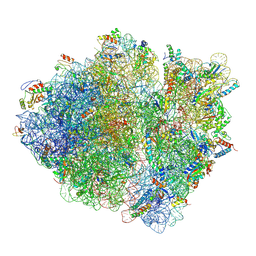
5JPQ
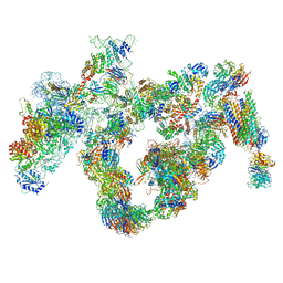 | | Cryo-EM structure of the 90S pre-ribosome | | 分子名称: | 18S ribosomal RNA, Bms1, Emg1, ... | | 著者 | Turk, M, Cheng, J, Berninghausen, O, Kornprobst, M, Flemming, D, Kos-Braun, I.C, Kos, M, Thoms, M, Hurt, E, Beckmann, R. | | 登録日 | 2016-05-04 | | 公開日 | 2016-07-27 | | 最終更新日 | 2024-05-08 | | 実験手法 | ELECTRON MICROSCOPY (7.3 Å) | | 主引用文献 | Architecture of the 90S Pre-ribosome: A Structural View on the Birth of the Eukaryotic Ribosome.
Cell, 166, 2016
|
|
2KW9
 | | Solution NMR Structure of SAP domain of MKL/myocardin-like protein 1 from H.sapiens, Northeast Structural Genomics Consortium Target HR4547E | | 分子名称: | MKL/myocardin-like protein 1 | | 著者 | Liu, G, Shastry, R, Ciccosanti, C, Janjua, H, Acton, T.B, Xiao, R, Mao, B, Everett, J.K, Montelione, G.T, Northeast Structural Genomics Consortium (NESG) | | 登録日 | 2010-03-31 | | 公開日 | 2010-06-16 | | 最終更新日 | 2024-05-15 | | 実験手法 | SOLUTION NMR | | 主引用文献 | Northeast Structural Genomics Consortium Target HR4547E
To be Published
|
|
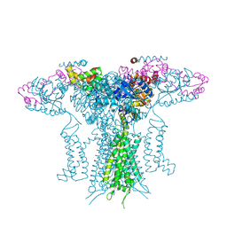
3RZE
 | | Structure of the human histamine H1 receptor in complex with doxepin | | 分子名称: | (2R)-2,3-dihydroxypropyl (9Z)-octadec-9-enoate, (3E)-3-(dibenzo[b,e]oxepin-11(6H)-ylidene)-N,N-dimethylpropan-1-amine, (3Z)-3-(dibenzo[b,e]oxepin-11(6H)-ylidene)-N,N-dimethylpropan-1-amine, ... | | 著者 | Shimamura, T, Han, G.W, Shiroishi, M, Weyand, S, Tsujimoto, H, Winter, G, Katritch, V, Abagyan, R, Cherezov, V, Liu, W, Kobayashi, T, Stevens, R, Iwata, S, GPCR Network (GPCR) | | 登録日 | 2011-05-11 | | 公開日 | 2011-06-15 | | 最終更新日 | 2023-09-13 | | 実験手法 | X-RAY DIFFRACTION (3.1 Å) | | 主引用文献 | Structure of the human histamine H1 receptor complex with doxepin.
Nature, 475, 2011
|
|
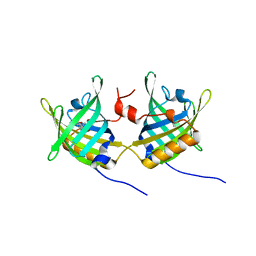
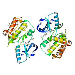
1GT1
 | | Complex of Bovine Odorant Binding Protein with Aminoanthracene and pyrazine | | 分子名称: | (3S)-1-octen-3-ol, 2-ISOBUTYL-3-METHOXYPYRAZINE, ANTHRACEN-1-YLAMINE, ... | | 著者 | Vincent, F, Ramoni, R, Spinelli, S, Grolli, S, Conti, V, Cambillau, C, Tegoni, M. | | 登録日 | 2002-01-10 | | 公開日 | 2003-10-03 | | 最終更新日 | 2023-12-13 | | 実験手法 | X-RAY DIFFRACTION (1.71 Å) | | 主引用文献 | Crystal Structures of Bovine Odorant-Binding Protein in Complex with Odorant Molecules.
Eur.J.Biochem., 271, 2004
|
|
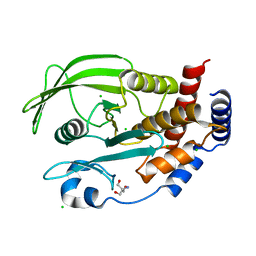
5JWQ
 | | Crystal structure of KaiC S431E in complex with foldswitch-stabilized KaiB from Thermosynechococcus elongatus | | 分子名称: | ADENOSINE-5'-DIPHOSPHATE, Circadian clock protein KaiB, Circadian clock protein kinase KaiC | | 著者 | Tseng, R, Goularte, N.F, Chavan, A, Luu, J, Chang, Y, Heilser, J, Tripathi, S, LiWang, A, Partch, C.L. | | 登録日 | 2016-05-12 | | 公開日 | 2017-03-29 | | 最終更新日 | 2024-03-06 | | 実験手法 | X-RAY DIFFRACTION (3.871 Å) | | 主引用文献 | Structural basis of the day-night transition in a bacterial circadian clock.
Science, 355, 2017
|
|


