2BEK
 
 | |
2ODY
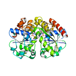
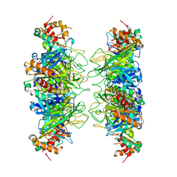
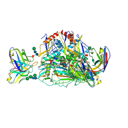
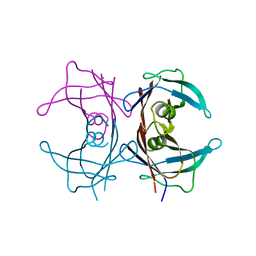
 | | Thrombin-bound boophilin displays a functional and accessible reactive-site loop | | 分子名称: | 2-acetamido-2-deoxy-beta-D-glucopyranose, Boophilin, PHOSPHATE ION, ... | | 著者 | Macedo-Ribeiro, S, Fuentes-Prior, P, Pereira, P.J.B. | | 登録日 | 2006-12-27 | | 公開日 | 2008-01-22 | | 最終更新日 | 2023-08-30 | | 実験手法 | X-RAY DIFFRACTION (2.35 Å) | | 主引用文献 | Isolation, cloning and structural characterisation of boophilin, a multifunctional kunitz-type proteinase inhibitor from the cattle tick.
Plos One, 3, 2008
|
|
3L5I
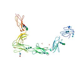
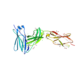 | | Crystal structure of FnIII domains of human GP130 (Domains 4-6) | | 分子名称: | 1,2-ETHANEDIOL, CHLORIDE ION, Interleukin-6 receptor subunit beta | | 著者 | Kershaw, N.J, Zhang, J.-G, Garrett, T.P.J, Czabotar, P.E. | | 登録日 | 2009-12-22 | | 公開日 | 2010-05-12 | | 最終更新日 | 2017-11-01 | | 実験手法 | X-RAY DIFFRACTION (1.9 Å) | | 主引用文献 | Crystal structure of the entire ectodomain of gp130: insights into the molecular assembly of the tall cytokine receptor complexes.
J.Biol.Chem., 285, 2010
|
|
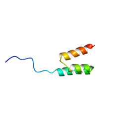
1POG
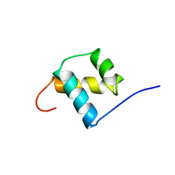 | | SOLUTION STRUCTURE OF THE OCT-1 POU-HOMEO DOMAIN DETERMINED BY NMR AND RESTRAINED MOLECULAR DYNAMICS | | 分子名称: | OCT-1 POU HOMEODOMAIN DNA-BINDING PROTEIN | | 著者 | Cox, M, Van Tilborg, P.J.A, De Laat, W, Boelens, R, Van Leeuwen, H.C, Van Der Vliet, P.C, Kaptein, R. | | 登録日 | 1994-10-12 | | 公開日 | 1995-07-31 | | 最終更新日 | 2024-05-22 | | 実験手法 | SOLUTION NMR | | 主引用文献 | Solution structure of the Oct-1 POU homeodomain determined by NMR and restrained molecular dynamics.
J.Biomol.NMR, 6, 1995
|
|
3V4R
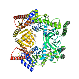 | | Crystal structure of a UvrB dimer-DNA complex | | 分子名称: | ADENOSINE-5'-DIPHOSPHATE, DNA: 5 -TACTGTTT-3, UvrABC system protein B | | 著者 | Webster, M.P.J, Jukes, R, Barrett, T. | | 登録日 | 2011-12-15 | | 公開日 | 2012-07-04 | | 最終更新日 | 2023-09-13 | | 実験手法 | X-RAY DIFFRACTION (3.25 Å) | | 主引用文献 | Crystal structure of the UvrB dimer: insights into the nature and functioning of the UvrAB damage engagement and UvrB-DNA complexes.
Nucleic Acids Res., 40, 2012
|
|
3L5J
 | | Crystal structure of FnIII domains of human GP130 (Domains 4-6) | | 分子名称: | 1,2-ETHANEDIOL, CHLORIDE ION, Interleukin-6 receptor subunit beta | | 著者 | Kershaw, N.J, Zhang, J.-G, Garrett, T.P.J, Czabotar, P.E. | | 登録日 | 2009-12-22 | | 公開日 | 2010-05-12 | | 最終更新日 | 2017-11-01 | | 実験手法 | X-RAY DIFFRACTION (3.042 Å) | | 主引用文献 | Crystal structure of the entire ectodomain of gp130: insights into the molecular assembly of the tall cytokine receptor complexes.
J.Biol.Chem., 285, 2010
|
|
3LL4
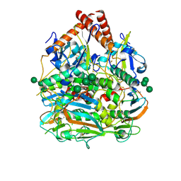
 | | Structure of the H13A mutant of Ykr043C in complex with fructose-1,6-bisphosphate | | 分子名称: | 1,6-FRUCTOSE DIPHOSPHATE (LINEAR FORM), Uncharacterized protein YKR043C | | 著者 | Singer, A, Xu, X, Cui, H, Dong, A, Stogios, P.J, Edwards, A.M, Joachimiak, A, Savchenko, A, Yakunin, A.F, Midwest Center for Structural Genomics (MCSG) | | 登録日 | 2010-01-28 | | 公開日 | 2010-03-09 | | 最終更新日 | 2023-09-06 | | 実験手法 | X-RAY DIFFRACTION (2.49 Å) | | 主引用文献 | Structure and activity of the metal-independent fructose-1,6-bisphosphatase YK23 from Saccharomyces cerevisiae.
J.Biol.Chem., 285, 2010
|
|
4IS4
 | |
4JRN
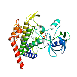 | | ROP18 kinase domain in complex with AMP-PNP and sucrose | | 分子名称: | MAGNESIUM ION, PHOSPHOAMINOPHOSPHONIC ACID-ADENYLATE ESTER, Rhoptry kinase family protein, ... | | 著者 | Lim, D, Gold, D.A, Lindsay, J, Rosowski, E.E, Niedelman, W, Yaffe, M.B, Saeij, J.P.J. | | 登録日 | 2013-03-21 | | 公開日 | 2013-10-23 | | 最終更新日 | 2020-07-29 | | 実験手法 | X-RAY DIFFRACTION (2.71 Å) | | 主引用文献 | Structure of the Toxoplasma gondii ROP18 kinase domain reveals a second ligand binding pocket required for acute virulence.
J.Biol.Chem., 288, 2013
|
|
3L5H
 | | Crystal structure of the full ectodomain of human gp130: New insights into the molecular assembly of receptor complexes | | 分子名称: | 2-acetamido-2-deoxy-beta-D-glucopyranose-(1-4)-2-acetamido-2-deoxy-beta-D-glucopyranose, Interleukin-6 receptor subunit beta, SULFATE ION, ... | | 著者 | Xu, Y, Garrett, T.P.J, Zhang, J.G. | | 登録日 | 2009-12-21 | | 公開日 | 2010-05-19 | | 最終更新日 | 2020-07-29 | | 実験手法 | X-RAY DIFFRACTION (3.6 Å) | | 主引用文献 | Crystal structure of the entire ectodomain of gp130: insights into the molecular assembly of the tall cytokine receptor complexes.
J.Biol.Chem., 285, 2010
|
|
2F9S
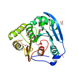
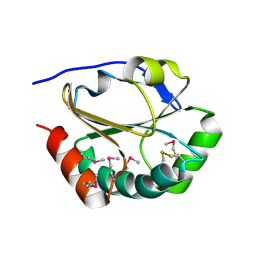 | | 2nd Crystal Structure Of A Soluble Domain Of ResA In The Oxidised Form | | 分子名称: | Thiol-disulfide oxidoreductase resA | | 著者 | Colbert, C.L, Wu, Q, Erbel, P.J.A, Gardner, K.H, Deisenhofer, J. | | 登録日 | 2005-12-06 | | 公開日 | 2006-04-18 | | 最終更新日 | 2011-07-13 | | 実験手法 | X-RAY DIFFRACTION (1.401 Å) | | 主引用文献 | Mechanism of substrate specificity in Bacillus subtilis ResA, a thioredoxin-like protein involved in cytochrome c maturation
Proc.Natl.Acad.Sci.USA, 103, 2006
|
|
4BAI
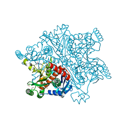 | | Mycobacterium tuberculosis Chorismate synthase before exposure to 266 nm UV laser | | 分子名称: | ACETATE ION, CHORISMATE SYNTHASE | | 著者 | Pereira, P.J.B, Royant, A, Panjikar, S, de Sanctis, D. | | 登録日 | 2012-09-14 | | 公開日 | 2013-04-17 | | 最終更新日 | 2023-11-15 | | 実験手法 | X-RAY DIFFRACTION (2.3 Å) | | 主引用文献 | In-house UV radiation-damage-induced phasing of selenomethionine-labeled protein structures.
J. Struct. Biol., 181, 2013
|
|
4BAJ
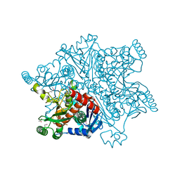 | | MYCOBACTERIUM TUBERCULOSIS CHORISMATE SYNTHASE after exposure to 266nm UV laser | | 分子名称: | ACETATE ION, CHORISMATE SYNTHASE | | 著者 | Pereira, P.J.B, Royant, A, Panjikar, S, de Sanctis, D. | | 登録日 | 2012-09-14 | | 公開日 | 2013-04-17 | | 最終更新日 | 2019-03-06 | | 実験手法 | X-RAY DIFFRACTION (2.3 Å) | | 主引用文献 | In-house UV radiation-damage-induced phasing of selenomethionine-labeled protein structures.
J. Struct. Biol., 181, 2013
|
|
4BAG
 | |
1WCV
 | |
1WYY
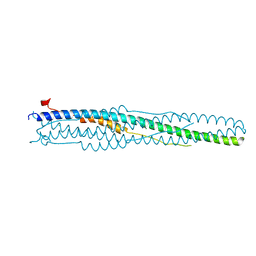 | | Post-fusion hairpin conformation of the sars coronavirus spike glycoprotein | | 分子名称: | CHLORIDE ION, E2 Glycoprotein | | 著者 | Duquerroy, S, Vigouroux, A, Rottier, P.J.M, Rey, F.A, Bosch, B.J. | | 登録日 | 2005-02-18 | | 公開日 | 2005-05-17 | | 最終更新日 | 2023-10-25 | | 実験手法 | X-RAY DIFFRACTION (2.2 Å) | | 主引用文献 | Central ions and lateral asparagine/glutamine zippers stabilize the post-fusion hairpin conformation of the SARS coronavirus spike glycoprotein
Virology, 335, 2005
|
|
7SGF
 | | Lassa virus glycoprotein construct (Josiah GPC-I53-50A) in complex with LAVA01 antibody | | 分子名称: | 2-acetamido-2-deoxy-beta-D-glucopyranose, 2-acetamido-2-deoxy-beta-D-glucopyranose-(1-4)-2-acetamido-2-deoxy-beta-D-glucopyranose, 2-acetamido-2-deoxy-beta-D-glucopyranose-(1-4)-[alpha-L-fucopyranose-(1-6)]2-acetamido-2-deoxy-beta-D-glucopyranose, ... | | 著者 | Antanasijevic, A, Brouwer, P.J.M, Ward, A.B. | | 登録日 | 2021-10-05 | | 公開日 | 2022-10-12 | | 最終更新日 | 2022-12-28 | | 実験手法 | ELECTRON MICROSCOPY (4.41 Å) | | 主引用文献 | Lassa virus glycoprotein nanoparticles elicit neutralizing antibody responses and protection.
Cell Host Microbe, 30, 2022
|
|
7SGE
 | |
7SGD
 | | Lassa virus glycoprotein construct(Josiah GPCysR4) recovered from GPC-I53-50 nanoparticle by localized reconstruction | | 分子名称: | 2-acetamido-2-deoxy-beta-D-glucopyranose, 2-acetamido-2-deoxy-beta-D-glucopyranose-(1-4)-2-acetamido-2-deoxy-beta-D-glucopyranose, 2-acetamido-2-deoxy-beta-D-glucopyranose-(1-4)-[alpha-L-fucopyranose-(1-6)]2-acetamido-2-deoxy-beta-D-glucopyranose, ... | | 著者 | Antanasijevic, A, Brouwer, P.J.M, Ward, A.B. | | 登録日 | 2021-10-05 | | 公開日 | 2022-10-12 | | 最終更新日 | 2022-12-28 | | 実験手法 | ELECTRON MICROSCOPY (3.97 Å) | | 主引用文献 | Lassa virus glycoprotein nanoparticles elicit neutralizing antibody responses and protection.
Cell Host Microbe, 30, 2022
|
|
1X7T
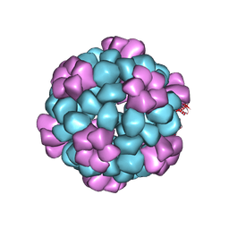
 | | Structure of TTR R104H: a non-amyloidogenic variant with protective clinical effects | | 分子名称: | Transthyretin | | 著者 | Neto-Silva, R.M, Macedo-Ribeiro, S, Pereira, P.J.B, Coll, M, Saraiva, M.J, Damas, A.M. | | 登録日 | 2004-08-16 | | 公開日 | 2005-03-22 | | 最終更新日 | 2023-08-23 | | 実験手法 | X-RAY DIFFRACTION (1.6 Å) | | 主引用文献 | X-ray crystallographic studies of two transthyretin variants: further insights into amyloidogenesis.
Acta Crystallogr.,Sect.D, 61, 2005
|
|
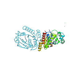
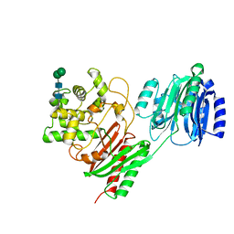
1S63
 | | Human protein farnesyltransferase complexed with L-778,123 and FPP | | 分子名称: | 4-[(5-{[4-(3-CHLOROPHENYL)-3-OXOPIPERAZIN-1-YL]METHYL}-1H-IMIDAZOL-1-YL)METHYL]BENZONITRILE, FARNESYL DIPHOSPHATE, Protein farnesyltransferase beta subunit, ... | | 著者 | Long, S.B, Casey, P.J, Beese, L.S. | | 登録日 | 2004-01-22 | | 公開日 | 2004-07-27 | | 最終更新日 | 2023-08-23 | | 実験手法 | X-RAY DIFFRACTION (1.9 Å) | | 主引用文献 | Crystallographic Analysis Reveals that Anticancer Clinical Candidate L-778,123 Inhibits Protein Farnesyltransferase and Geranylgeranyltransferase-I by Different Binding Modes.
Biochemistry, 43, 2004
|
|
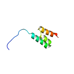
1GJS
 | | Solution structure of the Albumin binding domain of Streptococcal Protein G | | 分子名称: | IMMUNOGLOBULIN G BINDING PROTEIN G | | 著者 | Johansson, M.U, Frick, I.M, Nilsson, H, Kraulis, P.J, Hober, S, Jonasson, P, Nygren, A.P, Uhlen, M, Bjorck, L, Drakenberg, T, Forsen, S, Wikstrom, M. | | 登録日 | 2001-08-02 | | 公開日 | 2001-08-09 | | 最終更新日 | 2024-05-15 | | 実験手法 | SOLUTION NMR | | 主引用文献 | Structure, Specificity, and Mode of Interaction for Bacterial Albumin-Binding Modules
J.Biol.Chem., 277, 2002
|
|
1GJT
 | | Solution structure of the Albumin binding domain of Streptococcal Protein G | | 分子名称: | IMMUNOGLOBULIN G BINDING PROTEIN G | | 著者 | Johansson, M.U, Frick, I.M, Nilsson, H, Kraulis, P.J, Hober, S, Jonasson, P, Nygren, A.P, Uhlen, M, Bjorck, L, Drakenberg, T, Forsen, S, Wikstrom, M. | | 登録日 | 2001-08-02 | | 公開日 | 2001-08-09 | | 最終更新日 | 2024-05-15 | | 実験手法 | SOLUTION NMR | | 主引用文献 | Structure, Specificity, and Mode of Interaction for Bacterial Albumin-Binding Modules
J.Biol.Chem., 277, 2002
|
|
1GKA
 | | The molecular basis of the coloration mechanism in lobster shell. beta-crustacyanin at 3.2 A resolution | | 分子名称: | 2-AMINO-2-HYDROXYMETHYL-PROPANE-1,3-DIOL, 4-(2-HYDROXYETHYL)-1-PIPERAZINE ETHANESULFONIC ACID, ASTAXANTHIN, ... | | 著者 | Cianci, M, Rizkallah, P.J, Olczak, A, Raftery, J, Chayen, N.E, Zagalsky, P.F, Helliwell, J.R. | | 登録日 | 2001-08-10 | | 公開日 | 2002-08-08 | | 最終更新日 | 2023-12-13 | | 実験手法 | X-RAY DIFFRACTION (3.23 Å) | | 主引用文献 | The Molecular Basis of the Coloration Mechanism in Lobster Shell: Beta -Crustacyanin at 3.2-A Resolution
Proc.Natl.Acad.Sci.USA, 99, 2002
|
|
7TPU
 | | Crystal structure of a chitinase-modifying protein from Fusarium vanettenii (Fvan-cmp) | | 分子名称: | 2-acetamido-2-deoxy-beta-D-glucopyranose-(1-4)-2-acetamido-2-deoxy-beta-D-glucopyranose, Beta-lactamase domain-containing protein, alpha-D-mannopyranose-(1-3)-beta-D-mannopyranose-(1-4)-2-acetamido-2-deoxy-beta-D-glucopyranose-(1-4)-2-acetamido-2-deoxy-beta-D-glucopyranose | | 著者 | Dowling, N.V, Naumann, T.A, Price, N.P.J, Rose, D.R. | | 登録日 | 2022-01-26 | | 公開日 | 2023-02-15 | | 最終更新日 | 2024-04-03 | | 実験手法 | X-RAY DIFFRACTION (2.194 Å) | | 主引用文献 | Crystal structure of a polyglycine hydrolase determined using a RoseTTAFold model.
Acta Crystallogr D Struct Biol, 79, 2023
|
|


