6XM2
 
 | |
5D6C
 | |
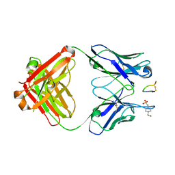
3LB6
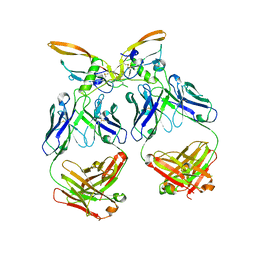
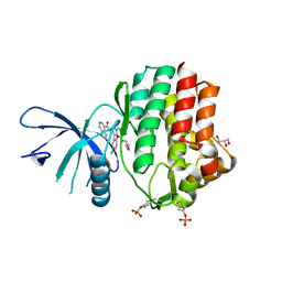
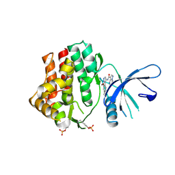
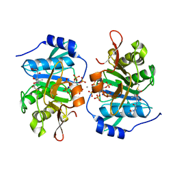
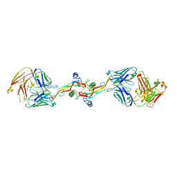
 | | The structure of IL-13 in complex with IL-13Ralpha2 | | 分子名称: | 2-acetamido-2-deoxy-beta-D-glucopyranose, CALCIUM ION, Interleukin-13, ... | | 著者 | Lupardus, P.J, Garcia, K.C, Birnbaum, M.E. | | 登録日 | 2010-01-07 | | 公開日 | 2010-03-16 | | 最終更新日 | 2024-04-03 | | 実験手法 | X-RAY DIFFRACTION (3.05 Å) | | 主引用文献 | Molecular basis for shared cytokine recognition revealed in the structure of an unusually high affinity complex between IL-13 and IL-13Ralpha2.
Structure, 18, 2010
|
|
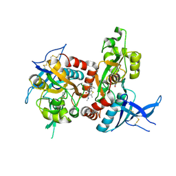
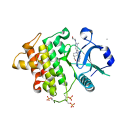
4EHZ
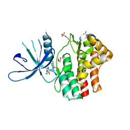
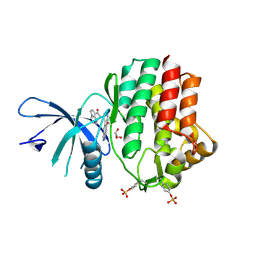
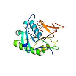
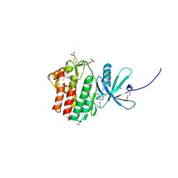 | | The Jak1 kinase domain in complex with inhibitor | | 分子名称: | 1,2-ETHANEDIOL, 2-methyl-1-(piperidin-4-yl)-1,6-dihydroimidazo[4,5-d]pyrrolo[2,3-b]pyridine, Tyrosine-protein kinase JAK1 | | 著者 | Lupardus, P.J, Steffek, M. | | 登録日 | 2012-04-04 | | 公開日 | 2012-07-04 | | 最終更新日 | 2013-01-23 | | 実験手法 | X-RAY DIFFRACTION (2.174 Å) | | 主引用文献 | Discovery and optimization of C-2 methyl imidazopyrrolopyridines as potent and orally bioavailable JAK1 inhibitors with selectivity over JAK2.
J.Med.Chem., 55, 2012
|
|
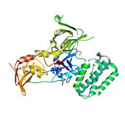
6N7B
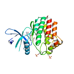
 | | Structure of the human JAK1 kinase domain with compound 38 | | 分子名称: | GLYCEROL, N-[3-(5-chloro-2-methoxyphenyl)-1-methyl-1H-pyrazol-4-yl]-1H-pyrazolo[4,3-c]pyridine-7-carboxamide, Tyrosine-protein kinase JAK1 | | 著者 | Lupardus, P.J, Brown, D. | | 登録日 | 2018-11-27 | | 公開日 | 2019-04-24 | | 最終更新日 | 2019-05-15 | | 実験手法 | X-RAY DIFFRACTION (1.81 Å) | | 主引用文献 | Discovery of a class of highly potent Janus Kinase 1/2 (JAK1/2) inhibitors demonstrating effective cell-based blockade of IL-13 signaling.
Bioorg.Med.Chem.Lett., 29, 2019
|
|
6N79
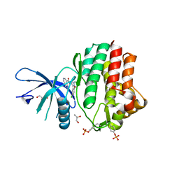
 | | Structure of the human JAK1 kinase domain with compound 20 | | 分子名称: | GLYCEROL, N-{5-[5-chloro-2-(difluoromethoxy)phenyl]-1H-pyrazol-4-yl}pyrazolo[1,5-a]pyrimidine-3-carboxamide, Tyrosine-protein kinase JAK1 | | 著者 | Lupardus, P.J, Brown, D. | | 登録日 | 2018-11-27 | | 公開日 | 2019-04-24 | | 最終更新日 | 2019-05-15 | | 実験手法 | X-RAY DIFFRACTION (2.27 Å) | | 主引用文献 | Discovery of a class of highly potent Janus Kinase 1/2 (JAK1/2) inhibitors demonstrating effective cell-based blockade of IL-13 signaling.
Bioorg.Med.Chem.Lett., 29, 2019
|
|
6N78
 | | Structure of the human JAK1 kinase domain with compound 21 | | 分子名称: | GLYCEROL, N-{3-[5-chloro-2-(difluoromethoxy)phenyl]-1-methyl-1H-pyrazol-4-yl}pyrazolo[1,5-a]pyrimidine-3-carboxamide, Tyrosine-protein kinase JAK1 | | 著者 | Lupardus, P.J, Brown, D. | | 登録日 | 2018-11-27 | | 公開日 | 2019-04-24 | | 最終更新日 | 2019-05-15 | | 実験手法 | X-RAY DIFFRACTION (1.83 Å) | | 主引用文献 | Discovery of a class of highly potent Janus Kinase 1/2 (JAK1/2) inhibitors demonstrating effective cell-based blockade of IL-13 signaling.
Bioorg.Med.Chem.Lett., 29, 2019
|
|
6N7A
 | | Structure of the human JAK1 kinase domain with compound 39 | | 分子名称: | GLYCEROL, N-[3-(5-chloro-2-methoxyphenyl)-1-methyl-1H-pyrazol-4-yl]-2-methyl-2H-pyrazolo[4,3-c]pyridine-7-carboxamide, Tyrosine-protein kinase JAK1 | | 著者 | Lupardus, P.J, Brown, D. | | 登録日 | 2018-11-27 | | 公開日 | 2019-04-24 | | 最終更新日 | 2019-05-15 | | 実験手法 | X-RAY DIFFRACTION (1.33 Å) | | 主引用文献 | Discovery of a class of highly potent Janus Kinase 1/2 (JAK1/2) inhibitors demonstrating effective cell-based blockade of IL-13 signaling.
Bioorg.Med.Chem.Lett., 29, 2019
|
|
6N7C
 | | Structure of the human JAK1 kinase domain with compound 56 | | 分子名称: | GLYCEROL, N-[5-(3-methoxynaphthalen-2-yl)-1H-pyrazol-4-yl]pyrazolo[1,5-a]pyrimidine-3-carboxamide, Tyrosine-protein kinase JAK1 | | 著者 | Lupardus, P.J, Brown, D. | | 登録日 | 2018-11-27 | | 公開日 | 2019-04-24 | | 最終更新日 | 2019-05-15 | | 実験手法 | X-RAY DIFFRACTION (1.69 Å) | | 主引用文献 | Discovery of a class of highly potent Janus Kinase 1/2 (JAK1/2) inhibitors demonstrating effective cell-based blockade of IL-13 signaling.
Bioorg.Med.Chem.Lett., 29, 2019
|
|
6N77
 | | Structure of the human JAK1 kinase domain with compound 15 | | 分子名称: | GLYCEROL, N-[3-(5-chloro-2-methoxyphenyl)-1-methyl-1H-pyrazol-4-yl]pyrazolo[1,5-a]pyrimidine-3-carboxamide, Tyrosine-protein kinase JAK1 | | 著者 | Lupardus, P.J, Brown, D. | | 登録日 | 2018-11-27 | | 公開日 | 2019-04-24 | | 最終更新日 | 2019-05-15 | | 実験手法 | X-RAY DIFFRACTION (1.64 Å) | | 主引用文献 | Discovery of a class of highly potent Janus Kinase 1/2 (JAK1/2) inhibitors demonstrating effective cell-based blockade of IL-13 signaling.
Bioorg.Med.Chem.Lett., 29, 2019
|
|
6N7D
 | | Structure of the human JAK1 kinase domain with compound 54 | | 分子名称: | GLYCEROL, N-[5-(6-methoxy-1H-indazol-5-yl)-1H-pyrazol-4-yl]pyrazolo[1,5-a]pyrimidine-3-carboxamide, Tyrosine-protein kinase JAK1 | | 著者 | Lupardus, P.J, Brown, D. | | 登録日 | 2018-11-27 | | 公開日 | 2019-04-24 | | 最終更新日 | 2019-05-15 | | 実験手法 | X-RAY DIFFRACTION (1.78 Å) | | 主引用文献 | Discovery of a class of highly potent Janus Kinase 1/2 (JAK1/2) inhibitors demonstrating effective cell-based blockade of IL-13 signaling.
Bioorg.Med.Chem.Lett., 29, 2019
|
|
3GCD
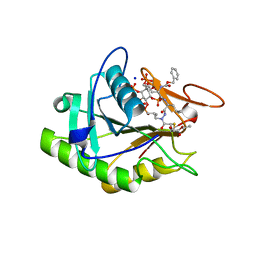 | | Structure of the V. cholerae RTX cysteine protease domain in complex with an aza-Leucine peptide inhibitor | | 分子名称: | INOSITOL HEXAKISPHOSPHATE, RTX toxin RtxA, SODIUM ION, ... | | 著者 | Lupardus, P.J, Garcia, K.C, Shen, A, Bogyo, M. | | 登録日 | 2009-02-21 | | 公開日 | 2009-05-26 | | 最終更新日 | 2023-09-06 | | 実験手法 | X-RAY DIFFRACTION (2.35 Å) | | 主引用文献 | Mechanistic and structural insights into the proteolytic activation of Vibrio cholerae MARTX toxin.
Nat.Chem.Biol., 5, 2009
|
|
3PEE
 | |
3PA8
 | |
3EEB
 | | Structure of the V. cholerae RTX cysteine protease domain | | 分子名称: | INOSITOL HEXAKISPHOSPHATE, RTX toxin RtxA, SODIUM ION | | 著者 | Lupardus, P.J, Shen, A, Bogyo, M, Garcia, K.C. | | 登録日 | 2008-09-04 | | 公開日 | 2008-10-21 | | 最終更新日 | 2024-02-21 | | 実験手法 | X-RAY DIFFRACTION (2.1 Å) | | 主引用文献 | Small molecule-induced allosteric activation of the Vibrio cholerae RTX cysteine protease domain
Science, 322, 2008
|
|
3DUH
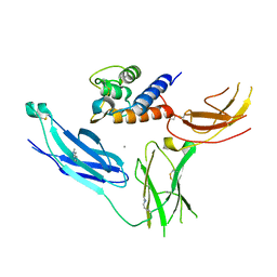
 | | Structure of Interleukin-23 | | 分子名称: | 2-acetamido-2-deoxy-beta-D-glucopyranose, Interleukin-12 subunit beta, Interleukin-23 subunit alpha | | 著者 | Lupardus, P.J, Garcia, K.C. | | 登録日 | 2008-07-17 | | 公開日 | 2008-08-19 | | 最終更新日 | 2023-08-30 | | 実験手法 | X-RAY DIFFRACTION (2.3 Å) | | 主引用文献 | The structure of interleukin-23 reveals the molecular basis of p40 subunit sharing with interleukin-12.
J.Mol.Biol., 382, 2008
|
|
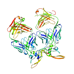
3E0G
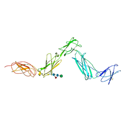 | | Structure of the Leukemia Inhibitory Factor Receptor (LIF-R) domains D1-D5 | | 分子名称: | 2-acetamido-2-deoxy-beta-D-glucopyranose-(1-4)-2-acetamido-2-deoxy-beta-D-glucopyranose, 2-acetamido-2-deoxy-beta-D-glucopyranose-(1-4)-[alpha-L-fucopyranose-(1-6)]2-acetamido-2-deoxy-beta-D-glucopyranose, Leukemia inhibitory factor receptor, ... | | 著者 | Lupardus, P.J, Garcia, K.C. | | 登録日 | 2008-07-31 | | 公開日 | 2008-09-23 | | 最終更新日 | 2021-10-20 | | 実験手法 | X-RAY DIFFRACTION (3.1 Å) | | 主引用文献 | Structural organization of a full-length gp130/LIF-R cytokine receptor transmembrane complex.
Mol.Cell, 31, 2008
|
|
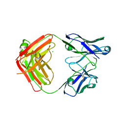
7S4G
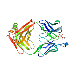
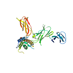
 | | Fab fragment bound to the Cter peptide of Ly6G6D | | 分子名称: | 4-(2-HYDROXYETHYL)-1-PIPERAZINE ETHANESULFONIC ACID, GLYCEROL, Lymphocyte antigen 6 complex locus protein G6d, ... | | 著者 | Rouge, L, Lupardus, P. | | 登録日 | 2021-09-08 | | 公開日 | 2022-04-13 | | 最終更新日 | 2023-10-18 | | 実験手法 | X-RAY DIFFRACTION (2.2 Å) | | 主引用文献 | Novel Anti-LY6G6D/CD3 T-Cell-Dependent Bispecific Antibody for the Treatment of Colorectal Cancer.
Mol.Cancer Ther., 21, 2022
|
|
5I2K
 | | Structure of the human GluN1/GluN2A LBD in complex with 7-{[ethyl(4-fluorophenyl)amino]methyl}-N,2-dimethyl-5-oxo-5H-[1,3]thiazolo[3,2-a]pyrimidine-3-carboxamide (compound 19) | | 分子名称: | 7-{[ethyl(4-fluorophenyl)amino]methyl}-N,2-dimethyl-5-oxo-5H-[1,3]thiazolo[3,2-a]pyrimidine-3-carboxamide, GLUTAMIC ACID, GLYCINE, ... | | 著者 | Wallweber, H.J.A, Lupardus, P.J. | | 登録日 | 2016-02-09 | | 公開日 | 2016-03-16 | | 最終更新日 | 2023-09-27 | | 実験手法 | X-RAY DIFFRACTION (2.86 Å) | | 主引用文献 | Discovery of GluN2A-Selective NMDA Receptor Positive Allosteric Modulators (PAMs): Tuning Deactivation Kinetics via Structure-Based Design.
J.Med.Chem., 59, 2016
|
|
4PO6
 | |
7RCO
 | |
8V52
 | |
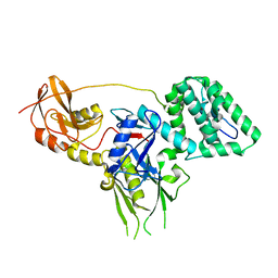
6O94
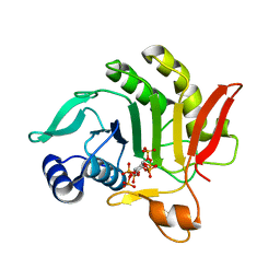
 | | Structure of the IRAK4 kinase domain with compound 17 | | 分子名称: | CALCIUM ION, Interleukin-1 receptor-associated kinase 4, N-{5-[4-(hydroxymethyl)piperidin-1-yl]-1-methyl-2-(morpholin-4-yl)-1H-benzimidazol-6-yl}pyrazolo[1,5-a]pyrimidine-3-carboxamide | | 著者 | Yu, C, Drobnick, J, Bryan, M.C, Kiefer, J, Lupardus, P.J. | | 登録日 | 2019-03-13 | | 公開日 | 2019-05-22 | | 最終更新日 | 2024-04-03 | | 実験手法 | X-RAY DIFFRACTION (1.98 Å) | | 主引用文献 | Development of Potent and Selective Pyrazolopyrimidine IRAK4 Inhibitors.
J.Med.Chem., 62, 2019
|
|
6DWI
 | |
6E2Q
 | |


