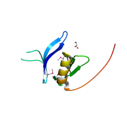
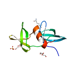
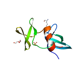
Loading
PDBj
MenuPDBj@FacebookPDBj@TwitterPDBj@YouTubewwPDB FoundationwwPDB
RCSB PDBPDBeBMRBAdv. SearchSearch help
PDB: 75 results

8E4V
DownloadVisualize
BU of 8e4v by Molmil
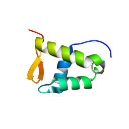
Solution structure of the WH domain of MORF
Descriptor: Isoform 3 of Histone acetyltransferase KAT6B
Authors:Zhang, Y, Kutateladze, T.G.
Deposit date:2022-08-19
Release date:2023-05-10
Last modified:2024-05-15
Method:SOLUTION NMR
Cite:MORF and MOZ acetyltransferases target unmethylated CpG islands through the winged helix domain.
Nat Commun, 14, 2023
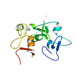
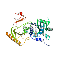
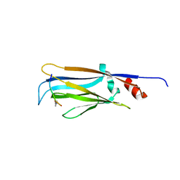
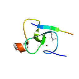
5D7E
DownloadVisualize
BU of 5d7e by Molmil
Crystal structure of Taf14 YEATS domain in complex with H3K9ac
Descriptor: 2-(N-MORPHOLINO)-ETHANESULFONIC ACID, DI(HYDROXYETHYL)ETHER, H3K9ac, ...
Authors:Andrews, F.H, Shanle, E.K, Strahl, B.D, Kutateladze, T.G.
Deposit date:2015-08-13
Release date:2015-09-23
Last modified:2019-12-11
Method:X-RAY DIFFRACTION (1.9 Å)
Cite:Association of Taf14 with acetylated histone H3 directs gene transcription and the DNA damage response.
Genes Dev., 29, 2015
5ERC
DownloadVisualize
BU of 5erc by Molmil
X-ray crystal structure of BRPF1 PZP domain
Descriptor: CALCIUM ION, Peregrin, ZINC ION
Authors:Klein, B.J, Andrews, F.H, Kutateladze, T.G.
Deposit date:2015-11-13
Release date:2015-12-30
Last modified:2024-03-06
Method:X-RAY DIFFRACTION (2.05 Å)
Cite:Bivalent interaction of the PZP domain of BRPF1 with the nucleosome impacts chromatin dynamics and acetylation.
Nucleic Acids Res., 44, 2016
4L7X
DownloadVisualize
BU of 4l7x by Molmil
Crystal structure of the DIDO PHD finger in complex with H3K4me3
Descriptor: Death-inducer obliterator 1, Histone H3 peptide, ZINC ION
Authors:Tong, Q, Gatchalian, J, Kutateladze, T.G.
Deposit date:2013-06-14
Release date:2013-07-24
Method:X-RAY DIFFRACTION (1.35 Å)
Cite:Dido3 PHD Modulates Cell Differentiation and Division.
Cell Rep, 4, 2013
7TV0
DownloadVisualize
BU of 7tv0 by Molmil
Crystal structure of BRD4 bromodomain 1 in complex with dual-acetylated SARS-CoV-2 E
Descriptor: Bromodomain-containing protein 4, Envelope small membrane protein
Authors:Vann, K.R, Holt, T.A, Kutateladze, T.G.
Deposit date:2022-02-03
Release date:2022-07-06
Last modified:2023-11-15
Method:X-RAY DIFFRACTION (2.6 Å)
Cite:Binding of the SARS-CoV-2 envelope E protein to human BRD4 is essential for infection.
Structure, 30, 2022
6DS6
DownloadVisualize
BU of 6ds6 by Molmil
Crystal structure of p300 ZZ domain in complex with histone H3 peptide
Descriptor: CHLORIDE ION, Histone H3 peptide-Histone acetyltransferase p300 Chimeric protein, ZINC ION
Authors:Zhang, Y, Kutateladze, T.G.
Deposit date:2018-06-13
Release date:2018-08-29
Last modified:2024-03-13
Method:X-RAY DIFFRACTION (1.95 Å)
Cite:The ZZ domain of p300 mediates specificity of the adjacent HAT domain for histone H3.
Nat. Struct. Mol. Biol., 25, 2018
7TUQ
DownloadVisualize
BU of 7tuq by Molmil
Crystal structure of BRD4 bromodomain 1 in complex with monoacetylated SARS-CoV-2 E
Descriptor: Bromodomain-containing protein 4, Envelope small membrane protein
Authors:Vann, K.R, Kutateladze, T.G.
Deposit date:2022-02-03
Release date:2022-07-06
Last modified:2023-11-15
Method:X-RAY DIFFRACTION (2.68 Å)
Cite:Binding of the SARS-CoV-2 envelope E protein to human BRD4 is essential for infection.
Structure, 30, 2022
7LKY
DownloadVisualize
BU of 7lky by Molmil
Crystal Structure of PHF1 Tudor domain in complex with a peptidomimetic ligand UNC6641
Descriptor: DIMETHYL SULFOXIDE, Isoform 1 of PHD finger protein 1, Peptidomimetic inhibitor UNC6641, ...
Authors:Liu, J, Kutateladze, T.G.
Deposit date:2021-02-03
Release date:2021-12-15
Last modified:2023-10-18
Method:X-RAY DIFFRACTION (1.85 Å)
Cite:Discovery of an H3K36me3-Derived Peptidomimetic Ligand with Enhanced Affinity for Plant Homeodomain Finger Protein 1 (PHF1).
J.Med.Chem., 64, 2021
4L58
DownloadVisualize
BU of 4l58 by Molmil
Crystal structure of the MLL5 PHD finger in complex with H3K4me3
Descriptor: Histone H3 peptide, Histone-lysine N-methyltransferase MLL5, ZINC ION
Authors:Tong, Q, Ali, M, Kutateladze, T.G.
Deposit date:2013-06-10
Release date:2013-06-26
Last modified:2013-07-24
Method:X-RAY DIFFRACTION (1.48 Å)
Cite:Molecular basis for chromatin binding and regulation of MLL5.
Proc.Natl.Acad.Sci.USA, 110, 2013
3RCP
DownloadVisualize
BU of 3rcp by Molmil
Crystal structure of the FAPP1 pleckstrin homology domain
Descriptor: GLYCEROL, Pleckstrin homology domain-containing family A member 3
Authors:Roy, S, He, J, Kutateladze, T.G.
Deposit date:2011-03-31
Release date:2011-04-20
Last modified:2017-11-08
Method:X-RAY DIFFRACTION (1.9 Å)
Cite:Molecular Basis of Phosphatidylinositol 4-Phosphate and ARF1 GTPase Recognition by the FAPP1 Pleckstrin Homology (PH) Domain.
J.Biol.Chem., 286, 2011
6WXK
DownloadVisualize
BU of 6wxk by Molmil
PHF23 PHD Domain Apo
Descriptor: PHD finger protein 23, ZINC ION
Authors:Vann, K.R, Zhang, J, Zhang, Y, Kutateladze, T.
Deposit date:2020-05-11
Release date:2020-07-15
Last modified:2023-10-18
Method:X-RAY DIFFRACTION (2.9 Å)
Cite:Mechanistic insights into chromatin targeting by leukemic NUP98-PHF23 fusion.
Nat Commun, 11, 2020
7K7T
DownloadVisualize
BU of 7k7t by Molmil
Crystal structure of human MORC4 ATPase-CW in complex with AMPPNP
Descriptor: Isoform 3 of MORC family CW-type zinc finger protein 4, MAGNESIUM ION, PHOSPHOAMINOPHOSPHONIC ACID-ADENYLATE ESTER, ...
Authors:Klein, B.J, Tencer, A.H, Kutateladze, T.G.
Deposit date:2020-09-24
Release date:2020-11-11
Last modified:2023-10-18
Method:X-RAY DIFFRACTION (2.94 Å)
Cite:Molecular mechanism of the MORC4 ATPase activation.
Nat Commun, 11, 2020
7MJU
DownloadVisualize
BU of 7mju by Molmil
Crystal structure of human AF10 PZP bound to histone H3 tail
Descriptor: Histone H3.1,Protein AF-10, ZINC ION
Authors:Klein, B.J, Kutateladze, T.G.
Deposit date:2021-04-20
Release date:2021-08-04
Last modified:2024-05-22
Method:X-RAY DIFFRACTION (2.1 Å)
Cite:The role of the PZP domain of AF10 in acute leukemia driven by AF10 translocations.
Nat Commun, 12, 2021
3LH0
DownloadVisualize
BU of 3lh0 by Molmil
Crystal structure of the 53BP1 tandem tudor domain in complex with p53K372me2
Descriptor: DIMETHYLATED p53 LYSINE 372 PEPTIDE, SULFATE ION, TRIETHYLENE GLYCOL, ...
Authors:Roy, S, Kutateladze, T.G.
Deposit date:2010-01-21
Release date:2010-03-23
Last modified:2023-09-06
Method:X-RAY DIFFRACTION (1.9 Å)
Cite:Structural insight into p53 recognition by the 53BP1 tandem Tudor domain.
J.Mol.Biol., 398, 2010
6MIN
DownloadVisualize
BU of 6min by Molmil
Crystal structure of Taf14 YEATS domain G82A mutant in complex with histone H3K9cr
Descriptor: Histone H3K9cr, Transcription initiation factor TFIID subunit 14
Authors:Klein, B.J, Andrews, F.H, Kutateladze, T.G.
Deposit date:2018-09-19
Release date:2018-11-14
Last modified:2023-11-15
Method:X-RAY DIFFRACTION (1.9 Å)
Cite:Structural insights into the pi-pi-pi stacking mechanism and DNA-binding activity of the YEATS domain.
Nat Commun, 9, 2018
6MIM
DownloadVisualize
BU of 6mim by Molmil
Crystal structure of AF9 YEATS domain Y78W mutant in complex with histone H3K9cr
Descriptor: Histone H3K9cr, Protein AF-9
Authors:Vann, K.R, Klein, B.J, Kutateladze, T.G.
Deposit date:2018-09-19
Release date:2018-11-14
Last modified:2023-11-15
Method:X-RAY DIFFRACTION (2.525 Å)
Cite:Structural insights into the pi-pi-pi stacking mechanism and DNA-binding activity of the YEATS domain.
Nat Commun, 9, 2018
6MIO
DownloadVisualize
BU of 6mio by Molmil
Crystal structure of Taf14 YEATS domain in complex with histone H3K9pr
Descriptor: Histone H3K9pr, Transcription initiation factor TFIID subunit 14
Authors:Klein, B.J, Andrews, F.H, Vann, K.R, Kutateladze, T.G.
Deposit date:2018-09-19
Release date:2018-11-14
Last modified:2023-10-11
Method:X-RAY DIFFRACTION (1.85 Å)
Cite:Structural insights into the pi-pi-pi stacking mechanism and DNA-binding activity of the YEATS domain.
Nat Commun, 9, 2018
6MIQ
DownloadVisualize
BU of 6miq by Molmil
Crystal structure of Taf14 YEATS domain in complex with histone H3K9bu
Descriptor: Histone H3K9bu, Transcription initiation factor TFIID subunit 14
Authors:Klein, B.J, Andrews, F.H, Vann, K.R, Kutateladze, T.G.
Deposit date:2018-09-19
Release date:2018-11-14
Last modified:2023-10-11
Method:X-RAY DIFFRACTION (1.75 Å)
Cite:Structural insights into the pi-pi-pi stacking mechanism and DNA-binding activity of the YEATS domain.
Nat Commun, 9, 2018
6MIP
DownloadVisualize
BU of 6mip by Molmil
Crystal structure of Taf14 YEATS domain G82A mutant
Descriptor: DI(HYDROXYETHYL)ETHER, Transcription initiation factor TFIID subunit 14
Authors:Klein, B.J, Andrews, F.H, Kutateladze, T.G.
Deposit date:2018-09-19
Release date:2018-11-14
Last modified:2023-10-11
Method:X-RAY DIFFRACTION (2 Å)
Cite:Structural insights into the pi-pi-pi stacking mechanism and DNA-binding activity of the YEATS domain.
Nat Commun, 9, 2018
6MIL
DownloadVisualize
BU of 6mil by Molmil
Crystal structure of AF9 YEATS domain in complex with histone H3K9bu
Descriptor: DI(HYDROXYETHYL)ETHER, Histone H3K9bu, MALONATE ION, ...
Authors:Vann, K.R, Klein, B.J, Kutateladze, T.G.
Deposit date:2018-09-19
Release date:2018-11-14
Last modified:2023-10-11
Method:X-RAY DIFFRACTION (1.93 Å)
Cite:Structural insights into the pi-pi-pi stacking mechanism and DNA-binding activity of the YEATS domain.
Nat Commun, 9, 2018
3LGF
DownloadVisualize
BU of 3lgf by Molmil
Crystal structure of the 53BP1 tandem tudor domain in complex with p53K370me2
Descriptor: DIMETHYLATED p53 Lysine 370 PEPTIDE, SULFATE ION, TRIETHYLENE GLYCOL, ...
Authors:Roy, S, Kutateladze, T.G.
Deposit date:2010-01-20
Release date:2010-03-23
Last modified:2023-09-06
Method:X-RAY DIFFRACTION (1.5 Å)
Cite:Structural insight into p53 recognition by the 53BP1 tandem Tudor domain.
J.Mol.Biol., 398, 2010
3LGL
DownloadVisualize
BU of 3lgl by Molmil
Crystal structure of the 53BP1 tandem tudor domain in complex with p53K382me2
Descriptor: DIMETHYLATED p53 LYSINE 382 PEPTIDE, SULFATE ION, TRIETHYLENE GLYCOL, ...
Authors:Roy, S, Kutateladze, T.G.
Deposit date:2010-01-20
Release date:2010-03-23
Last modified:2023-09-06
Method:X-RAY DIFFRACTION (1.6 Å)
Cite:Structural insight into p53 recognition by the 53BP1 tandem Tudor domain.
J.Mol.Biol., 398, 2010
2G6Q
DownloadVisualize
BU of 2g6q by Molmil
Crystal structure of ING2 PHD finger in complex with H3K4Me3 peptide
Descriptor: H3K4Me3 peptide, Inhibitor of growth protein 2, ZINC ION
Authors:Pena, P.V, Zhao, R, Kutateladze, T.G.
Deposit date:2006-02-24
Release date:2006-07-11
Last modified:2023-08-30
Method:X-RAY DIFFRACTION (2 Å)
Cite:Molecular mechanism of histone H3K4me3 recognition by plant homeodomain of ING2.
Nature, 442, 2006
2GP5
DownloadVisualize
BU of 2gp5 by Molmil
Crystal structure of catalytic core domain of jmjd2A complexed with alpha-Ketoglutarate
Descriptor: 2-OXOGLUTARIC ACID, FE (II) ION, Jumonji domain-containing protein 2A, ...
Authors:Chen, Z, Zang, J, Whetstine, J, Hong, X, Davrazou, F, Kutateladze, T.G, Simpson, M, Dai, S, Hagman, J, Shi, Y, Zhang, G.
Deposit date:2006-04-16
Release date:2006-05-23
Last modified:2024-02-14
Method:X-RAY DIFFRACTION (2.28 Å)
Cite:Structural insights into histone demethylation by JMJD2 family members
Cell(Cambridge,Mass.), 125, 2006
2GP3
DownloadVisualize
BU of 2gp3 by Molmil
Crystal structure of the catalytic core domain of jmjd2a
Descriptor: FE (II) ION, Jumonji domain-containing protein 2A, ZINC ION
Authors:Chen, Z, Zang, J, Whetstine, J, Hong, X, Davrazou, F, Kutateladze, T.G, Simpson, M, Dai, S, Hagman, J, Shi, Y, Zhang, G.
Deposit date:2006-04-16
Release date:2006-05-23
Last modified:2024-02-14
Method:X-RAY DIFFRACTION (2.35 Å)
Cite:Structural insights into histone demethylation by JMJD2 family members
Cell(Cambridge,Mass.), 125, 2006

224201

PDB entries from 2024-08-28

PDB statisticsPDBj update infoContact PDBjnumon