7TO9
 
 | |
7TO7
 | |
7T2Q
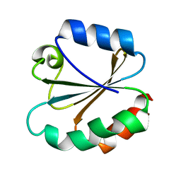
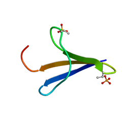
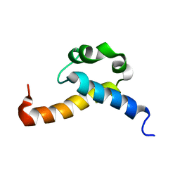
 | | PEGylated Calmodulin-1 (K148U) | | 分子名称: | CALCIUM ION, Calmodulin-1, MAGNESIUM ION, ... | | 著者 | Mackay, J.P, Payne, R.J, Patel, K, Dowman, L.J. | | 登録日 | 2021-12-06 | | 公開日 | 2022-10-26 | | 最終更新日 | 2023-10-25 | | 実験手法 | X-RAY DIFFRACTION (1.95 Å) | | 主引用文献 | Site-selective photocatalytic functionalization of peptides and proteins at selenocysteine.
Nat Commun, 13, 2022
|
|
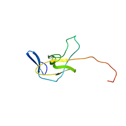
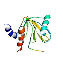
2HI3
 | | Solution structure of the homeodomain-only protein HOP | | 分子名称: | Homeodomain-only protein | | 著者 | Mackay, J.P, Kook, H, Epstein, J.A, Simpson, R.J, Yung, W.W. | | 登録日 | 2006-06-28 | | 公開日 | 2007-01-02 | | 最終更新日 | 2024-05-29 | | 実験手法 | SOLUTION NMR | | 主引用文献 | Analysis of the structure and function of the transcriptional coregulator HOP
Biochemistry, 45, 2006
|
|
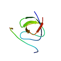
2MPL
 | |
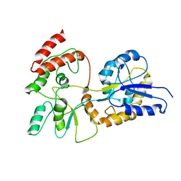
2RPN
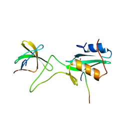
 | | A crucial role for high intrinsic specificity in the function of yeast SH3 domains | | 分子名称: | Actin-binding protein, Actin-regulating kinase 1 | | 著者 | Stollar, E.J, Garcia, B, Chong, A, Forman-Kay, J, Davidson, A. | | 登録日 | 2008-06-12 | | 公開日 | 2009-06-16 | | 最終更新日 | 2024-05-29 | | 実験手法 | SOLUTION NMR | | 主引用文献 | Structural, functional, and bioinformatic studies demonstrate the crucial role of an extended peptide binding site for the SH3 domain of yeast Abp1p
J.Biol.Chem., 284, 2009
|
|
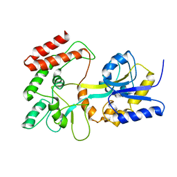
6UK1
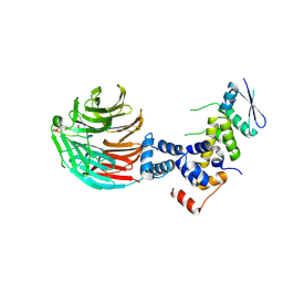
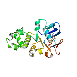 | | Crystal structure of nucleotide-binding domain 2 (NBD2) of the human Cystic Fibrosis Transmembrane Conductance Regulator (CFTR) | | 分子名称: | ADENOSINE-5'-TRIPHOSPHATE, Cystic fibrosis transmembrane conductance regulator, MAGNESIUM ION | | 著者 | Wang, C, Vorobiev, S.M, Vernon, R.M, Khazanov, N, Senderowitz, H, Forman-Kay, J.D, Hunt, J.F. | | 登録日 | 2019-10-03 | | 公開日 | 2020-10-07 | | 最終更新日 | 2023-10-11 | | 実験手法 | X-RAY DIFFRACTION (2.693 Å) | | 主引用文献 | A thermodynamically stabilized form of the second nucleotide binding domain from human CFTR shows a catalytically inactive conformation
To Be Published
|
|
4TRX
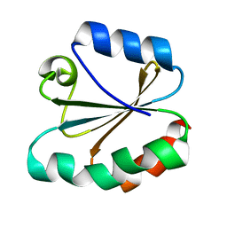 | |
3TRX
 | |
1EZP
 | | GLOBAL FOLD OF MALTODEXTRIN BINDING PROTEIN COMPLEXED WITH BETA-CYCLODEXTRIN USING PEPTIDE ORIENTATIONS FROM DIPOLAR COUPLINGS | | 分子名称: | MALTODEXTRIN BINDING PERIPLASMIC PROTEIN | | 著者 | Mueller, G.A, Choy, W.Y, Yang, D, Forman-Kay, J.D, Venters, R.A, Kay, L.E. | | 登録日 | 2000-05-11 | | 公開日 | 2001-05-08 | | 最終更新日 | 2024-05-22 | | 実験手法 | SOLUTION NMR | | 主引用文献 | Global folds of proteins with low densities of NOEs using residual dipolar couplings: application to the 370-residue maltodextrin-binding protein.
J.Mol.Biol., 300, 2000
|
|
1DDM
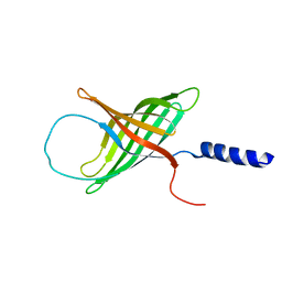
 | | SOLUTION STRUCTURE OF THE NUMB PTB DOMAIN COMPLEXED TO A NAK PEPTIDE | | 分子名称: | NUMB ASSOCIATE KINASE, NUMB PROTEIN | | 著者 | Zwahlen, C, Li, S.C, Kay, L.E, Pawson, T, Forman-Kay, J.D. | | 登録日 | 1999-11-11 | | 公開日 | 2000-04-10 | | 最終更新日 | 2024-05-22 | | 実験手法 | SOLUTION NMR | | 主引用文献 | Multiple modes of peptide recognition by the PTB domain of the cell fate determinant Numb.
EMBO J., 19, 2000
|
|
1EZO
 | | GLOBAL FOLD OF MALTODEXTRIN BINDING PROTEIN COMPLEXED WITH BETA-CYCLODEXTRIN | | 分子名称: | MALTOSE-BINDING PERIPLASMIC PROTEIN | | 著者 | Mueller, G.A, Choy, W.Y, Yang, D, Forman-Kay, J.D, Venters, R.A, Kay, L.E. | | 登録日 | 2000-05-11 | | 公開日 | 2001-05-03 | | 最終更新日 | 2024-05-22 | | 実験手法 | SOLUTION NMR | | 主引用文献 | Global folds of proteins with low densities of NOEs using residual dipolar couplings: application to the 370-residue maltodextrin-binding protein.
J.Mol.Biol., 300, 2000
|
|
1KA7
 | | SAP/SH2D1A bound to peptide n-Y-c | | 分子名称: | SH2 DOMAIN PROTEIN 1A, peptide n-Y-c | | 著者 | Hwang, P.M, Li, C, Morra, M, Lillywhite, J, Gertler, F, Terhorst, C, Kay, L.E, Pawson, T, Forman-Kay, J, Li, S.-C. | | 登録日 | 2001-10-31 | | 公開日 | 2001-11-07 | | 最終更新日 | 2024-05-22 | | 実験手法 | SOLUTION NMR | | 主引用文献 | A "three-pronged" binding mechanism for the SAP/SH2D1A SH2 domain: structural basis and relevance to the XLP syndrome.
EMBO J., 21, 2002
|
|
1KA6
 | | SAP/SH2D1A bound to peptide n-pY | | 分子名称: | SH2 DOMAIN PROTEIN 1A, peptide n-pY | | 著者 | Hwang, P.M, Li, C, Morra, M, Lillywhite, J, Gertler, F, Terhorst, C, Kay, L.E, Pawson, T, Forman-Kay, J, Li, S.-C. | | 登録日 | 2001-10-31 | | 公開日 | 2001-11-07 | | 最終更新日 | 2021-10-27 | | 実験手法 | SOLUTION NMR | | 主引用文献 | A "three-pronged" binding mechanism for the SAP/SH2D1A SH2 domain: structural basis and relevance to the XLP syndrome.
EMBO J., 21, 2002
|
|
2MX4
 | | NMR structure of Phosphorylated 4E-BP2 | | 分子名称: | Eukaryotic translation initiation factor 4E-binding protein 2 | | 著者 | Bah, A, Forman-Kay, J, Vernon, R, Siddiqui, Z, Krzeminski, M, Muhandiram, R, Zhao, C, Sonenberg, N, Kay, L. | | 登録日 | 2014-12-10 | | 公開日 | 2015-01-07 | | 最終更新日 | 2015-03-18 | | 実験手法 | SOLUTION NMR | | 主引用文献 | Folding of an intrinsically disordered protein by phosphorylation as a regulatory switch.
Nature, 519, 2015
|
|
2NMB
 | | DNUMB PTB DOMAIN COMPLEXED WITH A PHOSPHOTYROSINE PEPTIDE, NMR, ENSEMBLE OF STRUCTURES. | | 分子名称: | PROTEIN (GPPY PEPTIDE), PROTEIN (NUMB PROTEIN) | | 著者 | Li, S.-C, Zwahlen, C, Vincent, S.J.F, McGlade, C.J, Pawson, T, Forman-Kay, J.D. | | 登録日 | 1998-10-29 | | 公開日 | 1998-11-04 | | 最終更新日 | 2023-12-27 | | 実験手法 | SOLUTION NMR | | 主引用文献 | Structure of a Numb PTB domain-peptide complex suggests a basis for diverse binding specificity.
Nat.Struct.Biol., 5, 1998
|
|
2PLE
 | | NUCLEAR MAGNETIC RESONANCE STRUCTURE OF AN SH2 DOMAIN OF PHOSPHOLIPASE C-GAMMA1 COMPLEXED WITH A HIGH AFFINITY BINDING PEPTIDE | | 分子名称: | PHOSPHOLIPASE C GAMMA-1, C-TERMINAL SH2 DOMAIN, PHOSPHOPEPTIDE FROM PDGF | | 著者 | Pascal, S.M, Singer, A.U, Gish, G, Yamazaki, T, Shoelson, S.E, Pawson, T, Kay, L.E, Forman-Kay, J.D. | | 登録日 | 1994-08-19 | | 公開日 | 1995-01-26 | | 最終更新日 | 2017-11-29 | | 実験手法 | SOLUTION NMR | | 主引用文献 | Nuclear magnetic resonance structure of an SH2 domain of phospholipase C-gamma 1 complexed with a high affinity binding peptide.
Cell(Cambridge,Mass.), 77, 1994
|
|
2PLD
 | | NUCLEAR MAGNETIC RESONANCE STRUCTURE OF AN SH2 DOMAIN OF PHOSPHOLIPASE C-GAMMA1 COMPLEXED WITH A HIGH AFFINITY BINDING PEPTIDE | | 分子名称: | PHOSPHOLIPASE C GAMMA-1, C-TERMINAL SH2 DOMAIN, PHOSPHOPEPTIDE FROM PDGF | | 著者 | Pascal, S.M, Singer, A.U, Gish, G, Yamazaki, T, Shoelson, S.E, Pawson, T, Kay, L.E, Forman-Kay, J.D. | | 登録日 | 1994-08-19 | | 公開日 | 1995-01-26 | | 最終更新日 | 2017-11-29 | | 実験手法 | SOLUTION NMR | | 主引用文献 | Nuclear magnetic resonance structure of an SH2 domain of phospholipase C-gamma 1 complexed with a high affinity binding peptide.
Cell(Cambridge,Mass.), 77, 1994
|
|
2KIS
 | | Solution structure of CA150 FF1 domain and FF1-FF2 interdomain linker | | 分子名称: | Transcription elongation regulator 1 | | 著者 | Murphy, J.M, Hansen, D, Wiesner, S, Muhandiram, D, Borg, M, Smith, M.J, Sicheri, F, Kay, L.E, Forman-Kay, J.D, Pawson, T. | | 登録日 | 2009-05-08 | | 公開日 | 2009-09-08 | | 最終更新日 | 2024-05-08 | | 実験手法 | SOLUTION NMR | | 主引用文献 | Structural studies of FF domains of the transcription factor CA150 provide insights into the organization of FF domain tandem arrays.
J.Mol.Biol., 393, 2009
|
|
1JU5
 | | Ternary complex of an Crk SH2 domain, Crk-derived phophopeptide, and Abl SH3 domain by NMR spectroscopy | | 分子名称: | Abl, Crk | | 著者 | Donaldson, L.W, Pawson, T, Kay, L.E, Forman-Kay, J.D. | | 登録日 | 2001-08-23 | | 公開日 | 2002-11-06 | | 最終更新日 | 2021-11-10 | | 実験手法 | SOLUTION NMR | | 主引用文献 | Structure of a regulatory complex involving the Abl SH3 domain, the Crk
SH2 domain, and a Crk-derived phosphopeptide
Proc.Natl.Acad.Sci.USA, 99, 2002
|
|
3V7D
 | | Crystal Structure of ScSkp1-ScCdc4-pSic1 peptide complex | | 分子名称: | Cell division control protein 4, Protein SIC1, Suppressor of kinetochore protein 1 | | 著者 | Tang, X, Orlicky, S, Mittag, T, Csizmok, V, Pawson, T, Forman-Kay, J, Sicheri, F, Tyers, M. | | 登録日 | 2011-12-20 | | 公開日 | 2012-05-02 | | 最終更新日 | 2023-09-13 | | 実験手法 | X-RAY DIFFRACTION (2.306 Å) | | 主引用文献 | Composite low affinity interactions dictate recognition of the cyclin-dependent kinase inhibitor Sic1 by the SCFCdc4 ubiquitin ligase.
Proc.Natl.Acad.Sci.USA, 109, 2012
|
|
1MM5
 | | Solution NMR structure of the outer membrane enzyme PagP in OG micelles | | 分子名称: | CrcA protein | | 著者 | Hwang, P.M, Choy, W.-Y, Lo, E.I, Chen, L, Forman-Kay, J.D, Raetz, C.R.H, Prive, G.G, Bishop, R.E, Kay, L.E. | | 登録日 | 2002-09-03 | | 公開日 | 2002-09-13 | | 最終更新日 | 2024-05-22 | | 実験手法 | SOLUTION NMR | | 主引用文献 | Solution Structure and Dynamics of the Outer Membrane Enzyme PagP by NMR
Proc.Natl.Acad.Sci.USA, 99, 2002
|
|
1MM4
 | | Solution NMR structure of the outer membrane enzyme PagP in DPC micelles | | 分子名称: | CrcA protein | | 著者 | Hwang, P.M, Choy, W.-Y, Lo, E.I, Chen, L, Forman-Kay, J.D, Raetz, C.R.H, Prive, G.G, Bishop, R.E, Kay, L.E. | | 登録日 | 2002-09-03 | | 公開日 | 2002-09-13 | | 最終更新日 | 2024-05-22 | | 実験手法 | SOLUTION NMR | | 主引用文献 | Solution Structure and Dynamics of the Outer Membrane Enzyme PagP by NMR
Proc.Natl.Acad.Sci.USA, 99, 2002
|
|
2DJY
 | | Solution structure of Smurf2 WW3 domain-Smad7 PY peptide complex | | 分子名称: | Mothers against decapentaplegic homolog 7, Smad ubiquitination regulatory factor 2 | | 著者 | Chong, P.A, Lin, H, Wrana, J.L, Forman-Kay, J.D. | | 登録日 | 2006-04-06 | | 公開日 | 2006-05-02 | | 最終更新日 | 2024-05-29 | | 実験手法 | SOLUTION NMR | | 主引用文献 | An Expanded WW Domain Recognition Motif Revealed by the Interaction between Smad7 and the E3 Ubiquitin Ligase Smurf2.
J.Biol.Chem., 281, 2006
|
|
2AZV
 | | Solution structure of the T22G mutant of N-terminal SH3 domain of DRK (calculated without NOEs) | | 分子名称: | SH2-SH3 adapter protein drk | | 著者 | Bezsonova, I, Singer, A.U, Choy, W.-Y, Tollinger, M, Forman-Kay, J.D. | | 登録日 | 2005-09-12 | | 公開日 | 2005-12-13 | | 最終更新日 | 2024-05-22 | | 実験手法 | SOLUTION NMR | | 主引用文献 | Structural Comparison of the Unstable drkN SH3 Domain and a Stable Mutant
Biochemistry, 44, 2005
|
|


