Loading
PDBj
MenuPDBj@FacebookPDBj@TwitterPDBj@YouTubewwPDB FoundationwwPDB
RCSB PDBPDBeBMRBAdv. SearchSearch help
PDB: 156 results

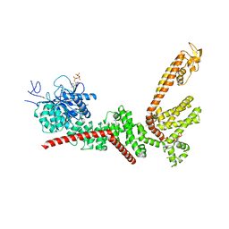
9B94
DownloadVisualize
BU of 9b94 by Molmil
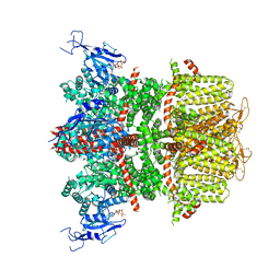
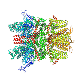
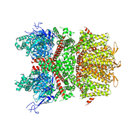
Cryo-EM structure of the E396A mutant of human TRPM4 in complex with calcium at 37 degrees Celsius
Descriptor: CALCIUM ION, Transient receptor potential cation channel subfamily M member 4
Authors:Hu, J, Lu, W, Du, J.
Deposit date:2024-04-01
Release date:2024-05-22
Last modified:2024-06-26
Method:ELECTRON MICROSCOPY (3.1 Å)
Cite:Physiological temperature drives TRPM4 ligand recognition and gating.
Nature, 630, 2024
9B91
DownloadVisualize
BU of 9b91 by Molmil
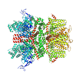
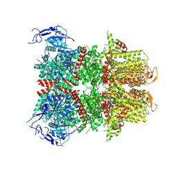
Cryo-EM structure of the human TRPM4 channel subunit in complex with calcium and ATP at 37 degrees Celsius
Descriptor: ADENOSINE-5'-TRIPHOSPHATE, CALCIUM ION, Transient receptor potential cation channel subfamily M member 4
Authors:Hu, J, Lu, W, Du, J.
Deposit date:2024-04-01
Release date:2024-05-15
Last modified:2024-06-26
Method:ELECTRON MICROSCOPY (3.3 Å)
Cite:Physiological temperature drives TRPM4 ligand recognition and gating.
Nature, 630, 2024
9B90
DownloadVisualize
BU of 9b90 by Molmil
Cryo-EM structure of the human TRPM4 channel in complex with calcium and ATP at 37 degrees Celsius
Descriptor: ADENOSINE-5'-TRIPHOSPHATE, CALCIUM ION, Transient receptor potential cation channel subfamily M member 4
Authors:Hu, J, Lu, W, Du, J.
Deposit date:2024-04-01
Release date:2024-05-15
Last modified:2024-06-26
Method:ELECTRON MICROSCOPY (3.4 Å)
Cite:Physiological temperature drives TRPM4 ligand recognition and gating.
Nature, 630, 2024
9B93
DownloadVisualize
BU of 9b93 by Molmil
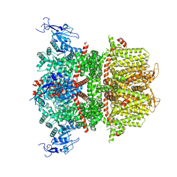
Cryo-EM structure of the human TRPM4 channel in the presence of EDTA at 37 degrees Celsius
Descriptor: Transient receptor potential cation channel subfamily M member 4
Authors:Hu, J, Lu, W, Du, J.
Deposit date:2024-04-01
Release date:2024-05-15
Last modified:2024-06-26
Method:ELECTRON MICROSCOPY (3.1 Å)
Cite:Physiological temperature drives TRPM4 ligand recognition and gating.
Nature, 630, 2024
9B8Z
DownloadVisualize
BU of 9b8z by Molmil
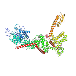
Cryo-EM structure of the human TRPM4 channel subunit in complex with calcium and decavanadate at 37 degrees Celsius
Descriptor: CALCIUM ION, DECAVANADATE, Transient receptor potential cation channel subfamily M member 4
Authors:Hu, J, Lu, W, Du, J.
Deposit date:2024-04-01
Release date:2024-05-15
Last modified:2024-10-16
Method:ELECTRON MICROSCOPY (3.4 Å)
Cite:Physiological temperature drives TRPM4 ligand recognition and gating.
Nature, 630, 2024
9B8W
DownloadVisualize
BU of 9b8w by Molmil
Cryo-EM structure of the human TRPM4 in complex with calcium at 37 degrees Celsius
Descriptor: CALCIUM ION, Transient receptor potential cation channel subfamily M member 4
Authors:Hu, J, Lu, W, Du, J.
Deposit date:2024-04-01
Release date:2024-05-15
Last modified:2024-10-16
Method:ELECTRON MICROSCOPY (3.1 Å)
Cite:Physiological temperature drives TRPM4 ligand recognition and gating.
Nature, 630, 2024
9B92
DownloadVisualize
BU of 9b92 by Molmil
Cryo-EM structure of the human TRPM4 in complex with calcium at 18 degrees Celsius
Descriptor: CALCIUM ION, Transient receptor potential cation channel subfamily M member 4
Authors:Hu, J, Lu, W, Du, J.
Deposit date:2024-04-01
Release date:2024-05-15
Last modified:2024-10-23
Method:ELECTRON MICROSCOPY (3.5 Å)
Cite:Physiological temperature drives TRPM4 ligand recognition and gating.
Nature, 630, 2024
9B8Y
DownloadVisualize
BU of 9b8y by Molmil
Cryo-EM structure of the human TRPM4 channel in complex with calcium and decavanadate at 37 degrees Celsius
Descriptor: CALCIUM ION, DECAVANADATE, Transient receptor potential cation channel subfamily M member 4
Authors:Hu, J, Lu, W, Du, J.
Deposit date:2024-04-01
Release date:2024-05-15
Last modified:2024-06-26
Method:ELECTRON MICROSCOPY (3.2 Å)
Cite:Physiological temperature drives TRPM4 ligand recognition and gating.
Nature, 630, 2024
9B8X
DownloadVisualize
BU of 9b8x by Molmil
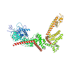
Cryo-EM structure of the human TRPM4 channel subunit in complex with calcium 37 degrees Celsius
Descriptor: CALCIUM ION, Transient receptor potential cation channel subfamily M member 4
Authors:Hu, J, Lu, W, Du, J.
Deposit date:2024-04-01
Release date:2024-05-15
Last modified:2024-06-26
Method:ELECTRON MICROSCOPY (3 Å)
Cite:Physiological temperature drives TRPM4 ligand recognition and gating.
Nature, 630, 2024
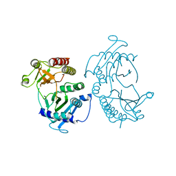
7X4B
DownloadVisualize
BU of 7x4b by Molmil
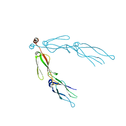
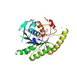
Crystal Structure of An Anti-CRISPR Protein
Descriptor: Anti-CRISPR protein (AcrIIC1), SULFATE ION
Authors:Hu, J, Zhang, S, Gao, J.Y, Liu, X, Liu, J.
Deposit date:2022-03-02
Release date:2022-10-26
Last modified:2024-10-23
Method:X-RAY DIFFRACTION (1.61 Å)
Cite:A redox switch regulates the assembly and anti-CRISPR activity of AcrIIC1.
Nat Commun, 13, 2022
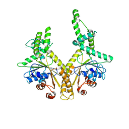
2QLD
DownloadVisualize
BU of 2qld by Molmil
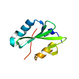
human Hsp40 Hdj1
Descriptor: DnaJ homolog subfamily B member 1
Authors:Hu, J, Wu, Y, Li, J, Fu, Z, Sha, B.
Deposit date:2007-07-12
Release date:2008-07-15
Last modified:2024-04-03
Method:X-RAY DIFFRACTION (2.7 Å)
Cite:The crystal structure of the putative peptide-binding fragment from the human Hsp40 protein Hdj1.
Bmc Struct.Biol., 8, 2008
1RQQ
DownloadVisualize
BU of 1rqq by Molmil
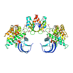
Crystal Structure of the Insulin Receptor Kinase in Complex with the SH2 Domain of APS
Descriptor: BISUBSTRATE INHIBITOR, Insulin receptor, MANGANESE (II) ION, ...
Authors:Hu, J, Liu, J, Ghirlando, R, Saltiel, A.R, Hubbard, S.R.
Deposit date:2003-12-06
Release date:2003-12-30
Last modified:2024-10-09
Method:X-RAY DIFFRACTION (2.6 Å)
Cite:Structural basis for recruitment of the adaptor protein APS to the activated insulin receptor.
Mol.Cell, 12, 2003
1RPY
DownloadVisualize
BU of 1rpy by Molmil
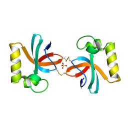
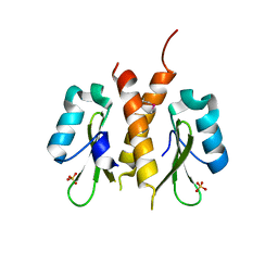
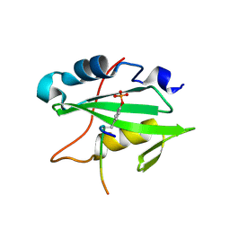
CRYSTAL STRUCTURE OF THE DIMERIC SH2 DOMAIN OF APS
Descriptor: SULFATE ION, adaptor protein APS
Authors:Hu, J, Liu, J, Ghirlando, R, Saltiel, A.R, Hubbard, S.R.
Deposit date:2003-12-03
Release date:2003-12-23
Last modified:2024-10-30
Method:X-RAY DIFFRACTION (2.3 Å)
Cite:Structural basis for recruitment of the adaptor protein APS to the activated insulin receptor.
Mol.Cell, 12, 2003
6PEP
DownloadVisualize
BU of 6pep by Molmil
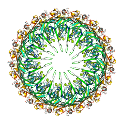
Focussed refinement of InvGN0N1:SpaPQR:PrgIJ from the Salmonella SPI-1 injectisome needle complex
Descriptor: Protein InvG, Protein PrgH, Protein PrgI, ...
Authors:Hu, J, Worrall, L.J, Strynadka, N.C.J.
Deposit date:2019-06-20
Release date:2019-10-23
Last modified:2024-10-23
Method:ELECTRON MICROSCOPY (3.8 Å)
Cite:T3S injectisome needle complex structures in four distinct states reveal the basis of membrane coupling and assembly.
Nat Microbiol, 4, 2019
6PEE
DownloadVisualize
BU of 6pee by Molmil
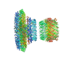
InvG secretin domain beta-barrel from Salmonella SPI-1 injectisome NC-base
Descriptor: LAURYL DIMETHYLAMINE-N-OXIDE, Protein InvG
Authors:Hu, J, Worrall, L.J, Strynadka, N.C.J.
Deposit date:2019-06-20
Release date:2019-10-23
Last modified:2024-03-13
Method:ELECTRON MICROSCOPY (3.42 Å)
Cite:T3S injectisome needle complex structures in four distinct states reveal the basis of membrane coupling and assembly.
Nat Microbiol, 4, 2019
6PEM
DownloadVisualize
BU of 6pem by Molmil
Focussed refinement of InvGN0N1:SpaPQR:PrgHK from Salmonella SPI-1 injectisome NC-base
Descriptor: Lipoprotein PrgK, Protein InvG, Protein PrgH, ...
Authors:Hu, J, Worrall, L.J, Strynadka, N.C.J.
Deposit date:2019-06-20
Release date:2019-10-23
Last modified:2024-10-30
Method:ELECTRON MICROSCOPY (3.5 Å)
Cite:T3S injectisome needle complex structures in four distinct states reveal the basis of membrane coupling and assembly.
Nat Microbiol, 4, 2019
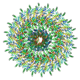
6Q15
DownloadVisualize
BU of 6q15 by Molmil
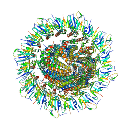
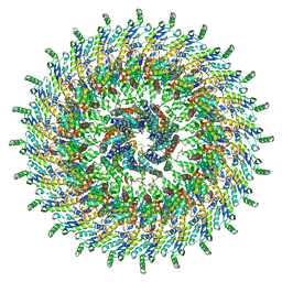
Structure of the Salmonella SPI-1 injectisome needle complex
Descriptor: Lipoprotein PrgK, Protein InvG, Protein PrgH, ...
Authors:Hu, J, Worrall, L.J, Strynadka, N.C.J.
Deposit date:2019-08-02
Release date:2019-10-23
Last modified:2024-11-06
Method:ELECTRON MICROSCOPY (5.15 Å)
Cite:T3S injectisome needle complex structures in four distinct states reveal the basis of membrane coupling and assembly.
Nat Microbiol, 4, 2019
6Q14
DownloadVisualize
BU of 6q14 by Molmil
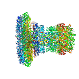
Structure of the Salmonella SPI-1 injectisome NC-base
Descriptor: Lipoprotein PrgK, Protein InvG, Protein PrgH, ...
Authors:Hu, J, Worrall, L.J, Strynadka, N.C.J.
Deposit date:2019-08-02
Release date:2019-10-23
Last modified:2024-10-16
Method:ELECTRON MICROSCOPY (3.8 Å)
Cite:T3S injectisome needle complex structures in four distinct states reveal the basis of membrane coupling and assembly.
Nat Microbiol, 4, 2019
6Q16
DownloadVisualize
BU of 6q16 by Molmil
Focussed refinement of InvGN0N1:PrgHK:SpaPQR:PrgIJ from Salmonella SPI-1 injectisome NC-base
Descriptor: Lipoprotein PrgK, Protein InvG, Protein PrgH, ...
Authors:Hu, J, Worrall, L.J, Strynadka, N.C.J.
Deposit date:2019-08-02
Release date:2019-10-23
Last modified:2024-11-06
Method:ELECTRON MICROSCOPY (4.1 Å)
Cite:T3S injectisome needle complex structures in four distinct states reveal the basis of membrane coupling and assembly.
Nat Microbiol, 4, 2019
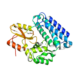
4TZ7
DownloadVisualize
BU of 4tz7 by Molmil
Crystal structure of type I phosphatidylinositol 4-phosphate 5-kinase alpha from Zebrafish
Descriptor: Phosphatidylinositol-4-phosphate 5-kinase, type I, alpha
Authors:Hu, J, Qin, Y, Wang, J, Li, L, Wu, D, Ha, Y.
Deposit date:2014-07-09
Release date:2015-09-02
Last modified:2023-12-27
Method:X-RAY DIFFRACTION (3.31 Å)
Cite:Resolution of structure of PIP5K1A reveals molecular mechanism for its regulation by dimerization and dishevelled.
Nat Commun, 6, 2015
3H84
DownloadVisualize
BU of 3h84 by Molmil
Crystal structure of GET3
Descriptor: ATPase GET3, CHLORIDE ION, MAGNESIUM ION, ...
Authors:Hu, J, Li, J, Qian, X, Sha, B.
Deposit date:2009-04-28
Release date:2009-12-22
Last modified:2024-02-21
Method:X-RAY DIFFRACTION (2.3 Å)
Cite:The crystal structures of yeast Get3 suggest a mechanism for tail-anchored protein membrane insertion.
Plos One, 4, 2009
1YVH
DownloadVisualize
BU of 1yvh by Molmil
Crystal Structure of the c-Cbl TKB Domain in Complex with the APS pTyr-618 Phosphopeptide
Descriptor: 13-mer fragment of SH2 and PH domain-containing adapter protein APS, CBL E3 ubiquitin protein ligase, MAGNESIUM ION
Authors:Hu, J, Hubbard, S.R.
Deposit date:2005-02-15
Release date:2005-03-08
Last modified:2024-10-16
Method:X-RAY DIFFRACTION (2.05 Å)
Cite:Structural Characterization of a Novel Cbl Phosphotyrosine Recognition Motif in the APS Family of Adapter Proteins
J.Biol.Chem., 280, 2005
3IO3
DownloadVisualize
BU of 3io3 by Molmil
GEt3 with ADP from D. Hansenii in Closed form
Descriptor: ADENOSINE-5'-DIPHOSPHATE, DEHA2D07832p, GLYCEROL, ...
Authors:Hu, J, Li, J, Qian, X, Sha, B.
Deposit date:2009-08-13
Release date:2009-12-22
Last modified:2023-09-06
Method:X-RAY DIFFRACTION (1.8 Å)
Cite:The crystal structures of yeast Get3 suggest a mechanism for tail-anchored protein membrane insertion
Plos One, 4, 2009
2HDV
DownloadVisualize
BU of 2hdv by Molmil
Crystal structure of the Src Homology-2 domain of the adapter protein SH2-B
Descriptor: SH2-B PH domain containing signaling mediator 1 gamma isoform
Authors:Hu, J, Hubbard, S.R.
Deposit date:2006-06-20
Release date:2006-08-08
Last modified:2023-08-30
Method:X-RAY DIFFRACTION (2 Å)
Cite:Structural Basis for Phosphotyrosine Recognition by the Src Homology-2 Domains of the Adapter Proteins SH2-B and APS.
J.Mol.Biol., 361, 2006
2HDX
DownloadVisualize
BU of 2hdx by Molmil
Crystal structure of the Src homology-2 domain of SH2-B in complex with Jak2 pTyr813 phosphopeptide
Descriptor: Jak2 protein, SH2-B PH domain containing signaling mediator 1 gamma isoform
Authors:Hu, J, Hubbard, S.R.
Deposit date:2006-06-21
Release date:2006-08-08
Last modified:2024-10-30
Method:X-RAY DIFFRACTION (2.35 Å)
Cite:Structural Basis for Phosphotyrosine Recognition by the Src Homology-2 Domains of the Adapter Proteins SH2-B and APS.
J.Mol.Biol., 361, 2006

227111

건을2024-11-06부터공개중

PDB statisticsPDBj update infoContact PDBjnumon