Loading
PDBj
MenuPDBj@FacebookPDBj@TwitterPDBj@YouTubewwPDB FoundationwwPDB
RCSB PDBPDBeBMRBAdv. SearchSearch help
PDB: 89 results

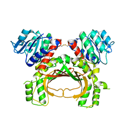
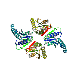
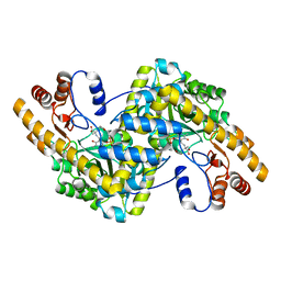
4TV7
DownloadVisualize
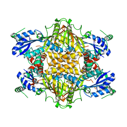
BU of 4tv7 by Molmil
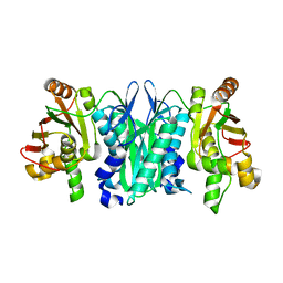
Crystal structure of Bacillus subtilis GabR at 2.05 Angstroms resolution
Descriptor: HTH-type transcriptional regulatory protein GabR
Authors:Goto, M, Okuda, K, Yoshimura, T.
Deposit date:2014-06-26
Release date:2014-12-24
Last modified:2020-01-29
Method:X-RAY DIFFRACTION (2.05 Å)
Cite:Role of the aminotransferase domain in Bacillus subtilis GabR, a pyridoxal 5'-phosphate-dependent transcriptional regulator
Mol. Microbiol., 95, 2015
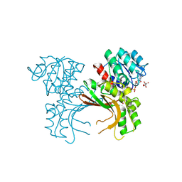
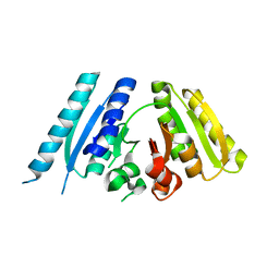
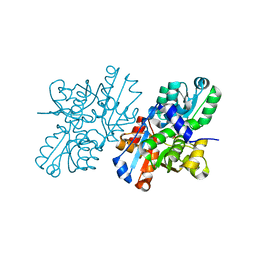
8I7P
DownloadVisualize
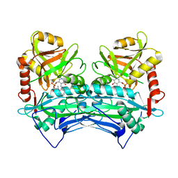
BU of 8i7p by Molmil
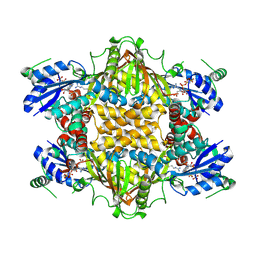
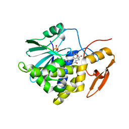
Crystal structure of Ricin A chain bound with N2-(2-amino-4-oxo-3,4-dihydropteridine-7-carbonyl)glycyl-L-tyrosine
Descriptor: 6-(2-ethyl-4-hydroxyphenyl)-1H-indazole-3-carboxamide, Ricin A chain, SULFATE ION
Authors:Goto, M, Sakamoto, N, Higashi, S, Kawata, R, Nagatsu, K, Saito, R.
Deposit date:2023-02-01
Release date:2023-09-20
Method:X-RAY DIFFRACTION (1.6 Å)
Cite:Crystal structure of ricin toxin A chain complexed with a highly potent pterin-based small-molecular inhibitor.
J Enzyme Inhib Med Chem, 38, 2023
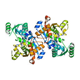
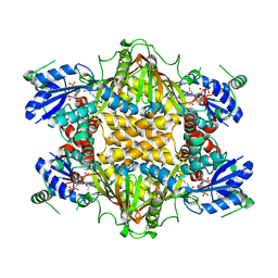
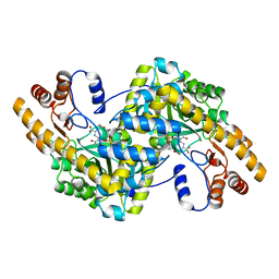
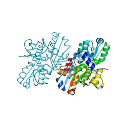
5AVO
DownloadVisualize
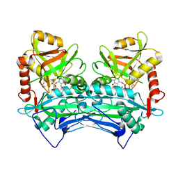
BU of 5avo by Molmil
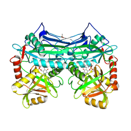
Crystal structure of the reduced form of homoserine dehydrogenase from Sulfolobus tokodaii.
Descriptor: Homoserine dehydrogenase
Authors:Goto, M, Yoshimune, K, Kaneko, R.
Deposit date:2015-06-25
Release date:2015-11-11
Last modified:2023-11-08
Method:X-RAY DIFFRACTION (1.8 Å)
Cite:Structural insight into activation of homoserine dehydrogenase from the archaeonSulfolobus tokodaiivia reduction.
Biochem Biophys Rep, 3, 2015
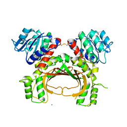
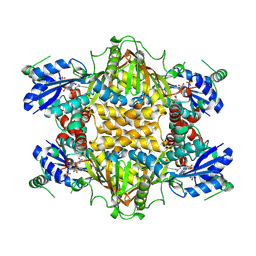
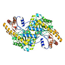
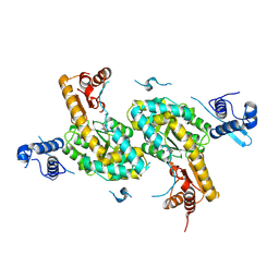
5X9D
DownloadVisualize
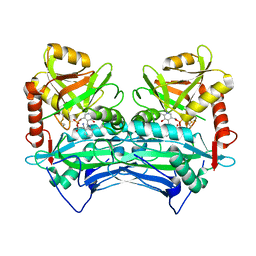
BU of 5x9d by Molmil
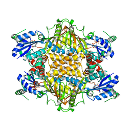
Crystal structure of homoserine dehydrogenase in complex with L-cysteine and NAD
Descriptor: (2R)-3-[[(4S)-3-aminocarbonyl-1-[(2R,3R,4S,5R)-5-[[[[(2R,3S,4R,5R)-5-(6-aminopurin-9-yl)-3,4-bis(oxidanyl)oxolan-2-yl]methoxy-oxidanyl-phosphoryl]oxy-oxidanyl-phosphoryl]oxymethyl]-3,4-bis(oxidanyl)oxolan-2-yl]-4H-pyridin-4-yl]sulfanyl]-2-azanyl-propanoic acid, Homoserine dehydrogenase, L(+)-TARTARIC ACID
Authors:Goto, M, Ogata, K, Kaneko, R, Yoshimune, K.
Deposit date:2017-03-06
Release date:2018-05-02
Last modified:2023-11-22
Method:X-RAY DIFFRACTION (2.1 Å)
Cite:Inhibition of homoserine dehydrogenase by formation of a cysteine-NAD covalent complex
Sci Rep, 8, 2018
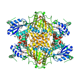
6LUT
DownloadVisualize
BU of 6lut by Molmil
Crystal structure of Serine Racemase from Dictyostelium discoideum.
Descriptor: Probable serine racemase
Authors:Goto, M, Mizobuchi, T, Yoshimura, T.
Deposit date:2020-01-31
Release date:2020-12-09
Last modified:2023-11-29
Method:X-RAY DIFFRACTION (1.35 Å)
Cite:Mechanism of eukaryotic serine racemase-catalyzed serine dehydration.
Biochim Biophys Acta Proteins Proteom, 1868, 2020
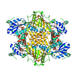
4YDR
DownloadVisualize
BU of 4ydr by Molmil
Crystal structure of oxidized homoserine dehydrogenase of Sulfolobus tokodaii
Descriptor: Homoserine dehydrogenase, SODIUM ION
Authors:Goto, M, Yoshimune, K, Kaneko, R.
Deposit date:2015-02-23
Release date:2015-11-11
Last modified:2019-12-18
Method:X-RAY DIFFRACTION (1.6 Å)
Cite:Structural insight into activation of homoserine dehydrogenase from the archaeonSulfolobus tokodaiivia reduction.
Biochem Biophys Rep, 3, 2015
1J21
DownloadVisualize
BU of 1j21 by Molmil
Crystal Structure of Thermus thermophilus HB8 Argininosuccinate Synthetase in complex with ATP and citrulline
Descriptor: ADENOSINE-5'-TRIPHOSPHATE, Argininosuccinate Synthetase, CITRULLINE
Authors:Goto, M, Hirotsu, K, RIKEN Structural Genomics/Proteomics Initiative (RSGI)
Deposit date:2002-12-24
Release date:2003-04-22
Last modified:2023-11-15
Method:X-RAY DIFFRACTION (2.2 Å)
Cite:Structures of argininosuccinate synthetase in enzyme-ATP-substrates and enzyme-AMP-product forms
J.Biol.Chem., 278, 2003
1J1Z
DownloadVisualize
BU of 1j1z by Molmil
Crystal Structure of Thermus thermophilus HB8 Argininosuccinate Synthetase in complex with substrate
Descriptor: ADENOSINE-5'-TRIPHOSPHATE, ASPARTIC ACID, Argininosuccinate Synthetase, ...
Authors:Goto, M, Hirotsu, K, RIKEN Structural Genomics/Proteomics Initiative (RSGI)
Deposit date:2002-12-24
Release date:2003-04-22
Last modified:2023-11-15
Method:X-RAY DIFFRACTION (2.1 Å)
Cite:Structures of argininosuccinate synthetase in enzyme-ATP-substrates and enzyme-AMP-product forms
J.Biol.Chem., 278, 2003
1J20
DownloadVisualize
BU of 1j20 by Molmil
Crystal Structure of Thermus thermophilus HB8 Argininosuccinate Synthetase in complex with product
Descriptor: ADENOSINE MONOPHOSPHATE, ARGININOSUCCINATE, Argininosuccinate Synthetase, ...
Authors:Goto, M, Hirotsu, K, RIKEN Structural Genomics/Proteomics Initiative (RSGI)
Deposit date:2002-12-24
Release date:2003-04-22
Last modified:2023-12-27
Method:X-RAY DIFFRACTION (2 Å)
Cite:Structures of argininosuccinate synthetase in enzyme-ATP-substrates and enzyme-AMP-product forms
J.Biol.Chem., 278, 2003
1KH3
DownloadVisualize
BU of 1kh3 by Molmil
Crystal Structure of Thermus thermophilus HB8 Argininosuccinate Synthetase in complex with inhibitor
Descriptor: ARGININE, ASPARTIC ACID, Argininosuccinate Synthetase, ...
Authors:goto, m, Hirotsu, k, miyahara, i, RIKEN Structural Genomics/Proteomics Initiative (RSGI)
Deposit date:2001-11-29
Release date:2003-04-22
Last modified:2024-03-13
Method:X-RAY DIFFRACTION (2.15 Å)
Cite:Structures of Argininosuccinate Synthetase in Enzyme-ATP Substrates and Enzyme-AMP Product Forms: STEREOCHEMISTRY OF THE CATALYTIC REACTION
J.Biol.Chem., 278, 2003
2YXZ
DownloadVisualize
BU of 2yxz by Molmil
Crystal structure of tt0281 from Thermus thermophilus HB8
Descriptor: Thiamin-monophosphate kinase
Authors:Goto, M.
Deposit date:2007-04-27
Release date:2008-04-29
Last modified:2024-03-13
Method:X-RAY DIFFRACTION (1.9 Å)
Cite:Crystal structure of tt0281 from Thermus thermophilus HB8
to be published
1KOR
DownloadVisualize
BU of 1kor by Molmil
Crystal Structure of Thermus thermophilus HB8 Argininosuccinate Synthetase in complex with inhibitors
Descriptor: ARGININE, Argininosuccinate Synthetase, PHOSPHOAMINOPHOSPHONIC ACID-ADENYLATE ESTER, ...
Authors:Goto, M, Nakajima, Y, Hirotsu, K, RIKEN Structural Genomics/Proteomics Initiative (RSGI)
Deposit date:2001-12-22
Release date:2002-04-03
Last modified:2011-07-13
Method:X-RAY DIFFRACTION (1.95 Å)
Cite:Crystal structure of argininosuccinate synthetase from Thermus thermophilus HB8. Structural basis for the catalytic action.
J.Biol.Chem., 277, 2002
2A1H
DownloadVisualize
BU of 2a1h by Molmil
X-ray crystal structure of human mitochondrial branched chain aminotransferase (BCATm) complexed with gabapentin
Descriptor: ACETIC ACID, PYRIDOXAL-5'-PHOSPHATE, [1-(AMINOMETHYL)CYCLOHEXYL]ACETIC ACID, ...
Authors:Goto, M, Miyahara, I, Hirotsu, K, Conway, M, Yennawar, N, Islam, M.M, Hutson, S.M.
Deposit date:2005-06-20
Release date:2005-09-06
Last modified:2017-10-11
Method:X-RAY DIFFRACTION (1.8 Å)
Cite:Structural determinants for branched-chain aminotransferase isozyme-specific inhibition by the anticonvulsant drug gabapentin.
J.Biol.Chem., 280, 2005
1KH2
DownloadVisualize
BU of 1kh2 by Molmil
Crystal Structure of Thermus thermophilus HB8 Argininosuccinate Synthetase in complex with ATP
Descriptor: ADENOSINE-5'-TRIPHOSPHATE, Argininosuccinate Synthetase
Authors:Goto, M, Nakajima, Y, Hirotsu, K, RIKEN Structural Genomics/Proteomics Initiative (RSGI)
Deposit date:2001-11-29
Release date:2002-04-03
Last modified:2024-03-13
Method:X-RAY DIFFRACTION (2.3 Å)
Cite:Crystal structure of argininosuccinate synthetase from Thermus thermophilus HB8. Structural basis for the catalytic action.
J.Biol.Chem., 277, 2002
1KH1
DownloadVisualize
BU of 1kh1 by Molmil
Crystal Structure of Thermus thermophilus HB8 Argininosuccinate Synthetase
Descriptor: Argininosuccinate Synthetase, SULFATE ION
Authors:Goto, M, Nakajima, Y, Hirotsu, K, RIKEN Structural Genomics/Proteomics Initiative (RSGI)
Deposit date:2001-11-29
Release date:2002-04-03
Last modified:2024-03-13
Method:X-RAY DIFFRACTION (2.3 Å)
Cite:Crystal structure of argininosuccinate synthetase from Thermus thermophilus HB8. Structural basis for the catalytic action.
J.Biol.Chem., 277, 2002
2COJ
DownloadVisualize
BU of 2coj by Molmil
Crystal structure of reduced human cytosolic branched-chain aminotransferase complexed with gabapentin
Descriptor: MAGNESIUM ION, PYRIDOXAL-5'-PHOSPHATE, [1-(AMINOMETHYL)CYCLOHEXYL]ACETIC ACID, ...
Authors:Goto, M.
Deposit date:2005-05-17
Release date:2005-08-30
Last modified:2021-11-10
Method:X-RAY DIFFRACTION (2.4 Å)
Cite:Structural determinants for branched-chain aminotransferase isozyme-specific inhibition by the anticonvulsant drug gabapentin
J.Biol.Chem., 280, 2005
2COG
DownloadVisualize
BU of 2cog by Molmil
Crystal structure of oxidized human cytosolic branched-chain aminotransferase complexed with 4-methylvalerate
Descriptor: 4-METHYL VALERIC ACID, MAGNESIUM ION, PYRIDOXAL-5'-PHOSPHATE, ...
Authors:Goto, M.
Deposit date:2005-05-17
Release date:2005-08-30
Last modified:2021-11-10
Method:X-RAY DIFFRACTION (2.1 Å)
Cite:Structural determinants for branched-chain aminotransferase isozyme-specific inhibition by the anticonvulsant drug gabapentin
J.Biol.Chem., 280, 2005
2COI
DownloadVisualize
BU of 2coi by Molmil
Crystal structure of oxidized human cytosolic branched-chain aminotransferase complexed with gabapentin
Descriptor: MAGNESIUM ION, PYRIDOXAL-5'-PHOSPHATE, [1-(AMINOMETHYL)CYCLOHEXYL]ACETIC ACID, ...
Authors:Goto, M.
Deposit date:2005-05-17
Release date:2005-08-30
Last modified:2021-11-10
Method:X-RAY DIFFRACTION (1.9 Å)
Cite:Structural determinants for branched-chain aminotransferase isozyme-specific inhibition by the anticonvulsant drug gabapentin
J.Biol.Chem., 280, 2005
3AB8
DownloadVisualize
BU of 3ab8 by Molmil
Crystal Structure of the Hypothetical Tandem-type Universal Stress Protein TTHA0350 complexed with ATPs.
Descriptor: ADENOSINE-5'-TRIPHOSPHATE, MAGNESIUM ION, Putative uncharacterized protein TTHA0350
Authors:Goto, M, Iino, H, Shimizu, N, Kuramitsu, S.
Deposit date:2009-12-01
Release date:2010-12-15
Last modified:2023-11-01
Method:X-RAY DIFFRACTION (1.7 Å)
Cite:Crystal Structure of the Hypothetical Tandem-type Universal Stress Protein TTHA0350 from Thermus thermophilus HB8
To be Published
3AB7
DownloadVisualize
BU of 3ab7 by Molmil
Crystal Structure of the Hypothetical Tandem-type Universal Stress Protein TTHA0350 from Thermus thermophilus HB8
Descriptor: Putative uncharacterized protein TTHA0350
Authors:Goto, M, Iino, H, Shimizu, N, Kuramitsu, S.
Deposit date:2009-12-01
Release date:2010-12-15
Last modified:2024-03-13
Method:X-RAY DIFFRACTION (2.52 Å)
Cite:Crystal Structure of the Hypothetical Tandem-type Universal Stress Protein TTHA0350 from Thermus thermophilus HB8
To be Published
1X2A
DownloadVisualize
BU of 1x2a by Molmil
Crystal Structure of e.coli AspAT complexed with N-phosphopyridoxyl-D-glutamic acid
Descriptor: Aspartate aminotransferase, N-({3-HYDROXY-2-METHYL-5-[(PHOSPHONOOXY)METHYL]PYRIDIN-4-YL}METHYL)-D-GLUTAMIC ACID
Authors:Goto, M.
Deposit date:2005-04-21
Release date:2005-06-14
Last modified:2024-03-13
Method:X-RAY DIFFRACTION (2.2 Å)
Cite:Binding of C5-dicarboxylic substrate to aspartate aminotransferase: implications for the conformational change at the transaldimination step.
Biochemistry, 44, 2005
1X28
DownloadVisualize
BU of 1x28 by Molmil
Crystal Structure of e.coli AspAT complexed with N-phosphopyridoxyl-L-glutamic acid
Descriptor: Aspartate aminotransferase, N-({3-hydroxy-2-methyl-5-[(phosphonooxy)methyl]pyridin-4-yl}methyl)-L-glutamic acid
Authors:Goto, M.
Deposit date:2005-04-21
Release date:2005-06-14
Last modified:2024-03-13
Method:X-RAY DIFFRACTION (2.4 Å)
Cite:Binding of C5-dicarboxylic substrate to aspartate aminotransferase: implications for the conformational change at the transaldimination step.
Biochemistry, 44, 2005
1X29
DownloadVisualize
BU of 1x29 by Molmil
Crystal Structure of e.coli AspAT complexed with N-phosphopyridoxyl-2-methyl-L-glutamic acid
Descriptor: Aspartate aminotransferase, N-({3-HYDROXY-2-METHYL-5-[(PHOSPHONOOXY)METHYL]PYRIDIN-4-YL}METHYL)-2-METHYL-L-GLUTAMIC ACID
Authors:Goto, M.
Deposit date:2005-04-21
Release date:2005-06-14
Last modified:2024-03-13
Method:X-RAY DIFFRACTION (2.2 Å)
Cite:Binding of C5-dicarboxylic substrate to aspartate aminotransferase: implications for the conformational change at the transaldimination step.
Biochemistry, 44, 2005
2ZR8
DownloadVisualize
BU of 2zr8 by Molmil
Crystal Structure of Modified Serine Racemase complexed with Serine
Descriptor: MAGNESIUM ION, N-(5'-PHOSPHOPYRIDOXYL)-D-ALANINE, SERINE, ...
Authors:Goto, M.
Deposit date:2008-08-25
Release date:2009-07-28
Last modified:2023-11-01
Method:X-RAY DIFFRACTION (2.2 Å)
Cite:Crystal structure of a homolog of mammalian serine racemase from Schizosaccharomyces pombe
J.Biol.Chem., 284, 2009
2ZPU
DownloadVisualize
BU of 2zpu by Molmil
Crystal Structure of Modified Serine Racemase from S.pombe.
Descriptor: MAGNESIUM ION, N-(5'-PHOSPHOPYRIDOXYL)-D-ALANINE, Uncharacterized protein C320.14
Authors:Goto, M.
Deposit date:2008-07-29
Release date:2009-04-28
Last modified:2023-11-01
Method:X-RAY DIFFRACTION (1.7 Å)
Cite:Serine racemase with catalytically active lysinoalanyl residue.
J.Biochem., 145, 2009


1234>

218853

PDB entries from 2024-04-24

PDB statisticsPDBj update infoContact PDBjnumon