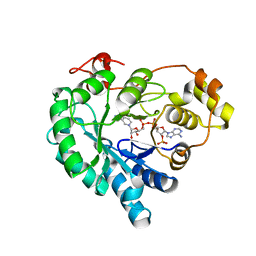
6J10
 
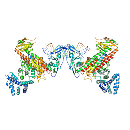
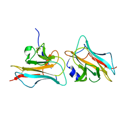
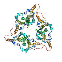 | | Ciclopirox inhibits Hepatitis B Virus secretion by blocking capsid assembly | | 分子名称: | 6-cyclohexyl-4-methyl-1-oxidanyl-pyridin-2-one, Capsid protein | | 著者 | Park, S, Jin, M.S, Cho, Y, Kang, J, Kim, S, Park, M, Park, H, Kim, J, Park, S, Hwang, J, Kim, Y, Kim, Y.J. | | 登録日 | 2018-12-27 | | 公開日 | 2019-04-17 | | 最終更新日 | 2019-05-29 | | 実験手法 | X-RAY DIFFRACTION (2.3 Å) | | 主引用文献 | Ciclopirox inhibits Hepatitis B Virus secretion by blocking capsid assembly.
Nat Commun, 10, 2019
|
|
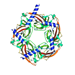
7KFU
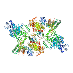 | | Cas6-RT-Cas1--Cas2 complex | | 分子名称: | Cas2, Cas6-RT-Cas1 | | 著者 | Hoel, C.M, Wang, J.Y, Doudna, J.A, Brohawn, S.G. | | 登録日 | 2020-10-14 | | 公開日 | 2021-03-31 | | 最終更新日 | 2024-03-06 | | 実験手法 | ELECTRON MICROSCOPY (3.9 Å) | | 主引用文献 | Structural coordination between active sites of a CRISPR reverse transcriptase-integrase complex.
Nat Commun, 12, 2021
|
|
7KFT
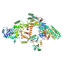 | | Partial Cas6-RT-Cas1--Cas2 complex | | 分子名称: | Cas2, Cas6-RT-Cas1 | | 著者 | Hoel, C.M, Wang, J.Y, Doudna, J.A, Brohawn, S.G. | | 登録日 | 2020-10-14 | | 公開日 | 2021-03-31 | | 最終更新日 | 2024-03-06 | | 実験手法 | ELECTRON MICROSCOPY (3.4 Å) | | 主引用文献 | Structural coordination between active sites of a CRISPR reverse transcriptase-integrase complex.
Nat Commun, 12, 2021
|
|
6J3N
 | |
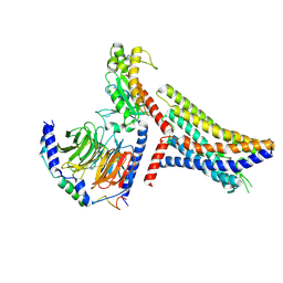
5YBB
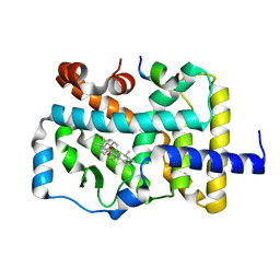
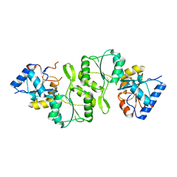
 | | Structural basis underlying complex assembly andconformational transition of the type I R-M system | | 分子名称: | DNA, Restriction endonuclease S subunits, S-ADENOSYLMETHIONINE, ... | | 著者 | Liu, Y.P, Tang, Q, Zhang, J.Z, Tian, L.F, Gao, P, Yan, X.X. | | 登録日 | 2017-09-04 | | 公開日 | 2017-11-29 | | 最終更新日 | 2018-02-07 | | 実験手法 | X-RAY DIFFRACTION (3.2 Å) | | 主引用文献 | Structural basis underlying complex assembly and conformational transition of the type I R-M system.
Proc. Natl. Acad. Sci. U.S.A., 114, 2017
|
|
4QPI
 | | Crystal structure of hepatitis A virus | | 分子名称: | CHLORIDE ION, Capsid protein VP1, Capsid protein VP2, ... | | 著者 | Wang, X, Ren, J, Gao, Q, Hu, Z, Sun, Y, Li, X, Rowlands, D.J, Yin, W, Wang, J, Stuart, D.I, Rao, Z, Fry, E.E. | | 登録日 | 2014-06-23 | | 公開日 | 2014-10-15 | | 最終更新日 | 2023-09-20 | | 実験手法 | X-RAY DIFFRACTION (3.01 Å) | | 主引用文献 | Hepatitis A virus and the origins of picornaviruses.
Nature, 517, 2015
|
|
6POM
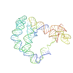 | | Cryo-EM structure of the full-length Bacillus subtilis glyQS T-box riboswitch in complex with tRNA-Gly | | 分子名称: | T-box GlyQS leader (155-MER), tRNAGly (75-MER) | | 著者 | Li, S, Su, Z, Zhang, J, Chiu, W. | | 登録日 | 2019-07-04 | | 公開日 | 2019-11-20 | | 最終更新日 | 2024-03-20 | | 実験手法 | ELECTRON MICROSCOPY (4.9 Å) | | 主引用文献 | Structural basis of amino acid surveillance by higher-order tRNA-mRNA interactions.
Nat.Struct.Mol.Biol., 26, 2019
|
|
7L7S
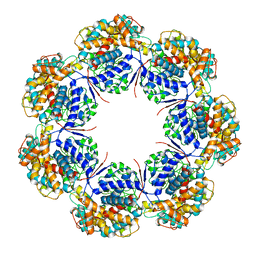 | | Human mitochondrial chaperonin mHsp60 | | 分子名称: | 60 kDa heat shock protein, mitochondrial | | 著者 | Chen, L, Wang, J.C.Y. | | 登録日 | 2020-12-30 | | 公開日 | 2021-08-25 | | 最終更新日 | 2024-05-29 | | 実験手法 | ELECTRON MICROSCOPY (3.5 Å) | | 主引用文献 | Structural basis for the structural dynamics of human mitochondrial chaperonin mHsp60.
Sci Rep, 11, 2021
|
|
6JAU
 | | The complex structure of Pseudomonas aeruginosa MucA/MucB. | | 分子名称: | CALCIUM ION, GLYCEROL, HEXAETHYLENE GLYCOL, ... | | 著者 | Li, T, He, L.H, Li, C.C, Liu, L, Peng, C.T, Shen, Y.L, Qin, X.F, Xiao, Q.J, Zhu, Y.B, Song, Y.J, Zhao, N.l, Zhao, C, Yang, J, Mu, X.Y, Huang, Q, Bao, R. | | 登録日 | 2019-01-25 | | 公開日 | 2020-01-29 | | 最終更新日 | 2020-08-19 | | 実験手法 | X-RAY DIFFRACTION (1.905 Å) | | 主引用文献 | Molecular basis of the lipid-induced MucA-MucB dissociation in Pseudomonas aeruginosa.
Commun Biol, 3, 2020
|
|
7C2Y
 | | The crystal structure of COVID-2019 main protease in the apo state | | 分子名称: | 3C-like proteinase | | 著者 | Zhou, X.L, Zhong, F.L, Lin, C, Zhou, H, Hu, X.H, Wang, Q.S, Li, J, Zhang, J. | | 登録日 | 2020-05-10 | | 公開日 | 2020-09-02 | | 最終更新日 | 2023-11-29 | | 実験手法 | X-RAY DIFFRACTION (1.91 Å) | | 主引用文献 | COVID-2019 main protease in the apo state
To Be Published
|
|
7CLT
 | | Crystal structure of the EFhd1/Swiprosin-2, a mitochondrial actin-binding protein | | 分子名称: | CALCIUM ION, EF-hand domain-containing protein D1, GLYCEROL, ... | | 著者 | Mun, S.A, Park, J, Park, K.R, Lee, Y, Kang, J.Y, Park, T, Jin, M, Yang, J, Jun, C.D, Eom, S.H. | | 登録日 | 2020-07-22 | | 公開日 | 2021-01-06 | | 最終更新日 | 2023-11-29 | | 実験手法 | X-RAY DIFFRACTION (2.07380986 Å) | | 主引用文献 | Structural and Biochemical Characterization of EFhd1/Swiprosin-2, an Actin-Binding Protein in Mitochondria.
Front Cell Dev Biol, 8, 2020
|
|
3COO
 | |
5NEF
 | | The structure of the G. violaceus guanidine II riboswitch P1 stem-loop with guanidine | | 分子名称: | GUANIDINE, RNA (5'-R(*GP*GP*UP*GP*GP*GP*GP*AP*CP*GP*AP*CP*CP*CP*CP*AP*(CBV)P*C)-3'), SODIUM ION, ... | | 著者 | Huang, L, Wang, J, Lilley, D.M.J. | | 登録日 | 2017-03-10 | | 公開日 | 2017-06-07 | | 最終更新日 | 2024-05-08 | | 実験手法 | X-RAY DIFFRACTION (1.91 Å) | | 主引用文献 | The Structure of the Guanidine-II Riboswitch.
Cell Chem Biol, 24, 2017
|
|
4IFT
 | | Crystal structure of double mutant thermostable NPPase from Geobacillus stearothermophilus | | 分子名称: | Thermostable NPPase | | 著者 | Guo, Z, Huang, J, Wang, F, Qiu, R, Wang, Y, Ji, C. | | 登録日 | 2012-12-15 | | 公開日 | 2013-12-18 | | 最終更新日 | 2023-11-08 | | 実験手法 | X-RAY DIFFRACTION (1.995 Å) | | 主引用文献 | Crystal structure of thermostable NPPase from Geobacillus stearothermophilus
To be Published
|
|
5NDH
 | | The structure of the G. violaceus guanidine II riboswitch P2 stem-loop | | 分子名称: | GUANIDINE, MAGNESIUM ION, RNA (5'-R(*GP*(CBV)P*GP*GP*GP*GP*AP*CP*GP*AP*CP*CP*CP*CP*GP*C)-3'), ... | | 著者 | Huang, L, Wang, J, Lilley, D.M.J. | | 登録日 | 2017-03-08 | | 公開日 | 2017-05-31 | | 最終更新日 | 2024-05-08 | | 実験手法 | X-RAY DIFFRACTION (1.81 Å) | | 主引用文献 | The Structure of the Guanidine-II Riboswitch.
Cell Chem Biol, 24, 2017
|
|
8T7C
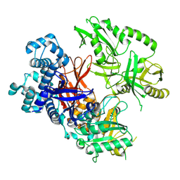 | | Crystal structure of human phospholipase C gamma 2 | | 分子名称: | 1,2-ETHANEDIOL, 1-phosphatidylinositol 4,5-bisphosphate phosphodiesterase gamma-2, CALCIUM ION | | 著者 | Chen, Y, Choi, H, Zhuang, N, Hu, L, Qian, D, Wang, J. | | 登録日 | 2023-06-20 | | 公開日 | 2024-06-26 | | 実験手法 | X-RAY DIFFRACTION (2.55 Å) | | 主引用文献 | The crystal and cryo-EM structures of PLCg2 reveal dynamic inter-domain recognitions in autoinhibition
To Be Published
|
|
7W1W
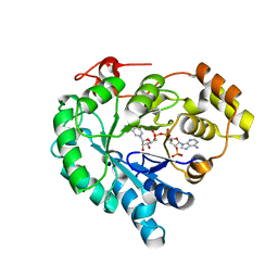 | | NADPH-bound AKR4C17 mutant F291D | | 分子名称: | AKR4-2, NADPH DIHYDRO-NICOTINAMIDE-ADENINE-DINUCLEOTIDE PHOSPHATE | | 著者 | Li, H, Yang, Y, Hu, Y, Chen, C.-C, Huang, J.-W, Min, J, Dai, L, Guo, R.-T. | | 登録日 | 2021-11-21 | | 公開日 | 2022-07-06 | | 最終更新日 | 2023-11-29 | | 実験手法 | X-RAY DIFFRACTION (1.82 Å) | | 主引用文献 | Structural analysis and engineering of aldo-keto reductase from glyphosate-resistant Echinochloa colona
J Hazard Mater, 436, 2022
|
|
7W1X
 | | Crystal structure of AKR4C16 bound with NADPH | | 分子名称: | AKR4-1, NADPH DIHYDRO-NICOTINAMIDE-ADENINE-DINUCLEOTIDE PHOSPHATE | | 著者 | Li, H, Yang, Y, Hu, Y, Chen, C.-C, Huang, J.-W, Min, J, Dai, L, Guo, R.-T. | | 登録日 | 2021-11-21 | | 公開日 | 2022-07-06 | | 最終更新日 | 2023-11-29 | | 実験手法 | X-RAY DIFFRACTION (1.9 Å) | | 主引用文献 | Structural analysis and engineering of aldo-keto reductase from glyphosate-resistant Echinochloa colona
J Hazard Mater, 436, 2022
|
|
4WV9
 | | Crystal structure of acetylcholine binding protein (AChBP) from Aplysia Californica in complex with click chemistry compound (3-exo)-8,8-dimethyl-3-[4-(pyridin-4-yl)-1H-1,2,3-triazol-1-yl]-8-azoniabicyclo[3.2.1]octane | | 分子名称: | (3-exo)-8,8-dimethyl-3-[4-(pyridin-4-yl)-1H-1,2,3-triazol-1-yl]-8-azoniabicyclo[3.2.1]octane, Soluble acetylcholine receptor | | 著者 | Talley, T.T, Bobango, J, Wu, J.M, Sankaran, B. | | 登録日 | 2014-11-04 | | 公開日 | 2015-04-22 | | 最終更新日 | 2023-12-27 | | 実験手法 | X-RAY DIFFRACTION (2 Å) | | 主引用文献 | Crystal structure of acetylcholine binding protein (AChBP) from Aplysia Californica in complex with click chemistry compound.
To Be Published
|
|
8JPP
 | | Cryo-EM structure of succinate receptor bound to succinate acid coupling MiniGsq | | 分子名称: | Guanine nucleotide-binding protein G(I)/G(S)/G(O) subunit gamma-2, Guanine nucleotide-binding protein G(I)/G(S)/G(T) subunit beta-1, Guanine nucleotide-binding protein G(s) subunit alpha isoforms short, ... | | 著者 | Wang, T.X, Tang, W.Q, Li, F.H, Wang, J.Y. | | 登録日 | 2023-06-12 | | 公開日 | 2024-05-22 | | 実験手法 | ELECTRON MICROSCOPY (3.2 Å) | | 主引用文献 | Molecular activation and G protein coupling selectivity of human succinate receptor SUCR1.
Cell Res., 2024
|
|
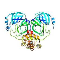
6JJP
 | | Crystal structure of Fab of a PD-1 monoclonal antibody MW11-h317 in complex with PD-1 | | 分子名称: | 2-acetamido-2-deoxy-beta-D-glucopyranose, Heavy chain of MW11-h317, Programmed cell death protein 1, ... | | 著者 | Wang, M, Wang, J, Wang, R, Jiao, S, Wang, S, Zhang, J, Zhang, M. | | 登録日 | 2019-02-26 | | 公開日 | 2019-10-30 | | 最終更新日 | 2023-11-22 | | 実験手法 | X-RAY DIFFRACTION (2.9 Å) | | 主引用文献 | Identification of a monoclonal antibody that targets PD-1 in a manner requiring PD-1 Asn58 glycosylation.
Commun Biol, 2, 2019
|
|
5NEX
 | |
6JIC
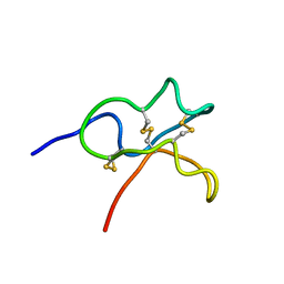 | | Identification and Characterization of a carboxypeptidase inhibitor from Lycium barbarum | | 分子名称: | WCI | | 著者 | Tan, W.L, Wong, K.H, Huang, J.Y, Tay, S.V, Wang, S.J. | | 登録日 | 2019-02-20 | | 公開日 | 2020-02-26 | | 最終更新日 | 2023-06-14 | | 実験手法 | SOLUTION NMR | | 主引用文献 | Identification and characterization of a wolfberry carboxypeptidase inhibitor from Lycium barbarum.
Food Chem, 351, 2021
|
|
2LEM
 | |
5YZ9
 | | zinc finger domain of METTL3-METTL14 N6-methyladenosine methyltransferase | | 分子名称: | N6-adenosine-methyltransferase catalytic subunit, ZINC ION | | 著者 | Dong, X, Tang, C, Gong, Z, Yin, P, Huang, J.B. | | 登録日 | 2017-12-13 | | 公開日 | 2018-03-28 | | 最終更新日 | 2024-05-01 | | 実験手法 | SOLUTION NMR, SOLUTION SCATTERING | | 主引用文献 | Solution structure of the RNA recognition domain of METTL3-METTL14 N6-methyladenosine methyltransferase.
Protein Cell, 10, 2019
|
|


