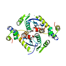
5JH5
 
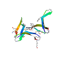
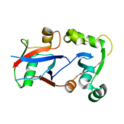
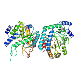
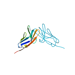
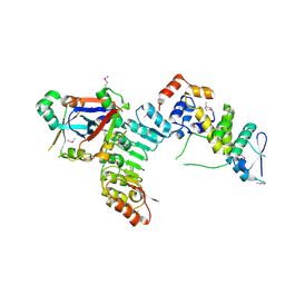 | | Structural Basis for the Hierarchical Assembly of the Core of PRC1.1 | | 分子名称: | BCL-6 corepressor-like protein 1, Lysine-specific demethylase 2B, Polycomb group RING finger protein 1, ... | | 著者 | Wong, S.J, Taylor, A.B, Hart, P.J, Kim, C.A. | | 登録日 | 2016-04-20 | | 公開日 | 2016-09-14 | | 最終更新日 | 2016-10-19 | | 実験手法 | X-RAY DIFFRACTION (2.55 Å) | | 主引用文献 | KDM2B Recruitment of the Polycomb Group Complex, PRC1.1, Requires Cooperation between PCGF1 and BCORL1.
Structure, 24, 2016
|
|
5JIE
 | |
5L1M
 | |
6DR5
 | |
6DR4
 | |
6DR6
 | |
5CZ1
 | |
5D7U
 | | Crystal structure of the C-terminal domain of MMTV integrase | | 分子名称: | ISOPROPYL ALCOHOL, Pr160 | | 著者 | Cook, N.J, Pye, V.E, Ballandras-Colas, A, Engelman, A, Cherepanov, P. | | 登録日 | 2015-08-14 | | 公開日 | 2016-02-17 | | 最終更新日 | 2024-01-10 | | 実験手法 | X-RAY DIFFRACTION (1.5 Å) | | 主引用文献 | Cryo-EM reveals a novel octameric integrase structure for betaretroviral intasome function.
Nature, 530, 2016
|
|
5CZ2
 | | Crystal structure of a two-domain fragment of MMTV integrase | | 分子名称: | MAGNESIUM ION, Pol polyprotein, ZINC ION | | 著者 | Cook, N, Ballandras-Colas, A, Engelman, A, Cherepanov, P. | | 登録日 | 2015-07-31 | | 公開日 | 2016-02-17 | | 最終更新日 | 2024-01-10 | | 実験手法 | X-RAY DIFFRACTION (2.72 Å) | | 主引用文献 | Cryo-EM reveals a novel octameric integrase structure for betaretroviral intasome function.
Nature, 530, 2016
|
|
3C7N
 | | Structure of the Hsp110:Hsc70 Nucleotide Exchange Complex | | 分子名称: | ADENOSINE-5'-DIPHOSPHATE, BERYLLIUM TRIFLUORIDE ION, CHLORIDE ION, ... | | 著者 | Schuermann, J.P, Jiang, J, Hart, P.J, Sousa, R. | | 登録日 | 2008-02-07 | | 公開日 | 2008-05-27 | | 最終更新日 | 2024-02-21 | | 実験手法 | X-RAY DIFFRACTION (3.115 Å) | | 主引用文献 | Structure of the Hsp110:Hsc70 nucleotide exchange machine
Mol.Cell, 31, 2008
|
|
4FQN
 | | Crystal structure of the CCM2 C-terminal Harmonin Homology Domain (HHD) | | 分子名称: | Malcavernin | | 著者 | Fisher, O.S, Zhang, R, Li, X, Murphy, J.W, Boggon, T.J. | | 登録日 | 2012-06-25 | | 公開日 | 2012-12-19 | | 最終更新日 | 2024-02-28 | | 実験手法 | X-RAY DIFFRACTION (1.9 Å) | | 主引用文献 | Structural studies of cerebral cavernous malformations 2 (CCM2) reveal a folded helical domain at its C-terminus.
Febs Lett., 587, 2013
|
|
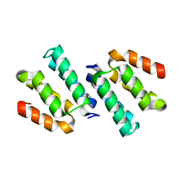
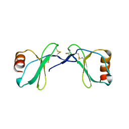
4HPM
 | | PCGF1 Ub fold (RAWUL)/BCORL1 PUFD Complex | | 分子名称: | BCL-6 corepressor-like protein 1, PHOSPHATE ION, Polycomb group RING finger protein 1 | | 著者 | Junco, S.E, Wang, R, Gaipa, J, Taylor, A.B, Gearhart, M.D, Bardwell, V.J, Hart, P.J, Kim, C.A. | | 登録日 | 2012-10-24 | | 公開日 | 2013-05-01 | | 最終更新日 | 2023-09-20 | | 実験手法 | X-RAY DIFFRACTION (1.85 Å) | | 主引用文献 | Structure of the Polycomb Group Protein PCGF1 in Complex with BCOR Reveals Basis for Binding Selectivity of PCGF Homologs.
Structure, 21, 2013
|
|
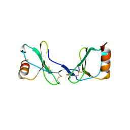
4HPL
 | | PCGF1 Ub fold (RAWUL)/BCOR PUFD Complex | | 分子名称: | BCL-6 corepressor, Polycomb group RING finger protein 1 | | 著者 | Junco, S.E, Wang, R, Gaipa, J, Taylor, A.B, Gearhart, M.D, Bardwell, V.J, Hart, P.J, Kim, C.A. | | 登録日 | 2012-10-24 | | 公開日 | 2013-05-01 | | 最終更新日 | 2024-02-28 | | 実験手法 | X-RAY DIFFRACTION (2 Å) | | 主引用文献 | Structure of the Polycomb Group Protein PCGF1 in Complex with BCOR Reveals Basis for Binding Selectivity of PCGF Homologs.
Structure, 21, 2013
|
|
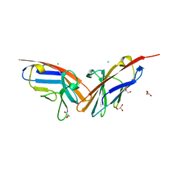
3K6B
 | | X-ray crystal structure of the E2 domain of APL-1 from C. elegans, in complex with sucrose octasulfate (SOS) | | 分子名称: | 2,3,4,6-tetra-O-sulfonato-alpha-D-glucopyranose, Beta-amyloid-like protein | | 著者 | Hoopes, J.T, Ha, Y. | | 登録日 | 2009-10-08 | | 公開日 | 2009-11-10 | | 最終更新日 | 2023-09-06 | | 実験手法 | X-RAY DIFFRACTION (2.8 Å) | | 主引用文献 | Structural characterization of the E2 domain of APL-1, a Caenorhabditis elegans homolog of human amyloid precursor protein, and its heparin binding site
J.Biol.Chem., 285, 2010
|
|
3K66
 | |
5VA4
 | |
3NYJ
 | | Crystal Structure Analysis of APP E2 domain | | 分子名称: | Amyloid beta A4 protein, OSMIUM ION | | 著者 | Ha, Y, Hu, J, Lee, S, Liu, X. | | 登録日 | 2010-07-15 | | 公開日 | 2011-06-01 | | 最終更新日 | 2017-11-08 | | 実験手法 | X-RAY DIFFRACTION (3.2 Å) | | 主引用文献 | The E2 Domains of APP and APLP1 Share a Conserved Mode of Dimerization.
Biochemistry, 50, 2011
|
|
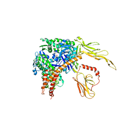
6BMC
 | | The structure of a dimeric type II DAH7PS associated with pyocyanin biosynthesis in Pseudomonas aeruginosa | | 分子名称: | CHLORIDE ION, COBALT (II) ION, PHOSPHOENOLPYRUVATE, ... | | 著者 | Sterritt, O.W, Jameson, G.B, Parker, E.J. | | 登録日 | 2017-11-14 | | 公開日 | 2018-10-03 | | 最終更新日 | 2023-10-04 | | 実験手法 | X-RAY DIFFRACTION (2.7 Å) | | 主引用文献 | Structural and functional characterisation of the entry point to pyocyanin biosynthesis inPseudomonas aeruginosadefines a new 3-deoxy-d-arabino-heptulosonate 7-phosphate synthase subclass.
Biosci. Rep., 38, 2018
|
|
2Q8T
 | |
2Q8R
 | |
2QSQ
 | | Crystal structure of the N-terminal domain of carcinoembryonic antigen (CEA) | | 分子名称: | CHLORIDE ION, Carcinoembryonic antigen-related cell adhesion molecule 5, GLYCEROL | | 著者 | Le Trong, I, Korotkova, N, Moseley, S.L, Stenkamp, R.E. | | 登録日 | 2007-07-31 | | 公開日 | 2008-01-01 | | 最終更新日 | 2024-02-21 | | 実験手法 | X-RAY DIFFRACTION (1.95 Å) | | 主引用文献 | Binding of Dr adhesins of Escherichia coli to carcinoembryonic antigen triggers receptor dissociation.
Mol.Microbiol., 67, 2008
|
|
2QST
 | |
3SLK
 | |
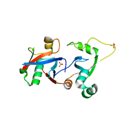
1VF6
 | | 2.1 Angstrom crystal structure of the PALS-1-L27N and PATJ L27 heterodimer complex | | 分子名称: | MAGUK p55 subfamily member 5, PALS1-associated tight junction protein | | 著者 | Li, Y, Lavie, A, Margolis, B, Karnak, D. | | 登録日 | 2004-04-09 | | 公開日 | 2004-04-20 | | 最終更新日 | 2023-12-27 | | 実験手法 | X-RAY DIFFRACTION (2.1 Å) | | 主引用文献 | Structural basis for L27 domain-mediated assembly of signaling and cell polarity complexes.
Embo J., 23, 2004
|
|
4QNB
 | |


