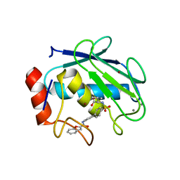
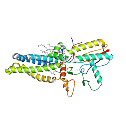
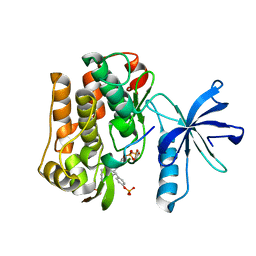
1BAI
 
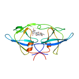 | | Crystal structure of Rous sarcoma virus protease in complex with inhibitor | | 分子名称: | N-[(2R)-2-({N~5~-[amino(iminio)methyl]-L-ornithyl-L-valyl}amino)-4-methylpentyl]-L-phenylalanyl-L-alpha-glutamyl-L-alanyl-L-norleucinamide, PROTEASE | | 著者 | Wu, J, Adomat, J.M, Ridky, T.W, Louis, J.M, Leis, J, Harrison, R.W, Weber, I.T. | | 登録日 | 1998-04-17 | | 公開日 | 1999-01-13 | | 最終更新日 | 2024-03-13 | | 実験手法 | X-RAY DIFFRACTION (2.4 Å) | | 主引用文献 | Structural basis for specificity of retroviral proteases.
Biochemistry, 37, 1998
|
|
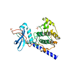
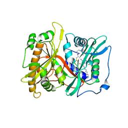
1A94
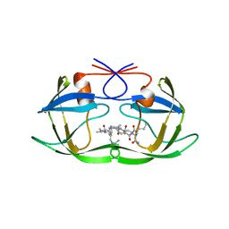 | | STRUCTURAL BASIS FOR SPECIFICITY OF RETROVIRAL PROTEASES | | 分子名称: | N-[(2R)-2-({N~5~-[amino(iminio)methyl]-L-ornithyl-L-valyl}amino)-4-methylpentyl]-L-phenylalanyl-L-alpha-glutamyl-L-alanyl-L-norleucinamide, PROTEASE | | 著者 | Wu, J, Adomat, J.M, Ridky, T.W, Louis, J.M, Leis, J, Harrison, R.W, Weber, I.T. | | 登録日 | 1998-04-16 | | 公開日 | 1999-01-13 | | 最終更新日 | 2024-02-07 | | 実験手法 | X-RAY DIFFRACTION (2 Å) | | 主引用文献 | Structural basis for specificity of retroviral proteases.
Biochemistry, 37, 1998
|
|
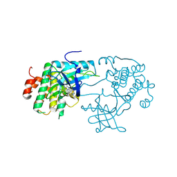
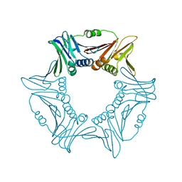
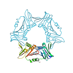
2WII
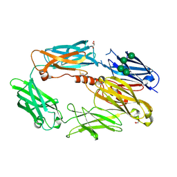 | | Complement C3b in complex with factor H domains 1-4 | | 分子名称: | 2-acetamido-2-deoxy-beta-D-glucopyranose, CALCIUM ION, COMPLEMENT C3 BETA CHAIN, ... | | 著者 | Wu, J, Janssen, B.J.C, Gros, P. | | 登録日 | 2009-05-12 | | 公開日 | 2009-06-09 | | 最終更新日 | 2023-12-13 | | 実験手法 | X-RAY DIFFRACTION (2.7 Å) | | 主引用文献 | Structure of complement fragment C3b-factor H and implications for host protection by complement regulators.
Nat. Immunol., 10, 2009
|
|
7O1Q
 | | Amyloid beta oligomer displayed on the alpha hemolysin scaffold | | 分子名称: | Alpha-hemolysin hybridized Abeta | | 著者 | Wu, J, Blum, T.B, Farrell, D.P, DiMaio, F, Abrahams, J.P, Luo, J. | | 登録日 | 2021-03-30 | | 公開日 | 2021-04-14 | | 最終更新日 | 2021-08-18 | | 実験手法 | ELECTRON MICROSCOPY (3.4 Å) | | 主引用文献 | Cryo-electron Microscopy Imaging of Alzheimer's Amyloid-beta 42 Oligomer Displayed on a Functionally and Structurally Relevant Scaffold.
Angew.Chem.Int.Ed.Engl., 60, 2021
|
|
1ZTQ
 | | Crystal structure of the catalytic domain of MMP-13 complexed with WAY-033 | | 分子名称: | CALCIUM ION, Collagenase 3, N-({4'-[(1-BENZOFURAN-2-YLCARBONYL)AMINO]-1,1'-BIPHENYL-4-YL}SULFONYL)-L-VALINE, ... | | 著者 | Wu, J, Rush III, T.S, Hotchandani, R, Du, X, Geck, M, Collins, E, Xu, Z.B, Skotnicki, J, Levin, J.I, Lovering, F. | | 登録日 | 2005-05-27 | | 公開日 | 2006-05-30 | | 最終更新日 | 2024-02-14 | | 実験手法 | X-RAY DIFFRACTION (2 Å) | | 主引用文献 | Identification of potent and selective MMP-13 inhibitors
Bioorg.Med.Chem.Lett., 15, 2005
|
|
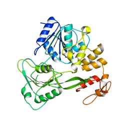
1SYK
 | | Crystal structure of E230Q mutant of cAMP-dependent protein kinase reveals unexpected apoenzyme conformation | | 分子名称: | cAMP-dependent protein kinase, alpha-catalytic subunit | | 著者 | Wu, J, Yang, J, Madhusudan, N, Xuong, N.H, Ten Eyck, L.F, Taylor, S.S. | | 登録日 | 2004-04-01 | | 公開日 | 2005-05-17 | | 最終更新日 | 2023-08-23 | | 実験手法 | X-RAY DIFFRACTION (2.8 Å) | | 主引用文献 | Crystal structure of the E230Q mutant of cAMP-dependent
protein kinase reveals an unexpected apoenzyme conformation and an
extended N-terminal A helix.
Protein Sci., 14, 2005
|
|
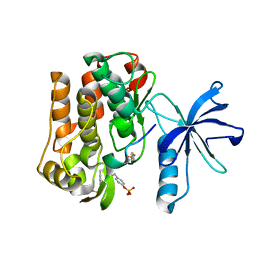
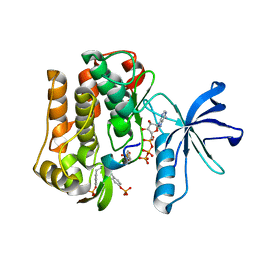
3D94
 | | Crystal structure of the insulin-like growth factor-1 receptor kinase in complex with PQIP | | 分子名称: | 3-[cis-3-(4-methylpiperazin-1-yl)cyclobutyl]-1-(2-phenylquinolin-7-yl)imidazo[1,5-a]pyrazin-8-amine, CALCIUM ION, Insulin-like growth factor 1 receptor beta chain | | 著者 | Wu, J, Li, W, Miller, W.T, Hubbard, S.R. | | 登録日 | 2008-05-26 | | 公開日 | 2008-07-29 | | 最終更新日 | 2023-08-30 | | 実験手法 | X-RAY DIFFRACTION (2.3 Å) | | 主引用文献 | Small-molecule inhibition and activation-loop trans-phosphorylation of the IGF1 receptor
Embo J., 27, 2008
|
|
8HFC
 | | Cryo-EM structure of yeast Erf2/Erf4 complex | | 分子名称: | PALMITIC ACID, Palmitoyltransferase ERF2, Ras modification protein ERF4, ... | | 著者 | Wu, J, Hu, Q, Zhang, Y, Yang, A, Liu, S. | | 登録日 | 2022-11-10 | | 公開日 | 2023-11-22 | | 実験手法 | ELECTRON MICROSCOPY (3.5 Å) | | 主引用文献 | Cryo-EM structure of Yeast Erf2/Erf4 complex
To Be Published
|
|
8HF3
 | | Cryo-EM structure of human ZDHHC9/GCP16 complex | | 分子名称: | 1,2-DILAUROYL-SN-GLYCERO-3-PHOSPHATE, Golgin subfamily A member 7, PALMITIC ACID, ... | | 著者 | Wu, J, Hu, Q, Zhang, Y, Liu, S, Yang, A. | | 登録日 | 2022-11-09 | | 公開日 | 2023-11-22 | | 実験手法 | ELECTRON MICROSCOPY (3.4 Å) | | 主引用文献 | Cryo-EM structure of human ZDHHC9/GCP16 complex
To Be Published
|
|
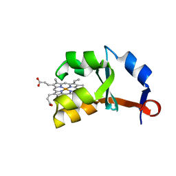
1ES1
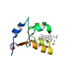 | | CRYSTAL STRUCTURE OF VAL61HIS MUTANT OF TRYPSIN-SOLUBILIZED FRAGMENT OF CYTOCHROME B5 | | 分子名称: | CYTOCHROME B5, PROTOPORPHYRIN IX CONTAINING FE | | 著者 | Wu, J, Gan, J.-H, Xia, Z.-X, Wang, Y.-H, Wang, W.-H, Xue, L.-L, Xie, Y, Huang, Z.-X. | | 登録日 | 2000-04-07 | | 公開日 | 2000-08-09 | | 最終更新日 | 2024-02-07 | | 実験手法 | X-RAY DIFFRACTION (2.1 Å) | | 主引用文献 | Crystal structure of recombinant trypsin-solubilized fragment of cytochrome b(5) and the structural comparison with Val61His mutant.
Proteins, 40, 2000
|
|
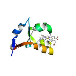
1EHB
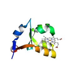 | | CRYSTAL STRUCTURE OF RECOMBINANT TRYPSIN-SOLUBILIZED FRAGMENT OF CYTOCHROME B5 | | 分子名称: | PROTEIN (CYTOCHROME B5), PROTOPORPHYRIN IX CONTAINING FE | | 著者 | Wu, J, Gan, J.-H, Xia, Z.-X, Wang, Y.-H, Wang, W.-H, Xue, L.-L, Xie, Y, Huang, Z.-X. | | 登録日 | 2000-02-20 | | 公開日 | 2000-08-09 | | 最終更新日 | 2024-02-07 | | 実験手法 | X-RAY DIFFRACTION (1.9 Å) | | 主引用文献 | Crystal structure of recombinant trypsin-solubilized fragment of cytochrome b(5) and the structural comparison with Val61His mutant.
Proteins, 40, 2000
|
|
8GYF
 | | Crystal structure of a bright green fluorescent protein (StayGold) with single mutation (K192Y) in jellyfish Cytaeis uchidae from Biortus | | 分子名称: | 1,2-ETHANEDIOL, staygold(K192Y) | | 著者 | Wu, J, Wang, F, Gui, W, Cheng, W, Yang, Y. | | 登録日 | 2022-09-22 | | 公開日 | 2023-10-04 | | 最終更新日 | 2023-11-15 | | 実験手法 | X-RAY DIFFRACTION (2 Å) | | 主引用文献 | Crystal structure of a bright green fluorescent protein (StayGold) with single mutation (K192Y) in jellyfish Cytaeis uchidae from Biortus
To Be Published
|
|
8I6G
 | |
8I6H
 | |
1YKS
 | | Crystal structure of yellow fever virus NS3 helicase | | 分子名称: | Genome polyprotein [contains: Flavivirin protease NS3 catalytic subunit] | | 著者 | Wu, J, Bera, A.K, Kuhn, R.J, Smith, J.L. | | 登録日 | 2005-01-18 | | 公開日 | 2005-08-23 | | 最終更新日 | 2024-02-14 | | 実験手法 | X-RAY DIFFRACTION (1.8 Å) | | 主引用文献 | Structure of the flavivirus helicase: implications for catalytic activity, protein interactions, and proteolytic processing.
J.Virol., 79, 2005
|
|
3BU3
 | |
3BU5
 | |
3BU6
 | |
2P6E
 | |
2P6G
 | |
2P6F
 | |
1M2M
 | | Crystal structure of E44A/E48A/E56A/D60A mutant of cytochrome b5 | | 分子名称: | PROTOPORPHYRIN IX CONTAINING FE, cytochrome b5 | | 著者 | Wu, J, Wang, Y.-H, Gan, J.-H, Wang, W.-H, Sun, B.-Y, Huang, Z.-X, Xia, Z.-X. | | 登録日 | 2002-06-24 | | 公開日 | 2003-03-18 | | 最終更新日 | 2023-10-25 | | 実験手法 | X-RAY DIFFRACTION (1.8 Å) | | 主引用文献 | Structures of Cytochrome b5 Mutated at the Charged Surface-Residues and Their Interactions with Cytochrome c
Chin.J.Chem., 20, 2002
|
|
1M2I
 | | Crystal structure of E44A/E56A mutant of cytochrome b5 | | 分子名称: | PROTOPORPHYRIN IX CONTAINING FE, cytochrome b5 | | 著者 | Wu, J, Wang, Y.-H, Gan, J.-H, Wang, W.-H, Sun, B.-Y, Huang, Z.-X, Xia, Z.-X. | | 登録日 | 2002-06-24 | | 公開日 | 2003-03-18 | | 最終更新日 | 2023-10-25 | | 実験手法 | X-RAY DIFFRACTION (1.8 Å) | | 主引用文献 | Structures of Cytochrome b5 Mutated at the Charged Surface-Residues and Their Interactions with Cytochrome c
Chin.J.Chem., 20, 2002
|
|
8J2H
 | | Crystal structure of a bright green fluorescent protein (StayGold) with single mutation (N137A) in jellyfish Cytaeis uchidae from Biortus | | 分子名称: | GLYCEROL, SODIUM ION, StayGold(N137A) | | 著者 | Wu, J, Wang, F, Gui, W, Cheng, W, Yang, Y. | | 登録日 | 2023-04-14 | | 公開日 | 2023-12-13 | | 実験手法 | X-RAY DIFFRACTION (1.7 Å) | | 主引用文献 | Crystal structure of a bright green fluorescent protein (StayGold) in jellyfish Cytaeis uchidae from Biortus
To Be Published
|
|
8J2J
 | | Crystal structure of a bright green fluorescent protein (StayGold) with single mutation (Y187F) in jellyfish Cytaeis uchidae from Biortus | | 分子名称: | 1,2-ETHANEDIOL, SULFATE ION, StayGold(Y187F) | | 著者 | Wu, J, Wang, F, Gui, W, Cheng, W, Yang, Y. | | 登録日 | 2023-04-14 | | 公開日 | 2023-12-13 | | 実験手法 | X-RAY DIFFRACTION (1.9 Å) | | 主引用文献 | Crystal structure of a bright green fluorescent protein (StayGold) in jellyfish Cytaeis uchidae from Biortus
To Be Published
|
|


