1FEC
 
 | |
3ADK
 | |
1K5G
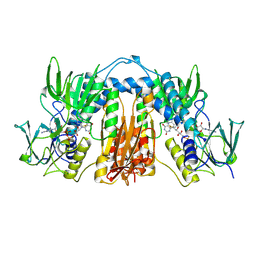
 | | Crystal structure of Ran-GDP-AlFx-RanBP1-RanGAP complex | | 分子名称: | ALUMINUM FLUORIDE, GTP-binding nuclear protein RAN, GUANOSINE-5'-DIPHOSPHATE, ... | | 著者 | Seewald, M.J, Koerner, C, Wittinghofer, A, Vetter, I.R. | | 登録日 | 2001-10-10 | | 公開日 | 2002-02-13 | | 最終更新日 | 2023-08-16 | | 実験手法 | X-RAY DIFFRACTION (3.1 Å) | | 主引用文献 | RanGAP mediates GTP hydrolysis without an arginine finger.
Nature, 415, 2002
|
|
1A2B
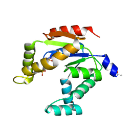
 | | HUMAN RHOA COMPLEXED WITH GTP ANALOGUE | | 分子名称: | 5'-GUANOSINE-DIPHOSPHATE-MONOTHIOPHOSPHATE, MAGNESIUM ION, TRANSFORMING PROTEIN RHOA | | 著者 | Ihara, K, Muraguchi, S, Kato, M, Shimizu, T, Shirakawa, M, Kuroda, S, Kaibuchi, K, Hakoshima, T. | | 登録日 | 1997-12-26 | | 公開日 | 1998-06-17 | | 最終更新日 | 2023-08-02 | | 実験手法 | X-RAY DIFFRACTION (2.4 Å) | | 主引用文献 | Crystal structure of human RhoA in a dominantly active form complexed with a GTP analogue.
J.Biol.Chem., 273, 1998
|
|
1K5D
 | | Crystal structure of Ran-GPPNHP-RanBP1-RanGAP complex | | 分子名称: | GTP-binding nuclear protein RAN, MAGNESIUM ION, PHOSPHOAMINOPHOSPHONIC ACID-GUANYLATE ESTER, ... | | 著者 | Seewald, M.J, Koerner, C, Wittinghofer, A, Vetter, I.R. | | 登録日 | 2001-10-10 | | 公開日 | 2002-02-13 | | 最終更新日 | 2023-08-16 | | 実験手法 | X-RAY DIFFRACTION (2.7 Å) | | 主引用文献 | RanGAP mediates GTP hydrolysis without an arginine finger.
Nature, 415, 2002
|
|
1MA9
 | | Crystal structure of the complex of human vitamin D binding protein and rabbit muscle actin | | 分子名称: | ADENOSINE-5'-TRIPHOSPHATE, Actin, Alpha Skeletal Muscle, ... | | 著者 | Verboven, C, Bogaerts, I, Waelkens, E, Rabijns, A, Van Baelen, H, Bouillon, R, De Ranter, C. | | 登録日 | 2002-08-02 | | 公開日 | 2003-02-04 | | 最終更新日 | 2011-07-13 | | 実験手法 | X-RAY DIFFRACTION (2.4 Å) | | 主引用文献 | Actin-DBP: the perfect structural fit?
Acta Crystallogr.,Sect.D, 59, 2003
|
|
2W3U
 | |
2W3T
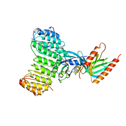
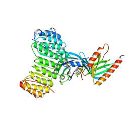
 | | Chloro complex of the Ni-Form of E.coli deformylase | | 分子名称: | CHLORIDE ION, ETHANOL, NICKEL (II) ION, ... | | 著者 | Ngo, Y.H.T, Palm, G.J, Hinrichs, W. | | 登録日 | 2008-11-14 | | 公開日 | 2009-12-15 | | 最終更新日 | 2023-12-13 | | 実験手法 | X-RAY DIFFRACTION (1.69 Å) | | 主引用文献 | Structure of the Ni(II) Complex of Escherichia Coli Peptide Deformylase and Suggestions on Deformylase Activities Depending on Different Metal(II) Centres.
J.Biol.Inorg.Chem., 15, 2010
|
|
8EH0
 | |
8EH1
 | |
8EGY
 | |
8EGZ
 | |
7ZFM
 | | Engineered Protein Targeting the Zika Viral Envelope Fusion Loop | | 分子名称: | 1,2-ETHANEDIOL, ACETIC ACID, HEXAETHYLENE GLYCOL, ... | | 著者 | Athayde, D, Archer, M, Viana, I.F.T, Adan, W.C.S, Xavier, L.S.S, Lins, R.D. | | 登録日 | 2022-04-01 | | 公開日 | 2022-08-17 | | 最終更新日 | 2024-01-31 | | 実験手法 | X-RAY DIFFRACTION (1.711 Å) | | 主引用文献 | In Vitro Neutralisation of Zika Virus by an Engineered Protein Targeting the Viral Envelope Fusion Loop
SSRN, 2022
|
|
1YAG
 | | STRUCTURE OF THE YEAST ACTIN-HUMAN GELSOLIN SEGMENT 1 COMPLEX | | 分子名称: | ADENOSINE-5'-TRIPHOSPHATE, CALCIUM ION, MAGNESIUM ION, ... | | 著者 | Vorobiev, S, Strokopytov, B, Frieden, C, Almo, S.C. | | 登録日 | 1998-10-09 | | 公開日 | 1999-10-09 | | 最終更新日 | 2024-04-03 | | 実験手法 | X-RAY DIFFRACTION (1.9 Å) | | 主引用文献 | The structure of nonvertebrate actin: Implications for the ATP
hydrolytic mechanism
Proc.Natl.Acad.Sci.USA, 100, 2003
|
|
1YVN
 | | THE YEAST ACTIN VAL 159 ASN MUTANT COMPLEX WITH HUMAN GELSOLIN SEGMENT 1. | | 分子名称: | ADENOSINE-5'-TRIPHOSPHATE, CALCIUM ION, MAGNESIUM ION, ... | | 著者 | Vorobiev, S.M, Belmont, L.D, Drubin, D.G, Almo, S.C. | | 登録日 | 1999-03-23 | | 公開日 | 2000-03-23 | | 最終更新日 | 2023-08-23 | | 実験手法 | X-RAY DIFFRACTION (2.1 Å) | | 主引用文献 | Crystal structure of yeast actin V159N mutant
To be Published
|
|
1D4X
 | | Crystal Structure of Caenorhabditis Elegans Mg-ATP Actin Complexed with Human Gelsolin Segment 1 at 1.75 A resolution. | | 分子名称: | ADENOSINE-5'-TRIPHOSPHATE, C. ELEGANS ACTIN 1/3, CALCIUM ION, ... | | 著者 | Vorobiev, S, Ono, S, Almo, S.C. | | 登録日 | 1999-10-06 | | 公開日 | 2001-05-02 | | 最終更新日 | 2024-02-07 | | 実験手法 | X-RAY DIFFRACTION (1.75 Å) | | 主引用文献 | The structure of nonvertebrate actin: implications for the ATP hydrolytic mechanism.
Proc.Natl.Acad.Sci.Usa, 100, 2003
|
|
7PQN
 | | Catalytic fragment of MASP-2 in complex with ecotin | | 分子名称: | Ecotin, GLYCEROL, Mannan-binding lectin serine protease 2 A chain, ... | | 著者 | Harmat, V, Fodor, K, Heja, D. | | 登録日 | 2021-09-17 | | 公開日 | 2022-05-18 | | 最終更新日 | 2024-01-31 | | 実験手法 | X-RAY DIFFRACTION (2.400015 Å) | | 主引用文献 | Synergy of protease-binding sites within the ecotin homodimer is crucial for inhibition of MASP enzymes and for blocking lectin pathway activation.
J.Biol.Chem., 298, 2022
|
|
7PQO
 | | Catalytic fragment of MASP-1 in complex with P1 site mutant ecotin | | 分子名称: | Ecotin, GLYCEROL, Mannan-binding lectin serine protease 1, ... | | 著者 | Harmat, V, Fodor, K, Heja, D. | | 登録日 | 2021-09-17 | | 公開日 | 2022-05-18 | | 最終更新日 | 2024-01-31 | | 実験手法 | X-RAY DIFFRACTION (3.39 Å) | | 主引用文献 | Synergy of protease-binding sites within the ecotin homodimer is crucial for inhibition of MASP enzymes and for blocking lectin pathway activation.
J.Biol.Chem., 298, 2022
|
|
8AMS
 | | Complex of human TRIM2 RING domain, UBCH5C, and Ubiquitin | | 分子名称: | 3,6,9,12,15,18,21-HEPTAOXATRICOSANE-1,23-DIOL, GLYCEROL, Polyubiquitin-C, ... | | 著者 | Perez-Borrajero, C, Kotova, I, Murciano, B, Hennig, J. | | 登録日 | 2022-08-04 | | 公開日 | 2023-11-15 | | 実験手法 | X-RAY DIFFRACTION (2.4 Å) | | 主引用文献 | Structural and biophysical studies of TRIM2 and TRIM3
To Be Published
|
|
2DNJ
 | |
6H6K
 | | The structure of the FKR mutant of the archaeal translation initiation factor 2 gamma subunit in complex with GDPCP, obtained in the absence of magnesium salts in the crystallization solution. | | 分子名称: | 1,2-ETHANEDIOL, PHOSPHOMETHYLPHOSPHONIC ACID GUANYLATE ESTER, SODIUM ION, ... | | 著者 | Nikonov, O, Kravchenko, O, Nevskaya, N, Stolboushkina, E, Gabdulkhakov, A, Garber, M, Nikonov, S. | | 登録日 | 2018-07-27 | | 公開日 | 2019-04-17 | | 最終更新日 | 2024-01-17 | | 実験手法 | X-RAY DIFFRACTION (2 Å) | | 主引用文献 | The third structural switch in the archaeal translation initiation factor 2 (aIF2) molecule and its possible role in the initiation of GTP hydrolysis and the removal of aIF2 from the ribosome.
Acta Crystallogr D Struct Biol, 75, 2019
|
|
6E5G
 | |
3DNI
 | | CRYSTALLOGRAPHIC REFINEMENT AND STRUCTURE OF DNASE I AT 2 ANGSTROMS RESOLUTION | | 分子名称: | CALCIUM ION, DEOXYRIBONUCLEASE I, alpha-D-galactopyranose-(1-6)-beta-D-mannopyranose-(1-3)-[beta-D-mannopyranose-(1-6)-alpha-D-mannopyranose-(1-6)]beta-D-mannopyranose-(1-4)-2-acetamido-2-deoxy-beta-D-glucopyranose-(1-4)-2-acetamido-2-deoxy-beta-D-glucopyranose | | 著者 | Oefner, C, Suck, D. | | 登録日 | 1992-08-20 | | 公開日 | 1994-01-31 | | 最終更新日 | 2020-07-29 | | 実験手法 | X-RAY DIFFRACTION (2 Å) | | 主引用文献 | Crystallographic refinement and structure of DNase I at 2 A resolution.
J.Mol.Biol., 192, 1986
|
|


