7NQB
 
 | |
4IRO
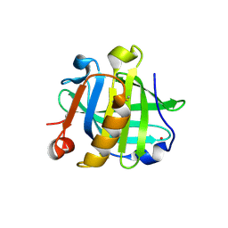
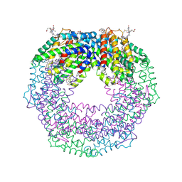
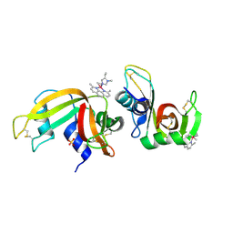
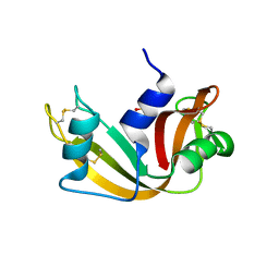
 | | Crystal structure of T-state carbonmonoxy hemoglobin from Trematomus bernacchii at pH 8.4 | | Descriptor: | CARBON MONOXIDE, Hemoglobin subunit alpha, Hemoglobin subunit beta, ... | | Authors: | Merlino, A, Balsamo, A, Mazzarella, L, Vergara, A. | | Deposit date: | 2013-01-15 | | Release date: | 2013-02-20 | | Last modified: | 2013-10-23 | | Method: | X-RAY DIFFRACTION (2.2 Å) | | Cite: | Role of tertiary structures on the Root effect in fish hemoglobins.
Biochim.Biophys.Acta, 1834, 2013
|
|
6XVX
 | |
6XW0
 | |
4S0Q
 | |
4RSZ
 | |
4S18
 | |
4S1Y
 | |
8B4N
 | |
6RJV
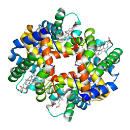
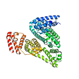 | | The X-ray structure of the Gold/Serum Albumin adduct obtained upon reaction of the protein with AuL12, a gold(III) dithiocarbamate complex | | Descriptor: | GOLD ION, MAGNESIUM ION, Serum albumin | | Authors: | Merlino, A, Giorgio, A, Ferraro, G. | | Deposit date: | 2019-04-29 | | Release date: | 2019-08-14 | | Last modified: | 2024-01-24 | | Method: | X-RAY DIFFRACTION (3.21 Å) | | Cite: | Structural Characterization of a Gold/Serum Albumin Complex.
Inorg.Chem., 58, 2019
|
|
7R1P
 | |
7R1Q
 | |
7OR6
 | |
7ORD
 | |
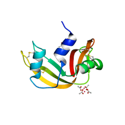
6F60
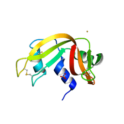
 | | The x-ray structure of bovine pancreatic ribonuclease in complex with a five-coordinate platinum(II) compound containing a sugar ligand | | Descriptor: | Ribonuclease pancreatic, SULFATE ION, five-coordinate platinum(II) compound | | Authors: | Merlino, A, Ferraro, G. | | Deposit date: | 2017-12-04 | | Release date: | 2018-04-25 | | Method: | X-RAY DIFFRACTION (1.14 Å) | | Cite: | Five-Coordinate Platinum(II) Compounds Containing Sugar Ligands: Synthesis, Characterization, Cytotoxic Activity, and Interaction with Biological Macromolecules.
Inorg Chem, 57, 2018
|
|
5E5E
 | |
4L2D
 | |
4L2C
 | |
5E5F
 | |
6FX8
 | |
6FX9
 | |
6FTV
 | |
6G5V
 | |
6G5Y
 | |
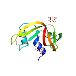
5ILC
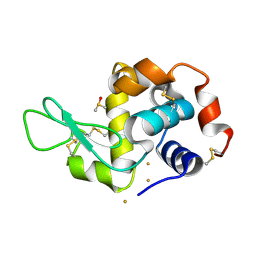
 | | The X-ray structure of the adduct formed in the reaction between hen egg white lysozyme a compound 2, a platin(II) compound containing a O, S bidentate ligand | | Descriptor: | ACETATE ION, CHLORIDE ION, DIMETHYL SULFOXIDE, ... | | Authors: | Merlino, A, Ferraro, G. | | Deposit date: | 2016-03-04 | | Release date: | 2016-12-07 | | Last modified: | 2024-01-10 | | Method: | X-RAY DIFFRACTION (1.75 Å) | | Cite: | Platinum(ii) O,S complexes as potential metallodrugs against Cisplatin resistance.
Dalton Trans, 45, 2016
|
|


