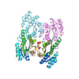
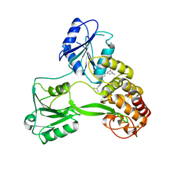
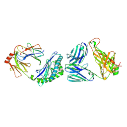
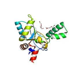
Loading
PDBj
MenuPDBj@FacebookPDBj@TwitterPDBj@YouTubewwPDB FoundationwwPDB
RCSB PDBPDBeBMRBAdv. SearchSearch help
Search by PDB author
5XTF
DownloadVisualize
BU of 5xtf by Molmil
Crystal structure of the cis-dihydrodiol naphthalene dehydrogenase NahB from Pseudomonas sp. MC1
Descriptor: 2,3-dihydroxy-2,3-dihydrophenylpropionate dehydrogenase
Authors:Park, A.K, Kim, H.-W.
Deposit date:2017-06-19
Release date:2017-08-09
Last modified:2023-11-22
Method:X-RAY DIFFRACTION (2.095 Å)
Cite:Crystal structure of cis-dihydrodiol naphthalene dehydrogenase (NahB) from Pseudomonas sp. MC1: Insights into the early binding process of the substrate
Biochem. Biophys. Res. Commun., 491, 2017
5ZHF
DownloadVisualize
BU of 5zhf by Molmil
Structure of VanYB unbound
Descriptor: D-alanyl-D-alanine carboxypeptidase, GLYCEROL, TETRAETHYLENE GLYCOL, ...
Authors:Kim, H.S, Hahn, H.
Deposit date:2018-03-13
Release date:2018-09-05
Method:X-RAY DIFFRACTION (1.65 Å)
Cite:Structural basis for the substrate recognition of peptidoglycan pentapeptides by Enterococcus faecalis VanYB.
Int. J. Biol. Macromol., 119, 2018
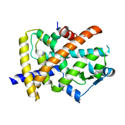
6W30
DownloadVisualize
BU of 6w30 by Molmil
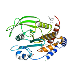
Protein Tyrosine Phosphatase 1B Bound to Amorphadiene
Descriptor: Amorphadiene, GLYCEROL, MAGNESIUM ION, ...
Authors:Sarkar, A, Kim, E.Y, Hongdusit, A, Sankaran, B, Fox, J.
Deposit date:2020-03-08
Release date:2021-05-26
Last modified:2023-10-18
Method:X-RAY DIFFRACTION (2.1 Å)
Cite:Microbially Guided Discovery and Biosynthesis of Biologically Active Natural Products.
Acs Synth Biol, 10, 2021
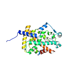
5Z5A
DownloadVisualize
BU of 5z5a by Molmil
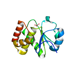
Crystal structure of Tk-PTP in the active form
Descriptor: Protein-tyrosine phosphatase, VANADATE ION
Authors:Ku, B, Yun, H.Y, Kim, S.J.
Deposit date:2018-01-17
Release date:2018-07-04
Last modified:2023-11-22
Method:X-RAY DIFFRACTION (1.8 Å)
Cite:Structural study reveals the temperature-dependent conformational flexibility of Tk-PTP, a protein tyrosine phosphatase from Thermococcus kodakaraensis KOD1
PLoS ONE, 13, 2018
4WXR
DownloadVisualize
BU of 4wxr by Molmil
X-ray crystal structure of NS3 Helicase from HCV with a bound inhibitor at 2.42 A resolution
Descriptor: NS3, {6-(3,5-diaminophenyl)-1-[4-(propan-2-yl)benzyl]-1H-indol-3-yl}acetic acid
Authors:Davies, D.R, Kim, H, Lorimer, D.
Deposit date:2014-11-14
Release date:2015-12-23
Last modified:2023-09-27
Method:X-RAY DIFFRACTION (2.42 Å)
Cite:crystal structure of NS3 Helicase from HCV with a bound inhibitor
TO BE PUBLISHED
1Y4U
DownloadVisualize
BU of 1y4u by Molmil
Conformation rearrangement of heat shock protein 90 upon ADP binding
Descriptor: Chaperone protein htpG
Authors:Huai, Q, Wang, H, Liu, Y, Kim, H, Toft, D, Ke, H.
Deposit date:2004-12-01
Release date:2005-04-19
Last modified:2024-04-03
Method:X-RAY DIFFRACTION (2.9 Å)
Cite:Structures of the N-terminal and middle domains of E. coli Hsp90 and conformation changes upon ADP binding.
Structure, 13, 2005
2G95
DownloadVisualize
BU of 2g95 by Molmil
Crystal Structure of Visfatin/Pre-B Cell Colony Enhancing Factor 1/Nicotinamide Phosphoribosyltransferase
Descriptor: Nicotinamide phosphoribosyltransferase
Authors:Kim, M.-K, Eom, S.H.
Deposit date:2006-03-05
Release date:2006-08-01
Last modified:2024-04-03
Method:X-RAY DIFFRACTION (1.9 Å)
Cite:Crystal Structure of Visfatin/Pre-B Cell Colony-enhancing Factor 1/Nicotinamide Phosphoribosyltransferase, Free and in Complex with the Anti-cancer Agent FK-866
J.Mol.Biol., 362, 2006
2G97
DownloadVisualize
BU of 2g97 by Molmil
Crystal Structure of Visfatin/Pre-B Cell Colony Enhancing Factor 1/Nicotinamide Phosphoribosyltransferase In Complex with the Specific Inhibitor FK-866
Descriptor: (2E)-N-{4-[1-(benzenecarbonyl)piperidin-4-yl]butyl}-3-(pyridin-3-yl)prop-2-enamide, Nicotinamide phosphoribosyltransferase
Authors:Kim, M.-K, Eom, S.H.
Deposit date:2006-03-05
Release date:2006-08-01
Last modified:2023-10-25
Method:X-RAY DIFFRACTION (2.9 Å)
Cite:Crystal Structure of Visfatin/Pre-B Cell Colony-enhancing Factor 1/Nicotinamide Phosphoribosyltransferase, Free and in Complex with the Anti-cancer Agent FK-866
J.Mol.Biol., 362, 2006
2G96
DownloadVisualize
BU of 2g96 by Molmil
Crystal Structure of Visfatin/Pre-B Cell Colony Enhancing Factor 1/Nicotinamide Phosphoribosyltransferase In Complex with Niconamide Mononucleotide
Descriptor: BETA-NICOTINAMIDE RIBOSE MONOPHOSPHATE, Nicotinamide phosphoribosyltransferase
Authors:Eom, S.H, Kim, M.-K.
Deposit date:2006-03-05
Release date:2006-08-01
Last modified:2023-10-25
Method:X-RAY DIFFRACTION (2.9 Å)
Cite:Crystal Structure of Visfatin/Pre-B Cell Colony-enhancing Factor 1/Nicotinamide Phosphoribosyltransferase, Free and in Complex with the Anti-cancer Agent FK-866
J.Mol.Biol., 362, 2006
7LAW
DownloadVisualize
BU of 7law by Molmil
crystal structure of GITR complex with GITR-L
Descriptor: 2-acetamido-2-deoxy-beta-D-glucopyranose, Tumor necrosis factor ligand superfamily member 18, Tumor necrosis factor receptor superfamily member 18
Authors:Longenecker, K.L, Rogers, B, Bigelow, L, Judge, R.A, Alvarez, H.
Deposit date:2021-01-07
Release date:2022-03-09
Last modified:2023-10-18
Method:X-RAY DIFFRACTION (2.752 Å)
Cite:An anti-PD-1-GITR-L bispecific agonist induces GITR clustering-mediated T cell activation for cancer immunotherapy.
Nat Cancer, 3, 2022
5K1A
DownloadVisualize
BU of 5k1a by Molmil
Crystal structure of the UAF1-USP12 complex in C2 space group
Descriptor: Ubiquitin carboxyl-terminal hydrolase 12, WD repeat-containing protein 48, ZINC ION
Authors:Li, H, D'Andrea, A.D, Zheng, N.
Deposit date:2016-05-18
Release date:2016-07-20
Last modified:2023-09-27
Method:X-RAY DIFFRACTION (2.3 Å)
Cite:Allosteric Activation of Ubiquitin-Specific Proteases by beta-Propeller Proteins UAF1 and WDR20.
Mol.Cell, 63, 2016
5K16
DownloadVisualize
BU of 5k16 by Molmil
Crystal structure of free Ubiquitin-specific protease 12
Descriptor: GLYCEROL, Ubiquitin carboxyl-terminal hydrolase 12, ZINC ION
Authors:Li, H, D'Andrea, A.D, Zheng, N.
Deposit date:2016-05-17
Release date:2016-07-20
Last modified:2023-09-27
Method:X-RAY DIFFRACTION (2.599 Å)
Cite:Allosteric Activation of Ubiquitin-Specific Proteases by beta-Propeller Proteins UAF1 and WDR20.
Mol.Cell, 63, 2016
5K1C
DownloadVisualize
BU of 5k1c by Molmil
Crystal structure of the UAF1/WDR20/USP12 complex
Descriptor: PHOSPHATE ION, TRIS(HYDROXYETHYL)AMINOMETHANE, Ubiquitin carboxyl-terminal hydrolase 12, ...
Authors:Li, H, D'Andrea, A.D, Zheng, N.
Deposit date:2016-05-18
Release date:2016-07-20
Last modified:2023-09-27
Method:X-RAY DIFFRACTION (3 Å)
Cite:Allosteric Activation of Ubiquitin-Specific Proteases by beta-Propeller Proteins UAF1 and WDR20.
Mol.Cell, 63, 2016
5K1B
DownloadVisualize
BU of 5k1b by Molmil
Crystal structure of the UAF1/USP12 complex in F222 space group
Descriptor: Ubiquitin carboxyl-terminal hydrolase 12, WD repeat-containing protein 48, ZINC ION
Authors:Li, H, D'Andrea, A.D, Zheng, N.
Deposit date:2016-05-18
Release date:2016-07-20
Last modified:2019-11-20
Method:X-RAY DIFFRACTION (3.3 Å)
Cite:Allosteric Activation of Ubiquitin-Specific Proteases by beta-Propeller Proteins UAF1 and WDR20.
Mol.Cell, 63, 2016
6YEJ
DownloadVisualize
BU of 6yej by Molmil
Cryo-EM structure of the Full-length disease type human Huntingtin
Descriptor: Huntingtin
Authors:Tame, G, Jung, T, Dal Perraro, M, Hebert, H, Song, J.
Deposit date:2020-03-24
Release date:2020-12-16
Method:ELECTRON MICROSCOPY (18.200001 Å)
Cite:The Polyglutamine Expansion at the N-Terminal of Huntingtin Protein Modulates the Dynamic Configuration and Phosphorylation of the C-Terminal HEAT Domain.
Structure, 28, 2020
5K19
DownloadVisualize
BU of 5k19 by Molmil
Crystal structure of WD repeat-containing protein 20
Descriptor: GLYCEROL, SULFATE ION, WD repeat-containing protein 20
Authors:Li, H, D'Andrea, A.D, Zheng, N.
Deposit date:2016-05-17
Release date:2016-07-20
Last modified:2023-09-27
Method:X-RAY DIFFRACTION (2.602 Å)
Cite:Allosteric Activation of Ubiquitin-Specific Proteases by beta-Propeller Proteins UAF1 and WDR20.
Mol.Cell, 63, 2016
5YSS
DownloadVisualize
BU of 5yss by Molmil
Crystal structure of aminocaproic acid cyclase in complex with NAD (+)
Descriptor: 3-hydroxybutyrate dehydrogenase, NICOTINAMIDE-ADENINE-DINUCLEOTIDE
Authors:Fu, Y, Yeom, S.J, Lee, S.G.
Deposit date:2017-11-15
Release date:2018-10-31
Last modified:2023-11-22
Method:X-RAY DIFFRACTION (2.276 Å)
Cite:A synthetic microbial biosensor for high-throughput screening of lactam biocatalysts
Nat Commun, 9, 2018
3P2F
DownloadVisualize
BU of 3p2f by Molmil
Crystal structure of TofI in an apo form
Descriptor: AHL synthase
Authors:Yu, S, Rhee, S.
Deposit date:2010-10-02
Release date:2011-07-06
Last modified:2024-03-20
Method:X-RAY DIFFRACTION (2.3 Å)
Cite:Small-molecule inhibitor binding to an N-acyl-homoserine lactone synthase
Proc.Natl.Acad.Sci.USA, 108, 2011
5X42
DownloadVisualize
BU of 5x42 by Molmil
Structure of DotL(590-659)-DotN derived from Legionella pneumophila
Descriptor: DotN, IcmJ (DotN), IcmO (DotL), ...
Authors:Kwak, M.J, Kim, J.D, Oh, B.H.
Deposit date:2017-02-09
Release date:2017-06-14
Last modified:2024-03-27
Method:X-RAY DIFFRACTION (1.8 Å)
Cite:Architecture of the type IV coupling protein complex of Legionella pneumophila
Nat Microbiol, 2, 2017
6XNG
DownloadVisualize
BU of 6xng by Molmil
MHC-like protein complex structure
Descriptor: (3R)-N-[(2S,3R)-1-(alpha-D-galactopyranosyloxy)-3-hydroxyheptadecan-2-yl]-3-hydroxyheptadecanamide, 2-acetamido-2-deoxy-beta-D-glucopyranose-(1-4)-2-acetamido-2-deoxy-beta-D-glucopyranose, Antigen-presenting glycoprotein CD1d1, ...
Authors:Thirunavukkarasu, P, Le Nours, J, Rossjohn, J.
Deposit date:2020-07-02
Release date:2021-11-10
Last modified:2023-10-18
Method:X-RAY DIFFRACTION (2.79 Å)
Cite:Host immunomodulatory lipids created by symbionts from dietary amino acids.
Nature, 600, 2021
3P2H
DownloadVisualize
BU of 3p2h by Molmil
Crystal structure of TofI in a ternary complex with an inhibitor and MTA
Descriptor: 5'-DEOXY-5'-METHYLTHIOADENOSINE, AHL synthase, N-(3-oxocyclohex-1-en-1-yl)octanamide
Authors:Yu, S, Rhee, S.
Deposit date:2010-10-02
Release date:2011-07-06
Last modified:2024-03-20
Method:X-RAY DIFFRACTION (2 Å)
Cite:Small-molecule inhibitor binding to an N-acyl-homoserine lactone synthase
Proc.Natl.Acad.Sci.USA, 108, 2011
6IJR
DownloadVisualize
BU of 6ijr by Molmil
Human PPARgamma ligand binding domain complexed with SB1495
Descriptor: 16 mer peptide from Nuclear receptor coactivator 1, N-{[3-({[(1S,2R)-2-{[(2E)-2-cyano-4,4-dimethylpent-2-enoyl]amino}cyclopentyl]oxy}methyl)phenyl]methyl}-4-[(4-methylpiperazin-1-yl)methyl]benzamide, Peroxisome proliferator-activated receptor gamma
Authors:Jang, J.Y, Han, B.W.
Deposit date:2018-10-11
Release date:2019-10-16
Last modified:2023-11-22
Method:X-RAY DIFFRACTION (2.85 Å)
Cite:Structural basis for the inhibitory effects of a novel reversible covalent ligand on PPAR gamma phosphorylation.
Sci Rep, 9, 2019
6IJS
DownloadVisualize
BU of 6ijs by Molmil
Human PPARgamma ligand binding domain complexed with SB1494
Descriptor: 16-mer peptide from Nuclear receptor coactivator 1, N-{[3-({[(1R,2S)-2-{[(2E)-2-cyano-4,4-dimethylpent-2-enoyl]amino}cyclopentyl]oxy}methyl)phenyl]methyl}-4-[(4-methylpiperazin-1-yl)methyl]benzamide, Peroxisome proliferator-activated receptor gamma
Authors:Jang, J.Y, Han, B.W.
Deposit date:2018-10-11
Release date:2019-10-16
Last modified:2023-11-22
Method:X-RAY DIFFRACTION (2.15 Å)
Cite:Structural basis for the inhibitory effects of a novel reversible covalent ligand on PPAR gamma phosphorylation.
Sci Rep, 9, 2019
5X1U
DownloadVisualize
BU of 5x1u by Molmil
Structure of the cytosolic domain of DotM derived from Legionella pneumophila
Descriptor: Uncharacterized protein
Authors:Kwak, M.J, Kim, Y.G, Oh, B.H.
Deposit date:2017-01-26
Release date:2017-06-14
Last modified:2018-06-20
Method:X-RAY DIFFRACTION (1.8 Å)
Cite:Architecture of the type IV coupling protein complex of Legionella pneumophila
Nat Microbiol, 2, 2017
6JQ7
DownloadVisualize
BU of 6jq7 by Molmil
The ligand-free structure of human PPARgamma LBD in the presence of the SRC-1 coactivator peptide
Descriptor: 16-mer peptide from Nuclear receptor coactivator 1, Peroxisome proliferator-activated receptor gamma
Authors:Jang, J.Y, Han, B.W.
Deposit date:2019-03-29
Release date:2019-10-16
Last modified:2023-11-22
Method:X-RAY DIFFRACTION (2.55 Å)
Cite:Structural basis for the inhibitory effects of a novel reversible covalent ligand on PPAR gamma phosphorylation.
Sci Rep, 9, 2019

224931

건을2024-09-11부터공개중

PDB statisticsPDBj update infoContact PDBjnumon