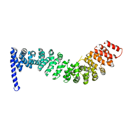
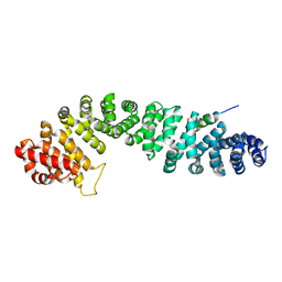
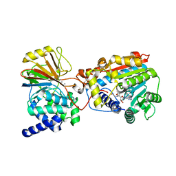
7EXS
 
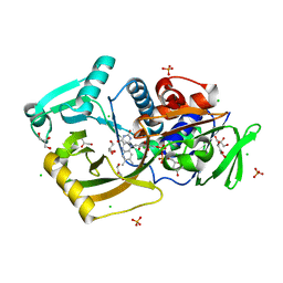 | | Thermomicrobium roseum sarcosine oxidase mutant - S320R | | 分子名称: | CHLORIDE ION, FLAVIN-ADENINE DINUCLEOTIDE, GLYCEROL, ... | | 著者 | Xin, Y, Shen, C, Tang, M.W, Shi, Y, Guo, Z.T, Gu, Z.H, Shao, J, Zhang, L. | | 登録日 | 2021-05-28 | | 公開日 | 2021-07-28 | | 最終更新日 | 2023-11-29 | | 実験手法 | X-RAY DIFFRACTION (1.42 Å) | | 主引用文献 | Recreating the natural evolutionary trend in key microdomains provides an effective strategy for engineering of a thermomicrobial N-demethylase.
J.Biol.Chem., 298, 2022
|
|
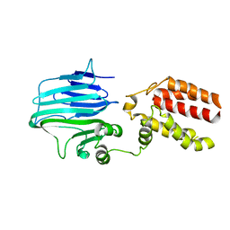
4XHJ
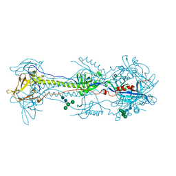
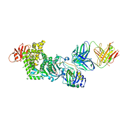 | | gHgL of Varicella-zoster virus in complex with human neutralizing antibodies. | | 分子名称: | 2-acetamido-2-deoxy-beta-D-glucopyranose, 2-acetamido-2-deoxy-beta-D-glucopyranose-(1-4)-2-acetamido-2-deoxy-beta-D-glucopyranose, Envelope glycoprotein H, ... | | 著者 | Xing, Y. | | 登録日 | 2015-01-05 | | 公開日 | 2015-05-13 | | 最終更新日 | 2023-09-27 | | 実験手法 | X-RAY DIFFRACTION (3.156 Å) | | 主引用文献 | A site of varicella-zoster virus vulnerability identified by structural studies of neutralizing antibodies bound to the glycoprotein complex gHgL.
Proc.Natl.Acad.Sci.USA, 112, 2015
|
|
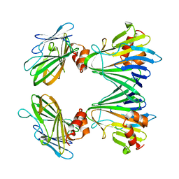
4XI5
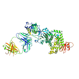 | | gHgL of varicella-zoster virus in complex with human neutralizing antibodies | | 分子名称: | 2-acetamido-2-deoxy-beta-D-glucopyranose, Envelope glycoprotein H, Envelope glycoprotein L, ... | | 著者 | Xing, Y. | | 登録日 | 2015-01-06 | | 公開日 | 2015-05-13 | | 最終更新日 | 2023-09-27 | | 実験手法 | X-RAY DIFFRACTION (3.9 Å) | | 主引用文献 | A site of varicella-zoster virus vulnerability identified by structural studies of neutralizing antibodies bound to the glycoprotein complex gHgL.
Proc.Natl.Acad.Sci.USA, 112, 2015
|
|
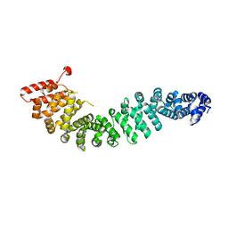
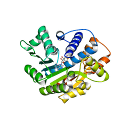
3C5W
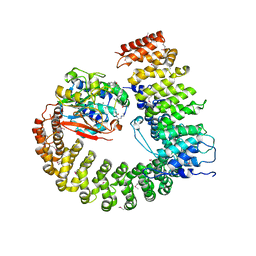
 | | Complex between PP2A-specific methylesterase PME-1 and PP2A core enzyme | | 分子名称: | PP2A A subunit, PP2A C subunit, PP2A-specific methylesterase PME-1 | | 著者 | Xing, Y, Li, Z, Chen, Y, Stock, J, Jeffrey, P.D, Shi, Y. | | 登録日 | 2008-02-01 | | 公開日 | 2008-04-15 | | 最終更新日 | 2024-04-03 | | 実験手法 | X-RAY DIFFRACTION (2.8 Å) | | 主引用文献 | Structural mechanism of demethylation and inactivation of protein phosphatase 2A.
Cell(Cambridge,Mass.), 133, 2008
|
|
3C5V
 | | PP2A-specific methylesterase apo form (PME) | | 分子名称: | Protein phosphatase methylesterase 1 | | 著者 | Xing, Y, Li, Z, Chen, Y, Stock, J, Jeffrey, P.D, Shi, Y. | | 登録日 | 2008-02-01 | | 公開日 | 2008-04-15 | | 最終更新日 | 2024-02-21 | | 実験手法 | X-RAY DIFFRACTION (2 Å) | | 主引用文献 | Structural mechanism of demethylation and inactivation of protein phosphatase 2A.
Cell(Cambridge,Mass.), 133, 2008
|
|
2IE4
 | | Structure of the Protein Phosphatase 2A Core Enzyme Bound to okadaic acid | | 分子名称: | MANGANESE (II) ION, OKADAIC ACID, Protein Phosphatase 2, ... | | 著者 | Xing, Y, Xu, Y, Chen, Y, Jeffrey, P.D, Chao, Y, Shi, Y. | | 登録日 | 2006-09-17 | | 公開日 | 2006-11-07 | | 最終更新日 | 2024-02-21 | | 実験手法 | X-RAY DIFFRACTION (2.6 Å) | | 主引用文献 | Structure of Protein Phosphatase 2A Core Enzyme Bound to Tumor-Inducing Toxins
Cell(Cambridge,Mass.), 127, 2006
|
|
2IE3
 | | Structure of the Protein Phosphatase 2A Core Enzyme Bound to Tumor-inducing Toxins | | 分子名称: | MANGANESE (II) ION, Protein Phosphatase 2, regulatory subunit A (PR 65), ... | | 著者 | Xing, Y, Xu, Y, Chen, Y, Jeffrey, P.D, Chao, Y, Shi, Y. | | 登録日 | 2006-09-17 | | 公開日 | 2006-11-07 | | 最終更新日 | 2023-11-15 | | 実験手法 | X-RAY DIFFRACTION (2.8 Å) | | 主引用文献 | Structure of Protein Phosphatase 2A Core Enzyme Bound to Tumor-Inducing Toxins
Cell(Cambridge,Mass.), 127, 2006
|
|
2Z6H
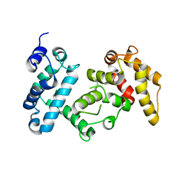
 | | Crystal Structure of Beta-Catenin Armadillo Repeat Region and Its C-Terminal domain | | 分子名称: | Catenin beta-1 | | 著者 | Xing, Y, Takemaru, K, Liu, J, Zheng, J, Moon, R, Xu, W. | | 登録日 | 2007-08-01 | | 公開日 | 2008-02-12 | | 最終更新日 | 2024-03-13 | | 実験手法 | X-RAY DIFFRACTION (2.2 Å) | | 主引用文献 | Crystal Structure of a Full-Length beta-Catenin
Structure, 16, 2008
|
|
2Z6G
 | | Crystal Structure of a Full-Length Zebrafish Beta-Catenin | | 分子名称: | B-catenin | | 著者 | Xing, Y, Takemaru, K, Liu, J, Zheng, J, Moon, R, Xu, W. | | 登録日 | 2007-08-01 | | 公開日 | 2008-02-12 | | 最終更新日 | 2023-11-01 | | 実験手法 | X-RAY DIFFRACTION (3.4 Å) | | 主引用文献 | Crystal Structure of a Full-Length beta-Catenin
Structure, 16, 2008
|
|
1ACI
 | |
1XTN
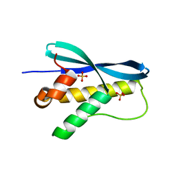 | | crystal structure of CISK-PX domain with sulfates | | 分子名称: | SULFATE ION, Serine/threonine-protein kinase Sgk3 | | 著者 | Xing, Y, Liu, D, Zhang, R, Joachimiak, A, Songyang, Z, Xu, W. | | 登録日 | 2004-10-22 | | 公開日 | 2004-11-02 | | 最終更新日 | 2023-08-23 | | 実験手法 | X-RAY DIFFRACTION (2.2 Å) | | 主引用文献 | Structural basis of membrane targeting by the Phox homology domain of cytokine-independent survival kinase (CISK-PX)
J.Biol.Chem., 279, 2004
|
|
1XTE
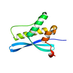 | | crystal structure of CISK-PX domain | | 分子名称: | Serine/threonine-protein kinase Sgk3 | | 著者 | Xing, Y, Liu, D, Zhang, R, Joachimiak, A, Songyang, Z, Xu, W. | | 登録日 | 2004-10-21 | | 公開日 | 2004-11-16 | | 最終更新日 | 2024-02-14 | | 実験手法 | X-RAY DIFFRACTION (1.6 Å) | | 主引用文献 | Structural basis of membrane targeting by the Phox homology domain of cytokine-independent survival kinase (CISK-PX)
J.Biol.Chem., 279, 2004
|
|
1QZ7
 | | Beta-catenin binding domain of Axin in complex with beta-catenin | | 分子名称: | Axin, Beta-catenin | | 著者 | Xing, Y, Clements, W.K, Kimelman, D, Xu, W. | | 登録日 | 2003-09-15 | | 公開日 | 2003-11-18 | | 最終更新日 | 2023-08-23 | | 実験手法 | X-RAY DIFFRACTION (2.2 Å) | | 主引用文献 | Crystal structure of a beta-catenin/Axin complex suggests a mechanism for the {beta}-catenin destruction complex
GENES DEV., 17, 2003
|
|
1TH1
 | | Beta-catenin in complex with a phosphorylated APC 20aa repeat fragment | | 分子名称: | Adenomatous polyposis coli protein, Beta-catenin | | 著者 | Xing, Y, Clements, W.K, Le Trong, I, Hinds, T.R, Stenkamp, R, Kimelman, D, Xu, W. | | 登録日 | 2004-05-31 | | 公開日 | 2004-09-07 | | 最終更新日 | 2023-08-23 | | 実験手法 | X-RAY DIFFRACTION (2.5 Å) | | 主引用文献 | Crystal Structure of a beta-Catenin/APC Complex Reveals a Critical Role for APC Phosphorylation in APC Function.
Mol.Cell, 15, 2004
|
|
5Y83
 | |
5Y82
 | |
3IEI
 | |
3P71
 | | Crystal structure of the complex of LCMT-1 and PP2A | | 分子名称: | 5'-{[(3S)-3-amino-3-carboxypropyl](ethyl)amino}-5'-deoxyadenosine, DI(HYDROXYETHYL)ETHER, Leucine carboxyl methyltransferase 1, ... | | 著者 | Xing, Y, Stanevich, V, Satyshur, K.A, Jiang, L. | | 登録日 | 2010-10-11 | | 公開日 | 2011-02-16 | | 最終更新日 | 2023-09-06 | | 実験手法 | X-RAY DIFFRACTION (2.7 Å) | | 主引用文献 | The Structural Basis for Tight Control of PP2A Methylation and Function by LCMT-1.
Mol.Cell, 41, 2011
|
|
4I5J
 | | PP2A PR70 Holoenzyme | | 分子名称: | CALCIUM ION, Serine/threonine-protein phosphatase 2A regulatory subunit B'' subunit alpha | | 著者 | Xing, Y, Jeffery, P.D, Shi, Y. | | 登録日 | 2012-11-28 | | 公開日 | 2013-05-08 | | 最終更新日 | 2024-02-28 | | 実験手法 | X-RAY DIFFRACTION (2.091 Å) | | 主引用文献 | Structure of the Ca(2+)-dependent PP2A heterotrimer and insights into Cdc6 dephosphorylation.
Cell Res., 23, 2013
|
|
4I5K
 | |
6VLK
 | | A varicella-zoster virus glycoprotein | | 分子名称: | 2-acetamido-2-deoxy-beta-D-glucopyranose, 2-acetamido-2-deoxy-beta-D-glucopyranose-(1-4)-2-acetamido-2-deoxy-beta-D-glucopyranose, Envelope glycoprotein B, ... | | 著者 | Xing, Y. | | 登録日 | 2020-01-24 | | 公開日 | 2020-07-15 | | 最終更新日 | 2023-10-11 | | 実験手法 | X-RAY DIFFRACTION (2.4529 Å) | | 主引用文献 | A glycoprotein B-neutralizing antibody structure at 2.8 angstrom uncovers a critical domain for herpesvirus fusion initiation.
Nat Commun, 11, 2020
|
|
2NYL
 | | Crystal structure of Protein Phosphatase 2A (PP2A) holoenzyme with the catalytic subunit carboxyl terminus truncated | | 分子名称: | MANGANESE (II) ION, Protein phosphatase 2, regulatory subunit A (PR 65), ... | | 著者 | Xing, Y, Xu, Y, Chen, Y, Chao, Y, Lin, Z, Shi, Y. | | 登録日 | 2006-11-20 | | 公開日 | 2006-12-12 | | 最終更新日 | 2023-11-15 | | 実験手法 | X-RAY DIFFRACTION (3.8 Å) | | 主引用文献 | Structure of the Protein Phosphatase 2A Holoenzyme.
Cell(Cambridge,Mass.), 127, 2006
|
|
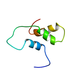
5DCF
 | | C-terminal domain of XerD recombinase in complex with gamma domain of FtsK | | 分子名称: | Tyrosine recombinase XerD,DNA translocase FtsK | | 著者 | Keller, A.N, Xin, Y, Lowe, J, Grainge, I. | | 登録日 | 2015-08-24 | | 公開日 | 2016-09-07 | | 最終更新日 | 2023-09-27 | | 実験手法 | X-RAY DIFFRACTION (2.3 Å) | | 主引用文献 | Activation of Xer-recombination at dif: structural basis of the FtsK gamma-XerD interaction.
Sci Rep, 6, 2016
|
|
6MG8
 | | Structural basis for cholesterol transport-like activity of the Hedgehog receptor Patched | | 分子名称: | CHOLESTEROL, Protein patched homolog 1 | | 著者 | Zhang, Y, Bulkley, D, Xin, Y, Roberts, K.J, Asarnow, D.E, Sharma, A, Myers, B.R, Cho, W, Cheng, Y, Beachy, P.A. | | 登録日 | 2018-09-13 | | 公開日 | 2018-11-28 | | 最終更新日 | 2022-03-16 | | 実験手法 | ELECTRON MICROSCOPY (3.6 Å) | | 主引用文献 | Structural Basis for Cholesterol Transport-like Activity of the Hedgehog Receptor Patched.
Cell, 175, 2018
|
|
8KGT
 | |


