1OS1
 
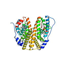
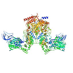
 | | Structure of Phosphoenolpyruvate Carboxykinase complexed with ATP,Mg, Ca and pyruvate. | | 分子名称: | ADENOSINE-5'-TRIPHOSPHATE, CALCIUM ION, MAGNESIUM ION, ... | | 著者 | Sudom, A, Walters, R, Pastushok, L, Goldie, D, Prasad, L, Delbaere, L.T, Goldie, H. | | 登録日 | 2003-03-18 | | 公開日 | 2003-09-23 | | 最終更新日 | 2023-11-15 | | 実験手法 | X-RAY DIFFRACTION (1.8 Å) | | 主引用文献 | Mechanisms of activation of phosphoenolpyruvate carboxykinase from Escherichia coli by Ca2+ and of desensitization by trypsin.
J.BACTERIOL., 185, 2003
|
|
8FTG
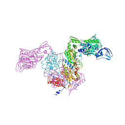
 | | Biophysical and Structural Characterization of an Anti-Caffeine VHH Antibody | | 分子名称: | 2-AMINO-2-HYDROXYMETHYL-PROPANE-1,3-DIOL, Anti-Caffeine VHH Antibody, CAFFEINE, ... | | 著者 | Horn, J.R, Smith, C.A, Sonneson, G.J, Walter, R. | | 登録日 | 2023-01-12 | | 公開日 | 2023-06-07 | | 最終更新日 | 2024-05-22 | | 実験手法 | X-RAY DIFFRACTION (1.13 Å) | | 主引用文献 | Molecular recognition requires dimerization of a VHH antibody.
Mabs, 15, 2023
|
|
2HC1
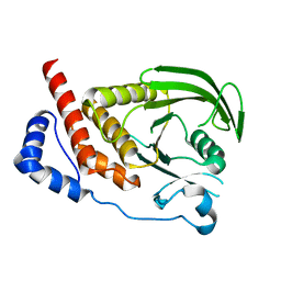
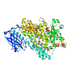
 | | Engineered catalytic domain of protein tyrosine phosphatase HPTPbeta. | | 分子名称: | ACETATE ION, CHLORIDE ION, Receptor-type tyrosine-protein phosphatase beta | | 著者 | Evdokimov, A.G, Pokross, M, Walter, R, Mekel, M. | | 登録日 | 2006-06-14 | | 公開日 | 2006-06-27 | | 最終更新日 | 2023-08-30 | | 実験手法 | X-RAY DIFFRACTION (1.3 Å) | | 主引用文献 | Engineering the catalytic domain of human protein tyrosine phosphatase beta for structure-based drug discovery.
Acta Crystallogr.,Sect.D, 62, 2006
|
|
2HC2
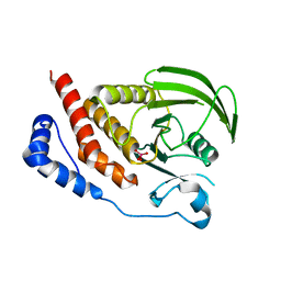
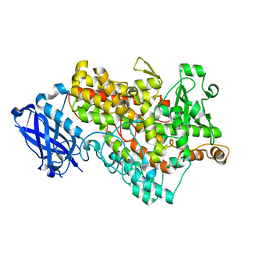
 | | Engineered protein tyrosine phosphatase beta catalytic domain | | 分子名称: | MAGNESIUM ION, Receptor-type tyrosine-protein phosphatase beta, SODIUM ION | | 著者 | Evdokimov, A.G, Pokross, M, Walter, R, Mekel, M. | | 登録日 | 2006-06-15 | | 公開日 | 2006-06-27 | | 最終更新日 | 2023-08-30 | | 実験手法 | X-RAY DIFFRACTION (1.4 Å) | | 主引用文献 | Engineering the catalytic domain of human protein tyrosine phosphatase beta for structure-based drug discovery.
Acta Crystallogr.,Sect.D, 62, 2006
|
|
4POU
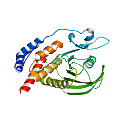
 | | VHH-metal in Complex with RNase A | | 分子名称: | Ribonuclease pancreatic, VHH Antibody | | 著者 | Fanning, S.W, Walter, R, Horn, J.R. | | 登録日 | 2014-02-26 | | 公開日 | 2014-09-24 | | 最終更新日 | 2014-10-22 | | 実験手法 | X-RAY DIFFRACTION (1.75 Å) | | 主引用文献 | Structural basis of an engineered dual-specific antibody: conformational diversity leads to a hypervariable loop metal-binding site.
Protein Eng.Des.Sel., 27, 2014
|
|
4POY
 | |
4PPT
 | |
1YGE
 | | LIPOXYGENASE-1 (SOYBEAN) AT 100K | | 分子名称: | FE (III) ION, LIPOXYGENASE-1 | | 著者 | Minor, W, Steczko, J, Stec, B, Otwinowski, Z, Bolin, J.T, Walter, R, Axelrod, B. | | 登録日 | 1996-06-04 | | 公開日 | 1997-07-23 | | 最終更新日 | 2024-02-14 | | 実験手法 | X-RAY DIFFRACTION (1.4 Å) | | 主引用文献 | Crystal structure of soybean lipoxygenase L-1 at 1.4 A resolution.
Biochemistry, 35, 1996
|
|
4XI3
 | | Estrogen Receptor Alpha Ligand Binding Domain in Complex with Bazedoxifene | | 分子名称: | Bazedoxifene, Estrogen receptor | | 著者 | Fanning, S.W, Mayne, C.G, Toy, W, Carlson, K, Greene, B, Nowak, J, Walter, R, Panchamukhi, S, Tajhorshid, E, Nettles, K.W, Chandarlapaty, S, Katzenellenbogen, J, Greene, G.L. | | 登録日 | 2015-01-06 | | 公開日 | 2016-01-13 | | 最終更新日 | 2024-03-06 | | 実験手法 | X-RAY DIFFRACTION (2.491 Å) | | 主引用文献 | The SERM/SERD bazedoxifene disrupts ESR1 helix 12 to overcome acquired hormone resistance in breast cancer cells.
Elife, 7, 2018
|
|
2I3U
 | | Structural studies of protein tyrosine phosphatase beta catalytic domain in complex with inhibitors | | 分子名称: | Receptor-type tyrosine-protein phosphatase beta | | 著者 | Evdokimov, A.G, Pokross, M.E, Walter, R.L, Mekel, M. | | 登録日 | 2006-08-20 | | 公開日 | 2006-08-29 | | 最終更新日 | 2023-08-30 | | 実験手法 | X-RAY DIFFRACTION (1.85 Å) | | 主引用文献 | Engineering the catalytic domain of human protein tyrosine phosphatase beta for structure-based drug discovery.
Acta Crystallogr.,Sect.D, 62, 2006
|
|
2I4E
 | | Structural studies of protein tyrosine phosphatase beta catalytic domain in complex with inhibitors | | 分子名称: | Receptor-type tyrosine-protein phosphatase beta, VANADATE ION | | 著者 | Evdokimov, A.G, Pokross, M.E, Walter, R.L, Mekel, M. | | 登録日 | 2006-08-21 | | 公開日 | 2006-08-29 | | 最終更新日 | 2023-08-30 | | 実験手法 | X-RAY DIFFRACTION (1.75 Å) | | 主引用文献 | Engineering the catalytic domain of human protein tyrosine phosphatase beta for structure-based drug discovery.
Acta Crystallogr.,Sect.D, 62, 2006
|
|
2I3R
 | | Engineered catalytic domain of protein tyrosine phosphatase HPTPbeta | | 分子名称: | CHLORIDE ION, Receptor-type tyrosine-protein phosphatase beta | | 著者 | Evdokimov, A.G, Pokross, M.E, Walter, R.L, Mekel, M. | | 登録日 | 2006-08-20 | | 公開日 | 2006-08-29 | | 最終更新日 | 2023-08-30 | | 実験手法 | X-RAY DIFFRACTION (1.85 Å) | | 主引用文献 | Engineering the catalytic domain of human protein tyrosine phosphatase beta for structure-based drug discovery.
Acta Crystallogr.,Sect.D, 62, 2006
|
|
2I4G
 | | Structural studies of protein tyrosine phosphatase beta catalytic domain in complex with a sulfamic acid (soaking experiment) | | 分子名称: | CHLORIDE ION, N-(TERT-BUTOXYCARBONYL)-L-TYROSYL-N-METHYL-4-(SULFOAMINO)-L-PHENYLALANINAMIDE, Receptor-type tyrosine-protein phosphatase beta | | 著者 | Evdokimov, A.G, Pokross, M.E, Walter, R.L, Mekel, M. | | 登録日 | 2006-08-21 | | 公開日 | 2006-08-29 | | 最終更新日 | 2023-08-30 | | 実験手法 | X-RAY DIFFRACTION (1.65 Å) | | 主引用文献 | Engineering the catalytic domain of human protein tyrosine phosphatase beta for structure-based drug discovery.
Acta Crystallogr.,Sect.D, 62, 2006
|
|
2I5X
 | | Engineering the PTPbeta catalytic domain with improved crystallization properties | | 分子名称: | (4-ETHYLPHENYL)SULFAMIC ACID, CHLORIDE ION, MAGNESIUM ION, ... | | 著者 | Evdokimov, A.G, Pokross, M.E, Walter, R.L, Mekel, M, Klopfenstein, S. | | 登録日 | 2006-08-26 | | 公開日 | 2006-09-05 | | 最終更新日 | 2023-08-30 | | 実験手法 | X-RAY DIFFRACTION (1.7 Å) | | 主引用文献 | Engineering the catalytic domain of human protein tyrosine phosphatase beta for structure-based drug discovery.
Acta Crystallogr.,Sect.D, 62, 2006
|
|
2I4H
 | | Structural studies of protein tyrosine phosphatase beta catalytic domain co-crystallized with a sulfamic acid inhibitor | | 分子名称: | CHLORIDE ION, MAGNESIUM ION, N-(TERT-BUTOXYCARBONYL)-L-TYROSYL-N-METHYL-4-(SULFOAMINO)-L-PHENYLALANINAMIDE, ... | | 著者 | Evdokimov, A.G, Pokross, M.E, Walter, R.L, Mekel, M. | | 登録日 | 2006-08-21 | | 公開日 | 2006-08-29 | | 最終更新日 | 2023-08-30 | | 実験手法 | X-RAY DIFFRACTION (2.15 Å) | | 主引用文献 | Engineering the catalytic domain of human protein tyrosine phosphatase beta for structure-based drug discovery.
Acta Crystallogr.,Sect.D, 62, 2006
|
|
2HFP
 | | Crystal Structure of PPAR Gamma with N-sulfonyl-2-indole carboxamide ligands | | 分子名称: | 3-(4-METHOXYPHENYL)-N-(PHENYLSULFONYL)-1-[3-(TRIFLUOROMETHYL)BENZYL]-1H-INDOLE-2-CARBOXAMIDE, Peroxisome proliferator-activated receptor gamma, SRC Peptide Fragment | | 著者 | Pokross, M.E, Evdokimov, A.G, Walter, R.L, Mekel, M.J, Hopkins, C.R. | | 登録日 | 2006-06-25 | | 公開日 | 2006-09-19 | | 最終更新日 | 2024-02-14 | | 実験手法 | X-RAY DIFFRACTION (2 Å) | | 主引用文献 | Design and synthesis of novel N-sulfonyl-2-indole carboxamides as potent PPAR-gamma binding agents with potential application to the treatment of osteoporosis.
Bioorg.Med.Chem.Lett., 16, 2006
|
|
2RHS
 | |
2RHQ
 | |
3D9A
 | | High Resolution Crystal Structure Structure of HyHel10 Fab Complexed to Hen Egg Lysozyme | | 分子名称: | Heavy Chain of HyHel10 Antibody Fragment (Fab), Light Chain of HyHel10 Antibody Fragment (Fab), Lysozyme C | | 著者 | DeSantis, M.E, Li, M, Shanmuganathan, A, Acchione, M, Walter, R, Wlodawer, A, Smith-Gill, S. | | 登録日 | 2008-05-27 | | 公開日 | 2008-06-10 | | 最終更新日 | 2023-08-30 | | 実験手法 | X-RAY DIFFRACTION (1.2 Å) | | 主引用文献 | Light chain somatic mutations change thermodynamics of binding and water coordination in the HyHEL-10 family of antibodies.
Mol.Immunol., 47, 2009
|
|
3PZW
 | |
1F8N
 | |
1FGT
 | |
1FGM
 | |
1FGQ
 | |
1FGR
 | |


