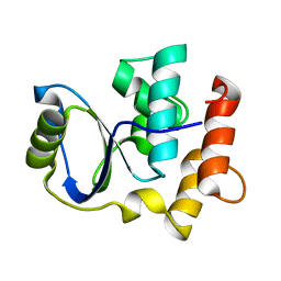
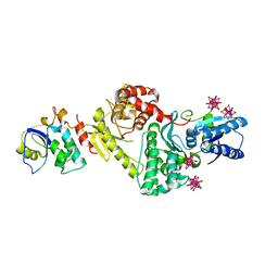
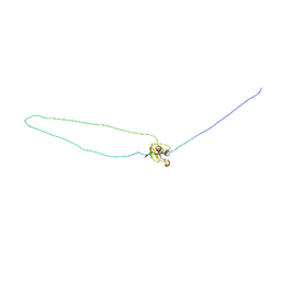
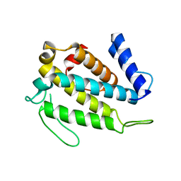
3ENP
 
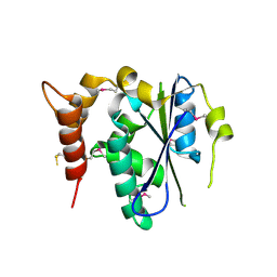 | | Crystal structure of human cgi121 | | 分子名称: | TP53RK-binding protein | | 著者 | Haffani, Y.Z, Ceccarelli, D.F, Neculai, D, Mao, D.Y, Sicheri, F. | | 登録日 | 2008-09-25 | | 公開日 | 2008-11-04 | | 最終更新日 | 2011-07-13 | | 実験手法 | X-RAY DIFFRACTION (2.48 Å) | | 主引用文献 | Atomic structure of the KEOPS complex: an ancient protein kinase-containing molecular machine.
Mol.Cell, 32, 2008
|
|
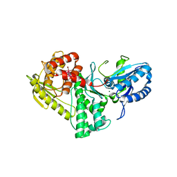
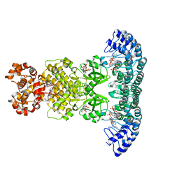
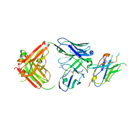
4K25
 | | Crystal Structure of yeast Qri7 homodimer | | 分子名称: | CALCIUM ION, Probable tRNA threonylcarbamoyladenosine biosynthesis protein QRI7, mitochondrial, ... | | 著者 | Neculai, D, Wan, L, Mao, D.Y, Sicheri, F. | | 登録日 | 2013-04-08 | | 公開日 | 2013-05-08 | | 最終更新日 | 2013-08-07 | | 実験手法 | X-RAY DIFFRACTION (2.88 Å) | | 主引用文献 | Reconstitution and characterization of eukaryotic N6-threonylcarbamoylation of tRNA using a minimal enzyme system.
Nucleic Acids Res., 41, 2013
|
|
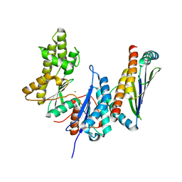
4JVG
 | | B-Raf Kinase in Complex with Birb796 | | 分子名称: | 1-(5-TERT-BUTYL-2-P-TOLYL-2H-PYRAZOL-3-YL)-3-[4-(2-MORPHOLIN-4-YL-ETHOXY)-NAPHTHALEN-1-YL]-UREA, Serine/threonine-protein kinase B-raf | | 著者 | Lavoie, H, Thevakumaran, N, Gavory, G, Li, J, Padeganeh, A, Guiral, S, Duchaine, J, Mao, D.Y.L, Bouvier, M, Sicheri, F, Therrien, M. | | 登録日 | 2013-03-25 | | 公開日 | 2013-05-29 | | 最終更新日 | 2024-02-28 | | 実験手法 | X-RAY DIFFRACTION (3.09 Å) | | 主引用文献 | Inhibitors that stabilize a closed RAF kinase domain conformation induce dimerization.
Nat.Chem.Biol., 9, 2013
|
|
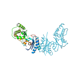
7KJT
 | | KEOPS tRNA modifying sub-complex of archaeal Cgi121 and tRNA | | 分子名称: | RNA (70-MER), Regulatory protein Cgi121 | | 著者 | Ceccarelli, D.F, Beenstock, J, Mao, D.Y.L, Sicheri, F. | | 登録日 | 2020-10-26 | | 公開日 | 2020-12-02 | | 最終更新日 | 2023-10-18 | | 実験手法 | X-RAY DIFFRACTION (3.34 Å) | | 主引用文献 | A substrate binding model for the KEOPS tRNA modifying complex.
Nat Commun, 11, 2020
|
|
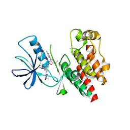
2K8Y
 | | Solution NMR Structure of Cgi121 from Methanococcus jannaschii. Northeast Structural Genomics Consortium Target MJ0187 | | 分子名称: | Uncharacterized protein MJ0187 | | 著者 | Rumpel, S, Fares, C, Neculai, D, Arrowsmith, C, Montelione, G.T, Sicheri, F, Northeast Structural Genomics Consortium (NESG) | | 登録日 | 2008-09-25 | | 公開日 | 2008-11-04 | | 最終更新日 | 2024-05-22 | | 実験手法 | SOLUTION NMR | | 主引用文献 | Atomic structure of the KEOPS complex: an ancient protein kinase-containing molecular machine.
Mol.Cell, 32, 2008
|
|
3EN9
 | |
3ENO
 | |
3ENC
 | |
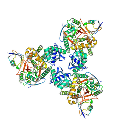
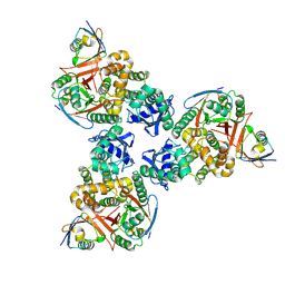
3ENH
 | | Crystal structure of Cgi121/Bud32/Kae1 complex | | 分子名称: | HEXATANTALUM DODECABROMIDE, Putative O-sialoglycoprotein endopeptidase, Uncharacterized protein MJ0187 | | 著者 | Neculai, D. | | 登録日 | 2008-09-25 | | 公開日 | 2008-10-28 | | 最終更新日 | 2024-04-03 | | 実験手法 | X-RAY DIFFRACTION (3.6 Å) | | 主引用文献 | Atomic Structure of the KEOPS Complex: An Ancient Protein Kinase-Containing Molecular Machine
Mol.Cell, 32, 2008
|
|
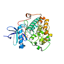
4O1O
 | | Crystal Structure of RNase L in complex with 2-5A | | 分子名称: | Ribonuclease L, [[(2R,3R,4R,5R)-5-(6-aminopurin-9-yl)-4-[[(2R,3R,4R,5R)-5-(6-aminopurin-9-yl)-4-[[(2R,3S,4R,5R)-5-(6-aminopurin-9-yl)-3,4-dihydroxy-oxolan-2-yl]methoxy-hydroxy-phosphoryl]oxy-3-hydroxy-oxolan-2-yl]methoxy-hydroxy-phosphoryl]oxy-3-hydroxy-oxolan-2-yl]methoxy-hydroxy-phosphoryl] phosphono hydrogen phosphate | | 著者 | Huang, H, Zeqiraj, E, Ceccarelli, D.F, Sicheri, F. | | 登録日 | 2013-12-16 | | 公開日 | 2014-02-05 | | 最終更新日 | 2023-09-20 | | 実験手法 | X-RAY DIFFRACTION (3.27 Å) | | 主引用文献 | Dimeric structure of pseudokinase RNase L bound to 2-5A reveals a basis for interferon-induced antiviral activity.
Mol.Cell, 53, 2014
|
|
4O1P
 | | Crystal Structure of RNase L in complex with 2-5A and AMP-PNP | | 分子名称: | MAGNESIUM ION, PHOSPHOAMINOPHOSPHONIC ACID-ADENYLATE ESTER, Ribonuclease L, ... | | 著者 | Huang, H, Zeqiraj, E, Ceccarelli, D.F, Sicheri, F. | | 登録日 | 2013-12-16 | | 公開日 | 2014-02-05 | | 最終更新日 | 2023-09-20 | | 実験手法 | X-RAY DIFFRACTION (2.5 Å) | | 主引用文献 | Dimeric structure of pseudokinase RNase L bound to 2-5A reveals a basis for interferon-induced antiviral activity.
Mol.Cell, 53, 2014
|
|
4DDG
 | |
4DDI
 | |
1MV3
 | |
1MUZ
 | |
1MV0
 | |
5TWG
 | |
5TWF
 | |
5TWH
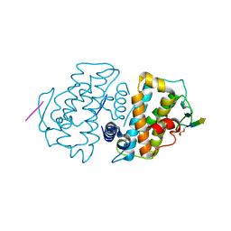 | |
6BXI
 | | X-ray crystal structure of NDR1 kinase domain | | 分子名称: | MAGNESIUM ION, PHOSPHOAMINOPHOSPHONIC ACID-ADENYLATE ESTER, Serine/threonine-protein kinase 38 | | 著者 | Xiong, S, Sicheri, F. | | 登録日 | 2017-12-18 | | 公開日 | 2018-08-29 | | 最終更新日 | 2024-03-13 | | 実験手法 | X-RAY DIFFRACTION (2.2 Å) | | 主引用文献 | Structural Basis for Auto-Inhibition of the NDR1 Kinase Domain by an Atypically Long Activation Segment.
Structure, 26, 2018
|
|
8T6I
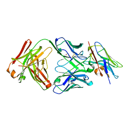 | | Structure of VHH-Fab complex with engineered Crystal Kappa region | | 分子名称: | Fab heavy chain, Fab light chain, GLYCEROL, ... | | 著者 | Filippova, E.V, Thompson, I, Kossiakoff, A.A. | | 登録日 | 2023-06-16 | | 公開日 | 2023-11-29 | | 最終更新日 | 2024-01-03 | | 実験手法 | X-RAY DIFFRACTION (2.55 Å) | | 主引用文献 | Engineered antigen-binding fragments for enhanced crystallization of antibody:antigen complexes.
Protein Sci., 33, 2024
|
|
8T7I
 | | Structure of the S1CE variant of Fab F1 (FabS1CE-F1) | | 分子名称: | 1,2-ETHANEDIOL, CHLORIDE ION, S1CE variant of Fab F1 heavy chain, ... | | 著者 | Singer, A.U, Bruce, H.A, Enderle, L, Blazer, L, Adams, J.J, Sicheri, F, Sidhu, S.S. | | 登録日 | 2023-06-20 | | 公開日 | 2023-11-22 | | 最終更新日 | 2024-01-10 | | 実験手法 | X-RAY DIFFRACTION (2.6 Å) | | 主引用文献 | Engineered antigen-binding fragments for enhanced crystallization of antibody:antigen complexes.
Protein Sci., 33, 2024
|
|
8T7G
 | | Structure of the CK variant of Fab F1 (FabC-F1) | | 分子名称: | 1,2-ETHANEDIOL, CHLORIDE ION, CK variant of Fab F1 heavy chain, ... | | 著者 | Singer, A.U, Bruce, H.A, Blazer, L, Adams, J.J, Sicheri, F, Sidhu, S.S. | | 登録日 | 2023-06-20 | | 公開日 | 2023-11-22 | | 最終更新日 | 2024-01-10 | | 実験手法 | X-RAY DIFFRACTION (2 Å) | | 主引用文献 | Engineered antigen-binding fragments for enhanced crystallization of antibody:antigen complexes.
Protein Sci., 33, 2024
|
|
8T7F
 | | Structure of the S1 variant of Fab F1 | | 分子名称: | 4-(2-HYDROXYETHYL)-1-PIPERAZINE ETHANESULFONIC ACID, S1 variant of Fab F1 heavy chain, S1 variant of Fab F1 light chain, ... | | 著者 | Singer, A.U, Bruce, H.A, Enderle, L, Blazer, L, Adams, J.J, Sicheri, F, Sidhu, S.S. | | 登録日 | 2023-06-20 | | 公開日 | 2023-11-22 | | 最終更新日 | 2024-01-10 | | 実験手法 | X-RAY DIFFRACTION (3.5 Å) | | 主引用文献 | Engineered antigen-binding fragments for enhanced crystallization of antibody:antigen complexes.
Protein Sci., 33, 2024
|
|
8T9Y
 | | Structure of VHH-Fab complex with engineered Elbow FNQIKG and Crystal Kappa regions | | 分子名称: | DI(HYDROXYETHYL)ETHER, Fab heavy chain, Fab light chain, ... | | 著者 | Filippova, E.V, Thompson, I, Kossiakoff, A.A. | | 登録日 | 2023-06-26 | | 公開日 | 2023-11-29 | | 最終更新日 | 2024-01-03 | | 実験手法 | X-RAY DIFFRACTION (2.52 Å) | | 主引用文献 | Engineered antigen-binding fragments for enhanced crystallization of antibody:antigen complexes.
Protein Sci., 33, 2024
|
|


