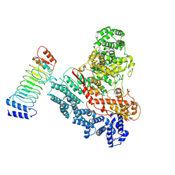
8J5R
 
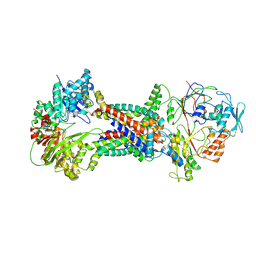 | | Cryo-EM structure of Mycobacterium tuberculosis OppABCD in the resting state | | 分子名称: | IRON/SULFUR CLUSTER, Putative peptide transport permease protein Rv1282c, Putative peptide transport permease protein Rv1283c, ... | | 著者 | Yang, X, Hu, T, Zhang, B, Rao, Z. | | 登録日 | 2023-04-24 | | 公開日 | 2024-04-03 | | 最終更新日 | 2024-07-31 | | 実験手法 | ELECTRON MICROSCOPY (3.28 Å) | | 主引用文献 | An oligopeptide permease, OppABCD, requires an iron-sulfur cluster domain for functionality.
Nat.Struct.Mol.Biol., 31, 2024
|
|
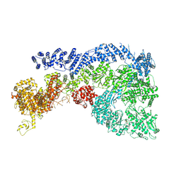
8J5T
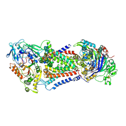 | | Cryo-EM structure of Mycobacterium tuberculosis OppABCD in the catalytic intermediate state | | 分子名称: | ADENOSINE-5'-TRIPHOSPHATE, IRON/SULFUR CLUSTER, MAGNESIUM ION, ... | | 著者 | Yang, X, Hu, T, Zhang, B, Rao, Z. | | 登録日 | 2023-04-24 | | 公開日 | 2024-04-03 | | 最終更新日 | 2024-07-31 | | 実験手法 | ELECTRON MICROSCOPY (2.98 Å) | | 主引用文献 | An oligopeptide permease, OppABCD, requires an iron-sulfur cluster domain for functionality.
Nat.Struct.Mol.Biol., 31, 2024
|
|
8J5Q
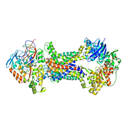 | | Cryo-EM structure of Mycobacterium tuberculosis OppABCD in the pre-translocation state | | 分子名称: | Endogenous oligopeptide, IRON/SULFUR CLUSTER, Putative peptide transport permease protein Rv1282c, ... | | 著者 | Yang, X, Hu, T, Zhang, B, Rao, Z. | | 登録日 | 2023-04-24 | | 公開日 | 2024-04-03 | | 最終更新日 | 2024-07-31 | | 実験手法 | ELECTRON MICROSCOPY (3.25 Å) | | 主引用文献 | An oligopeptide permease, OppABCD, requires an iron-sulfur cluster domain for functionality.
Nat.Struct.Mol.Biol., 31, 2024
|
|
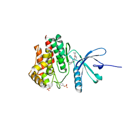
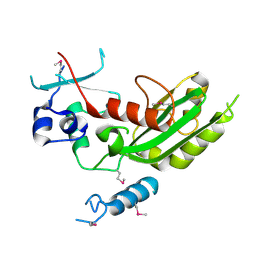
5YJG
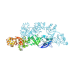 | | Structural insights into periostin functions | | 分子名称: | CALCIUM ION, CHLORIDE ION, CYSTEINE, ... | | 著者 | Liu, H, Liu, J, Xu, F. | | 登録日 | 2017-10-10 | | 公開日 | 2018-05-23 | | 最終更新日 | 2024-11-13 | | 実験手法 | X-RAY DIFFRACTION (2.399 Å) | | 主引用文献 | Structural characterizations of human periostin dimerization and cysteinylation.
FEBS Lett., 592, 2018
|
|
9IKQ
 | |
5VIF
 | | Electrophilic probes for deciphering substrate recognition by O-GlcNAc transferase | | 分子名称: | 2-{[(2E)-4-chlorobut-2-enoyl]amino}-2-deoxy-beta-D-glucopyranose, CKII, UDP-N-acetylglucosamine--peptide N-acetylglucosaminyltransferase 110 kDa subunit, ... | | 著者 | Jiang, J, Li, B, Hu, C.-W, Worth, M, Fan, D, Li, H. | | 登録日 | 2017-04-15 | | 公開日 | 2017-10-18 | | 最終更新日 | 2024-10-23 | | 実験手法 | X-RAY DIFFRACTION (2.25 Å) | | 主引用文献 | Electrophilic probes for deciphering substrate recognition by O-GlcNAc transferase.
Nat. Chem. Biol., 13, 2017
|
|
5VIE
 | | Electrophilic probes for deciphering substrate recognition by O-GlcNAc transferase | | 分子名称: | 2-{[(2E)-4-chlorobut-2-enoyl]amino}-2-deoxy-beta-D-glucopyranose, 2-{[(2E)-but-2-enoyl]amino}-2-deoxy-beta-D-glucopyranose, CKII, ... | | 著者 | Jiang, J, Li, B, Hu, C.-W, Worth, M, Fan, D, Li, H. | | 登録日 | 2017-04-15 | | 公開日 | 2017-10-18 | | 最終更新日 | 2024-11-20 | | 実験手法 | X-RAY DIFFRACTION (2.6 Å) | | 主引用文献 | Electrophilic probes for deciphering substrate recognition by O-GlcNAc transferase.
Nat. Chem. Biol., 13, 2017
|
|
5WAL
 | |
5WEV
 | |
3QWQ
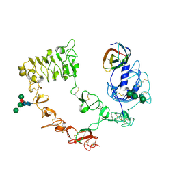 | |
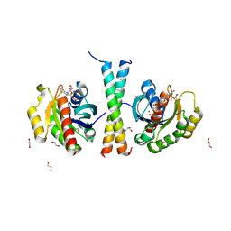
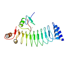
5EJL
 | | MrkH, A novel c-di-GMP dependence transcription regulatory factor. | | 分子名称: | 1,2-ETHANEDIOL, 9,9'-[(2R,3R,3aS,5S,7aR,9R,10R,10aS,12S,14aR)-3,5,10,12-tetrahydroxy-5,12-dioxidooctahydro-2H,7H-difuro[3,2-d:3',2'-j][1,3,7,9,2,8]tetraoxadiphosphacyclododecine-2,9-diyl]bis(2-amino-1,9-dihydro-6H-purin-6-one), Klebsiella pneumoniae genome assembly NOVST, ... | | 著者 | Wang, F, Zhu, D, Gu, L. | | 登録日 | 2015-11-02 | | 公開日 | 2016-10-19 | | 最終更新日 | 2024-03-20 | | 実験手法 | X-RAY DIFFRACTION (2.3 Å) | | 主引用文献 | The PilZ domain of MrkH represents a novel DNA binding motif
Protein Cell, 7, 2016
|
|
3QWR
 | |
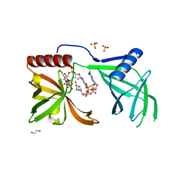
5WHG
 | | Vms1 mitochondrial localization core | | 分子名称: | Protein VMS1, ZINC ION | | 著者 | Fredrickson, E.K, Schubert, H.L, Rutter, J, Hill, C.P. | | 登録日 | 2017-07-17 | | 公開日 | 2017-11-15 | | 最終更新日 | 2024-11-20 | | 実験手法 | X-RAY DIFFRACTION (2.7 Å) | | 主引用文献 | Sterol Oxidation Mediates Stress-Responsive Vms1 Translocation to Mitochondria.
Mol. Cell, 68, 2017
|
|
9JWG
 | |
9JTA
 | |
9JW1
 | | Cryo-EM structure of Human RNF213 | | 分子名称: | ADENOSINE-5'-TRIPHOSPHATE, MAGNESIUM ION, Ring finger protein 213 | | 著者 | Zhang, H. | | 登録日 | 2024-10-09 | | 公開日 | 2025-05-07 | | 実験手法 | ELECTRON MICROSCOPY (3.46 Å) | | 主引用文献 | Shigella effector IpaH1.4 subverts host E3 ligase RNF213 to evade antibacterial immunity.
Nat Commun, 16, 2025
|
|
7V2Z
 | |
4H6H
 | |
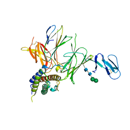
8STB
 | | The structure of abxF, an enzyme catalyzing the formation of the chiral spiroketal of an anthrabenzoxocinone antibiotic, (-)-ABX | | 分子名称: | GLYCEROL, Glyoxalase, SULFATE ION, ... | | 著者 | Luo, Z, Jia, X, Yan, X, Qu, X, Kobe, B. | | 登録日 | 2023-05-09 | | 公開日 | 2024-05-22 | | 最終更新日 | 2025-06-04 | | 実験手法 | X-RAY DIFFRACTION (2.22 Å) | | 主引用文献 | An enzymatic dual-oxa Diels-Alder reaction constructs the oxygen-bridged tricyclic acetal unit of (-)-anthrabenzoxocinone.
Nat.Chem., 2025
|
|
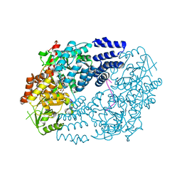
3BGL
 | | Hepatoselectivity of Statins: Design and synthesis of 4-sulfamoyl pyrroles as HMG-CoA reductase inhibitors | | 分子名称: | (3R,5R)-7-[2-(4-fluorophenyl)-5-(1-methylethyl)-4-(morpholin-4-ylsulfonyl)-3-phenyl-1H-pyrrol-1-yl]-3,5-dihydroxyheptanoic acid, 3-hydroxy-3-methylglutaryl-coenzyme A reductase | | 著者 | Finzel, B.C, Pavlovsky, A, Park, W.K.C. | | 登録日 | 2007-11-26 | | 公開日 | 2008-01-29 | | 最終更新日 | 2024-02-21 | | 実験手法 | X-RAY DIFFRACTION (2.225 Å) | | 主引用文献 | Hepatoselectivity of statins: design and synthesis of 4-sulfamoyl pyrroles as HMG-CoA reductase inhibitors.
Bioorg.Med.Chem.Lett., 18, 2008
|
|
8IS2
 | |
4H6I
 | |
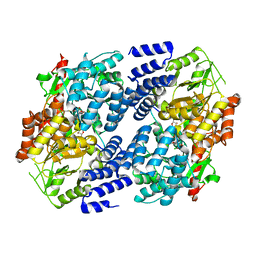
6AZU
 | | Holo IDO1 crystal structure | | 分子名称: | Indoleamine 2,3-dioxygenase 1, PROTOPORPHYRIN IX CONTAINING FE, SULFATE ION | | 著者 | Lewis, H.A, Yan, C. | | 登録日 | 2017-09-13 | | 公開日 | 2018-03-21 | | 最終更新日 | 2024-11-13 | | 実験手法 | X-RAY DIFFRACTION (2.822 Å) | | 主引用文献 | Immune-modulating enzyme indoleamine 2,3-dioxygenase is effectively inhibited by targeting its apo-form.
Proc. Natl. Acad. Sci. U.S.A., 115, 2018
|
|
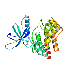
6AZV
 | | IDO1/BMS-978587 crystal structure | | 分子名称: | (1R,2S)-2-(4-[bis(2-methylpropyl)amino]-3-{[(4-methylphenyl)carbamoyl]amino}phenyl)cyclopropane-1-carboxylic acid, Indoleamine 2,3-dioxygenase 1 | | 著者 | Lewis, H.A. | | 登録日 | 2017-09-13 | | 公開日 | 2018-03-21 | | 最終更新日 | 2024-10-23 | | 実験手法 | X-RAY DIFFRACTION (2.755 Å) | | 主引用文献 | Immune-modulating enzyme indoleamine 2,3-dioxygenase is effectively inhibited by targeting its apo-form.
Proc. Natl. Acad. Sci. U.S.A., 115, 2018
|
|
1SZJ
 | |


