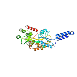
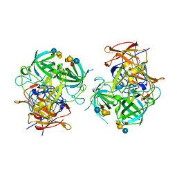
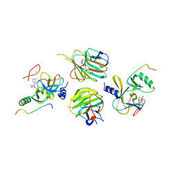
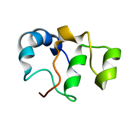
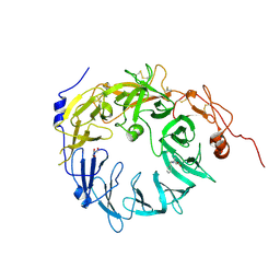
3HWJ
 
 | | Crystal structure of the second PHR domain of Mouse Myc-binding protein 2 (MYCBP-2) | | 分子名称: | DIMETHYL SULFOXIDE, E3 ubiquitin-protein ligase MYCBP2 | | 著者 | Sampathkumar, P, Ozyurt, S.A, Wasserman, S.R, Miller, S.A, Bain, K.T, Rutter, M.E, Gheyi, T, Klemke, R.L, Atwell, S, Sauder, J.M, Burley, S.K, New York SGX Research Center for Structural Genomics (NYSGXRC) | | 登録日 | 2009-06-17 | | 公開日 | 2009-07-21 | | 最終更新日 | 2023-12-27 | | 実験手法 | X-RAY DIFFRACTION (2.25 Å) | | 主引用文献 | Structures of PHR domains from Mus musculus Phr1 (Mycbp2) explain the loss-of-function mutation (Gly1092-->Glu) of the C. elegans ortholog RPM-1.
J.Mol.Biol., 397, 2010
|
|
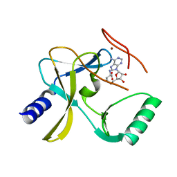
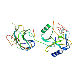
7MK7
 | | Augmentor domain of augmentor-beta | | 分子名称: | ALK and LTK ligand 1,Maltodextrin-binding protein, alpha-D-glucopyranose-(1-4)-alpha-D-glucopyranose | | 著者 | Krimmer, S.G, Reshetnyak, A.V, Puleo, D.E, Schlessinger, J. | | 登録日 | 2021-04-21 | | 公開日 | 2021-11-24 | | 最終更新日 | 2023-10-18 | | 実験手法 | X-RAY DIFFRACTION (2.42815185 Å) | | 主引用文献 | Structural basis for ligand reception by anaplastic lymphoma kinase.
Nature, 600, 2021
|
|
5F59
 | | The crystal structure of MLL3 SET domain | | 分子名称: | Histone-lysine N-methyltransferase 2C, S-ADENOSYL-L-HOMOCYSTEINE, ZINC ION | | 著者 | Li, Y, Lei, M, Chen, Y. | | 登録日 | 2015-12-04 | | 公開日 | 2016-02-24 | | 最終更新日 | 2024-03-20 | | 実験手法 | X-RAY DIFFRACTION (2.801 Å) | | 主引用文献 | Structural basis for activity regulation of MLL family methyltransferases.
Nature, 530, 2016
|
|
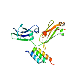
6IR5
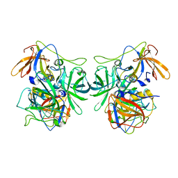 | | P domain of GII.3-TV24 | | 分子名称: | VP1 Capsid protein | | 著者 | Yang, Y. | | 登録日 | 2018-11-10 | | 公開日 | 2019-11-13 | | 最終更新日 | 2023-11-22 | | 実験手法 | X-RAY DIFFRACTION (2.6 Å) | | 主引用文献 | Structural basis of host ligand specificity change of GII porcine noroviruses from their closely related GII human noroviruses.
Emerg Microbes Infect, 8, 2019
|
|
5F5E
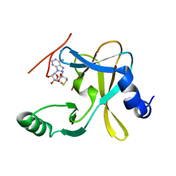 | |
6IS5
 | | P domain of GII.3-TV24 with A-tetrasaccharide complex | | 分子名称: | VP1 Capsid protein, alpha-L-fucopyranose-(1-2)-[2-acetamido-2-deoxy-alpha-D-galactopyranose-(1-3)]beta-D-galactopyranose-(1-4)-alpha-D-glucopyranose | | 著者 | Yang, Y. | | 登録日 | 2018-11-15 | | 公開日 | 2019-11-20 | | 最終更新日 | 2023-11-22 | | 実験手法 | X-RAY DIFFRACTION (2.501 Å) | | 主引用文献 | Structural basis of host ligand specificity change of GII porcine noroviruses from their closely related GII human noroviruses.
Emerg Microbes Infect, 8, 2019
|
|
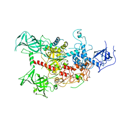
5F6L
 | | The crystal structure of MLL1 (N3861I/Q3867L) in complex with RbBP5 and Ash2L | | 分子名称: | Histone-lysine N-methyltransferase 2A, Retinoblastoma-binding protein 5, S-ADENOSYL-L-HOMOCYSTEINE, ... | | 著者 | Li, Y, Lei, M, Chen, Y. | | 登録日 | 2015-12-06 | | 公開日 | 2016-02-24 | | 最終更新日 | 2023-11-08 | | 実験手法 | X-RAY DIFFRACTION (1.9 Å) | | 主引用文献 | Structural basis for activity regulation of MLL family methyltransferases.
Nature, 530, 2016
|
|
6ITM
 | | Crystal structure of FXR in complex with agonist XJ034 | | 分子名称: | 1-adamantyl-[4-(5-chloranyl-2-methyl-phenyl)piperazin-1-yl]methanone, Bile acid receptor, HD3 Peptide from Nuclear receptor coactivator 1 | | 著者 | Zhang, H, Wang, Z. | | 登録日 | 2018-11-23 | | 公開日 | 2019-11-27 | | 最終更新日 | 2023-11-22 | | 実験手法 | X-RAY DIFFRACTION (2.5 Å) | | 主引用文献 | Pose Filter-Based Ensemble Learning Enables Discovery of Orally Active, Nonsteroidal Farnesoid X Receptor Agonists.
J.Chem.Inf.Model., 60, 2020
|
|
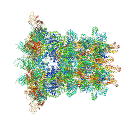
6J0F
 | | Cryo-EM Structure of an Extracellular Contractile Injection System, PVC sheath/tube terminator in extended state | | 分子名称: | Pvc1, Pvc16 | | 著者 | Jiang, F, Li, N, Wang, X, Cheng, J, Huang, Y, Yang, Y, Yang, J, Cai, B, Wang, Y, Jin, Q, Gao, N. | | 登録日 | 2018-12-24 | | 公開日 | 2019-04-10 | | 最終更新日 | 2024-03-27 | | 実験手法 | ELECTRON MICROSCOPY (3.8 Å) | | 主引用文献 | Cryo-EM Structure and Assembly of an Extracellular Contractile Injection System.
Cell, 177, 2019
|
|
5F6K
 | | Crystal structure of the MLL3-Ash2L-RbBP5 complex | | 分子名称: | Histone-lysine N-methyltransferase 2C, Retinoblastoma-binding protein 5, S-ADENOSYL-L-HOMOCYSTEINE, ... | | 著者 | Li, Y, Lei, M, Chen, Y. | | 登録日 | 2015-12-06 | | 公開日 | 2016-02-24 | | 最終更新日 | 2023-11-08 | | 実験手法 | X-RAY DIFFRACTION (2.411 Å) | | 主引用文献 | Structural basis for activity regulation of MLL family methyltransferases.
Nature, 530, 2016
|
|
6J0N
 | | Cryo-EM Structure of an Extracellular Contractile Injection System, baseplate in extended state, refined in C6 symmetry | | 分子名称: | Pvc1, Pvc11, Pvc12, ... | | 著者 | Jiang, F, Li, N, Wang, X, Cheng, J, Huang, Y, Yang, Y, Yang, J, Cai, B, Wang, Y, Jin, Q, Gao, N. | | 登録日 | 2018-12-25 | | 公開日 | 2019-04-10 | | 最終更新日 | 2024-03-27 | | 実験手法 | ELECTRON MICROSCOPY (3.5 Å) | | 主引用文献 | Cryo-EM Structure and Assembly of an Extracellular Contractile Injection System.
Cell, 177, 2019
|
|
8JC5
 | |
8JCA
 | |
6V9T
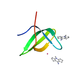 | | Tudor domain of TDRD3 in complex with a small molecule | | 分子名称: | 4-methyl-2,3,4,5,6,7-hexahydrodicyclopenta[b,e]pyridin-8(1H)-imine, Tudor domain-containing protein 3, UNKNOWN ATOM OR ION | | 著者 | Li, W, Tempel, W, Arrowsmith, C.H, Bountra, C, Edwards, A.M, Min, J, Structural Genomics Consortium (SGC) | | 登録日 | 2019-12-16 | | 公開日 | 2019-12-25 | | 最終更新日 | 2023-10-18 | | 実験手法 | X-RAY DIFFRACTION (2.154 Å) | | 主引用文献 | A small molecule antagonist of SMN disrupts the interaction between SMN and RNAP II.
Nat Commun, 13, 2022
|
|
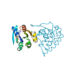
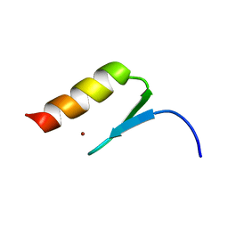
2MP0
 | | Protein Phosphorylation upon a Fleeting Encounter | | 分子名称: | Glucose-specific phosphotransferase enzyme IIA component, PHOSPHITE ION, Phosphoenolpyruvate-protein phosphotransferase | | 著者 | Xing, Q, Yang, J, Huang, P, Zhang, W, Tang, C. | | 登録日 | 2014-05-08 | | 公開日 | 2014-08-20 | | 最終更新日 | 2024-05-01 | | 実験手法 | SOLUTION NMR | | 主引用文献 | Visualizing an ultra-weak protein-protein interaction in phosphorylation signaling.
Angew.Chem.Int.Ed.Engl., 53, 2014
|
|
2MXP
 | |
4QQ6
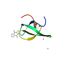 | | Crystal Structure of tudor domain of SMN1 in complex with a small organic molecule | | 分子名称: | 4-methyl-2,3,4,5,6,7-hexahydrodicyclopenta[b,e]pyridin-8(1H)-imine, Survival motor neuron protein, UNKNOWN ATOM OR ION | | 著者 | Liu, Y, Tempel, W, Iqbal, A, Walker, J.R, Bountra, C, Arrowsmith, C.H, Edwards, A.M, Brown, P.J, Min, J, Structural Genomics Consortium (SGC) | | 登録日 | 2014-06-26 | | 公開日 | 2014-08-06 | | 最終更新日 | 2023-09-20 | | 実験手法 | X-RAY DIFFRACTION (1.75 Å) | | 主引用文献 | A small molecule antagonist of SMN disrupts the interaction between SMN and RNAP II.
Nat Commun, 13, 2022
|
|
4QQD
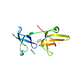 | | Crystal Structure of tandem tudor domains of UHRF1 in complex with a small organic molecule | | 分子名称: | 4-methyl-2,3,4,5,6,7-hexahydrodicyclopenta[b,e]pyridin-8(1H)-imine, E3 ubiquitin-protein ligase UHRF1, UNKNOWN ATOM OR ION | | 著者 | Liu, Y, Tempel, W, Iqbal, A, Walker, J.R, Bountra, C, Arrowsmith, C.H, Edwards, A.M, Brown, P.J, Min, J, Structural Genomics Consortium (SGC) | | 登録日 | 2014-06-27 | | 公開日 | 2014-08-06 | | 最終更新日 | 2024-04-03 | | 実験手法 | X-RAY DIFFRACTION (2.28 Å) | | 主引用文献 | A small molecule antagonist of SMN disrupts the interaction between SMN and RNAP II.
Nat Commun, 13, 2022
|
|
7BY7
 | | Bacteriophage SPO1 protein Gp46 | | 分子名称: | Putative gene 46 protein | | 著者 | Liu, B, Zhang, P. | | 登録日 | 2020-04-22 | | 公開日 | 2021-04-28 | | 最終更新日 | 2024-05-15 | | 実験手法 | SOLUTION NMR | | 主引用文献 | Bacteriophage protein Gp46 is a cross-species inhibitor of nucleoid-associated HU proteins
Proc.Natl.Acad.Sci.USA, 119, 2022
|
|
4WXX
 | |
4WWI
 | |
8K6P
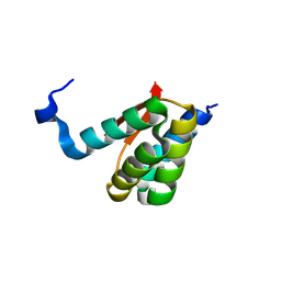 | |
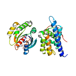
8K6Q
 | | Crystal structure of HOIL-1L LTM domain | | 分子名称: | RanBP-type and C3HC4-type zinc finger-containing protein 1 | | 著者 | Yan, Z, Pan, L.F. | | 登録日 | 2023-07-25 | | 公開日 | 2024-07-03 | | 実験手法 | X-RAY DIFFRACTION (1.59 Å) | | 主引用文献 | Mechanistic insights into the homo-dimerization of HOIL-1L and SHARPIN.
Biochem.Biophys.Res.Commun., 689, 2023
|
|
6X3L
 | | Sortilin-Progranulin Interaction With Compound 2 | | 分子名称: | 1-benzyl-3-tert-butyl-1H-pyrazole-5-carboxylic acid, 2-acetamido-2-deoxy-beta-D-glucopyranose-(1-4)-2-acetamido-2-deoxy-beta-D-glucopyranose, GLYCEROL, ... | | 著者 | Parthasarathy, G, Soisson, S.M. | | 登録日 | 2020-05-21 | | 公開日 | 2020-08-12 | | 最終更新日 | 2023-10-18 | | 実験手法 | X-RAY DIFFRACTION (2.7 Å) | | 主引用文献 | Identification of potent inhibitors of the sortilin-progranulin interaction.
Bioorg.Med.Chem.Lett., 30, 2020
|
|
6X48
 | | Sortilin-Progranulin Interaction With Compound 17 | | 分子名称: | GLYCEROL, N-(3,5-dichlorobenzene-1-carbonyl)-5,5-dimethyl-L-norleucine, Sortilin, ... | | 著者 | Parthasarathy, G, Soisson, S.M. | | 登録日 | 2020-05-22 | | 公開日 | 2020-08-12 | | 最終更新日 | 2023-10-18 | | 実験手法 | X-RAY DIFFRACTION (2.9 Å) | | 主引用文献 | Identification of potent inhibitors of the sortilin-progranulin interaction.
Bioorg.Med.Chem.Lett., 30, 2020
|
|


