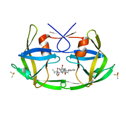
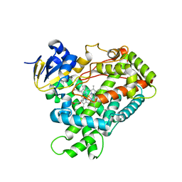
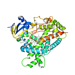
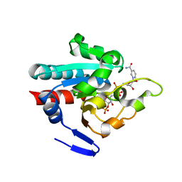
2HB2
 
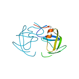 | | Structure of HIV protease 6X mutant in apo form | | 分子名称: | Protease | | 著者 | Heaslet, H, Tam, K, Elder, J.H, Stout, C.D. | | 登録日 | 2006-06-13 | | 公開日 | 2007-06-26 | | 最終更新日 | 2024-02-14 | | 実験手法 | X-RAY DIFFRACTION (2.3 Å) | | 主引用文献 | Conformational flexibility in the flap domains of ligand-free HIV protease.
Acta Crystallogr.,Sect.D, 63, 2007
|
|
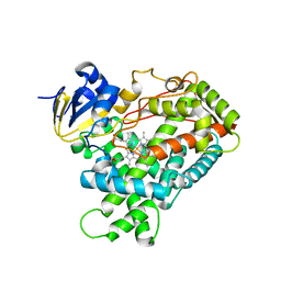
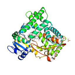
2HI4
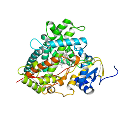 | | Crystal Structure of Human Microsomal P450 1A2 in complex with alpha-naphthoflavone | | 分子名称: | 2-PHENYL-4H-BENZO[H]CHROMEN-4-ONE, Cytochrome P450 1A2, PROTOPORPHYRIN IX CONTAINING FE | | 著者 | Sansen, S, Yano, J.K, Reynald, R.L, Schoch, G.S, Stout, C.D, Johnson, E.F. | | 登録日 | 2006-06-29 | | 公開日 | 2007-02-20 | | 最終更新日 | 2023-08-30 | | 実験手法 | X-RAY DIFFRACTION (1.95 Å) | | 主引用文献 | Adaptations for the oxidation of polycyclic aromatic hydrocarbons exhibited by the structure of human P450 1A2.
J.Biol.Chem., 282, 2007
|
|
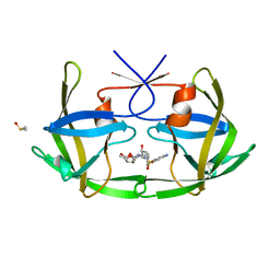
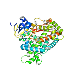
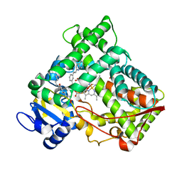
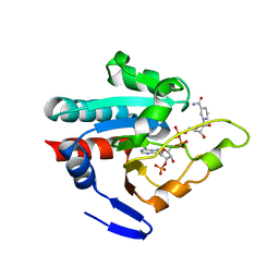
2HC0
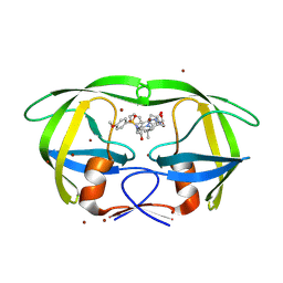 | | Structure of HIV protease 6X mutant in complex with AB-2. | | 分子名称: | BROMIDE ION, Protease, [1-((1S,2R)-1-BENZYL-2-HYDROXY-3-{ISOBUTYL[(4-METHOXYPHENYL)SULFONYL]AMINO}PROPYL)-1H-1,2,3-TRIAZOL-4-YL]METHYL (1R,2R)-2-HYDROXY-2,3-DIHYDRO-1H-INDEN-1-YLCARBAMATE | | 著者 | Heaslet, H, Brik, A, Lin, Y.-C, Elder, J.H, Stout, C.D. | | 登録日 | 2006-06-14 | | 公開日 | 2007-06-26 | | 最終更新日 | 2021-10-20 | | 実験手法 | X-RAY DIFFRACTION (1.3 Å) | | 主引用文献 | Structure of HIV Protease 6X Mutant in complex with AB-2
To be Published
|
|
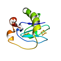
3LYN
 | |
3OGQ
 | | Crystal Structure of 6s-98S FIV Protease with Lopinavir bound | | 分子名称: | DIMETHYL SULFOXIDE, FIV protease, N-{1-BENZYL-4-[2-(2,6-DIMETHYL-PHENOXY)-ACETYLAMINO]-3-HYDROXY-5-PHENYL-PENTYL}-3-METHYL-2-(2-OXO-TETRAHYDRO-PYRIMIDIN-1-YL)-BUTYRAMIDE | | 著者 | Lin, Y.-C, Perryman, A.L, Elder, J.H, Stout, C.D. | | 登録日 | 2010-08-17 | | 公開日 | 2011-06-08 | | 最終更新日 | 2024-02-21 | | 実験手法 | X-RAY DIFFRACTION (1.8 Å) | | 主引用文献 | Structural basis for drug and substrate specificity exhibited by FIV encoding a chimeric FIV/HIV protease.
Acta Crystallogr.,Sect.D, 67, 2011
|
|
3OGP
 | | Crystal Structure of 6s-98S FIV Protease with Darunavir bound | | 分子名称: | (3R,3AS,6AR)-HEXAHYDROFURO[2,3-B]FURAN-3-YL(1S,2R)-3-[[(4-AMINOPHENYL)SULFONYL](ISOBUTYL)AMINO]-1-BENZYL-2-HYDROXYPROPYLCARBAMATE, DIMETHYL SULFOXIDE, FIV Protease | | 著者 | Lin, Y.-C, Perryman, A.L, Elder, J.H, Stout, C.D. | | 登録日 | 2010-08-17 | | 公開日 | 2011-06-08 | | 最終更新日 | 2023-09-06 | | 実験手法 | X-RAY DIFFRACTION (1.7 Å) | | 主引用文献 | Structural basis for drug and substrate specificity exhibited by FIV encoding a chimeric FIV/HIV protease.
Acta Crystallogr.,Sect.D, 67, 2011
|
|
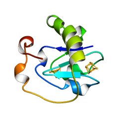
2LIS
 | |
2LYN
 | |
1D3W
 | | Crystal structure of ferredoxin 1 d15e mutant from azotobacter vinelandii at 1.7 angstrom resolution. | | 分子名称: | FE3-S4 CLUSTER, FERREDOXIN 1, IRON/SULFUR CLUSTER | | 著者 | Chen, K, Hirst, J, Camba, R, Bonagura, C.A, Stout, C.D, Burges, B.K, Armstrong, F.A. | | 登録日 | 1999-10-01 | | 公開日 | 1999-10-14 | | 最終更新日 | 2024-02-07 | | 実験手法 | X-RAY DIFFRACTION (1.7 Å) | | 主引用文献 | Atomically defined mechanism for proton transfer to a buried redox centre in a protein.
Nature, 405, 2000
|
|
1A6L
 | | T14C MUTANT OF AZOTOBACTER VINELANDII FDI | | 分子名称: | FE3-S4 CLUSTER, FERREDOXIN, IRON/SULFUR CLUSTER | | 著者 | Gao-Sheridan, H.S, Kemper, M.A, Khayat, R, Armstrong, F.A, Prasad, G.S, Sridhar, V, Stout, C.D, Burgess, B.K. | | 登録日 | 1998-02-26 | | 公開日 | 1998-05-27 | | 最終更新日 | 2024-05-22 | | 実験手法 | X-RAY DIFFRACTION (2.1 Å) | | 主引用文献 | A T14C variant of Azotobacter vinelandii ferredoxin I undergoes facile [3Fe-4S]0 to [4Fe-4S]2+ conversion in vitro but not in vivo.
J.Biol.Chem., 273, 1998
|
|
2P85
 | |
2PC0
 | | Apo Wild-type HIV Protease in the open conformation | | 分子名称: | MAGNESIUM ION, Protease, R-1,2-PROPANEDIOL | | 著者 | Heaslet, H, Rosenfeld, R, Giffin, M.J, Elder, J.H, McRee, D.E, Stout, C.D. | | 登録日 | 2007-03-29 | | 公開日 | 2007-06-26 | | 最終更新日 | 2023-08-30 | | 実験手法 | X-RAY DIFFRACTION (1.4 Å) | | 主引用文献 | Conformational flexibility in the flap domains of ligand-free HIV protease.
Acta Crystallogr.,Sect.D, 63, 2007
|
|
2PG5
 | | Crystal Structure of Human Microsomal P450 2A6 N297Q | | 分子名称: | 1,2-ETHANEDIOL, Cytochrome P450 2A6, PROTOPORPHYRIN IX CONTAINING FE | | 著者 | Sansen, S, Hsu, M.H, Stout, C.D, Johnson, E.F. | | 登録日 | 2007-04-06 | | 公開日 | 2007-07-24 | | 最終更新日 | 2023-08-30 | | 実験手法 | X-RAY DIFFRACTION (1.95 Å) | | 主引用文献 | Structural insight into the altered substrate specificity of human cytochrome P450 2A6 mutants.
Arch.Biochem.Biophys., 464, 2007
|
|
2PG7
 | | Crystal Structure of Human Microsomal P450 2A6 N297Q/I300V | | 分子名称: | Cytochrome P450 2A6, PROTOPORPHYRIN IX CONTAINING FE | | 著者 | Sansen, S, Hsu, M.H, Stout, C.D, Johnson, E.F. | | 登録日 | 2007-04-06 | | 公開日 | 2007-07-24 | | 最終更新日 | 2023-08-30 | | 実験手法 | X-RAY DIFFRACTION (2.8 Å) | | 主引用文献 | Structural insight into the altered substrate specificity of human cytochrome P450 2A6 mutants.
Arch.Biochem.Biophys., 464, 2007
|
|
2PG6
 | | Crystal Structure of Human Microsomal P450 2A6 L240C/N297Q | | 分子名称: | Cytochrome P450 2A6, PROTOPORPHYRIN IX CONTAINING FE | | 著者 | Sansen, S, Hsu, M.H, Stout, C.D, Johnson, E.F. | | 登録日 | 2007-04-06 | | 公開日 | 2007-07-24 | | 最終更新日 | 2023-08-30 | | 実験手法 | X-RAY DIFFRACTION (2.53 Å) | | 主引用文献 | Structural insight into the altered substrate specificity of human cytochrome P450 2A6 mutants.
Arch.Biochem.Biophys., 464, 2007
|
|
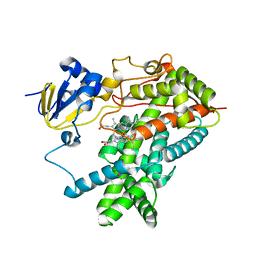
2O8V
 | | PAPS reductase in a covalent complex with thioredoxin C35A | | 分子名称: | Phosphoadenosine phosphosulfate reductase, Thioredoxin 1 | | 著者 | Chartron, J, Shiau, C, Stout, C.D, Carroll, K.S. | | 登録日 | 2006-12-12 | | 公開日 | 2007-03-27 | | 最終更新日 | 2023-08-30 | | 実験手法 | X-RAY DIFFRACTION (3 Å) | | 主引用文献 | 3'-Phosphoadenosine-5'-phosphosulfate Reductase in Complex with Thioredoxin: A Structural Snapshot in the Catalytic Cycle.
Biochemistry, 46, 2007
|
|
2Q6N
 | | Structure of Cytochrome P450 2B4 with Bound 1-(4-cholorophenyl)imidazole | | 分子名称: | 1-(4-CHLOROPHENYL)-1H-IMIDAZOLE, Cytochrome P450 2B4, PROTOPORPHYRIN IX CONTAINING FE | | 著者 | Zhao, Y, Sun, L, Muralidhara, B.K, Kumar, S, White, M.A, Stout, C.D, Halpert, J.R. | | 登録日 | 2007-06-05 | | 公開日 | 2007-11-06 | | 最終更新日 | 2023-08-30 | | 実験手法 | X-RAY DIFFRACTION (3.2 Å) | | 主引用文献 | Structural and thermodynamic consequences of 1-(4-chlorophenyl)imidazole binding to cytochrome P450 2B4.
Biochemistry, 46, 2007
|
|
2NNJ
 | | CYP2C8dH complexed with felodipine | | 分子名称: | Cytochrome P450 2C8, FELODIPINE, PALMITIC ACID, ... | | 著者 | Schoch, G.A, Yano, J.K, Stout, C.D, Johnson, E.F. | | 登録日 | 2006-10-24 | | 公開日 | 2007-10-23 | | 最終更新日 | 2023-08-30 | | 実験手法 | X-RAY DIFFRACTION (2.28 Å) | | 主引用文献 | Determinants of cytochrome P450 2C8 substrate binding: structures of complexes with montelukast, troglitazone, felodipine, and 9-cis-retinoic acid.
J.Biol.Chem., 283, 2008
|
|
2NNI
 | | CYP2C8dH complexed with montelukast | | 分子名称: | Cytochrome P450 2C8, MONTELUKAST, PALMITIC ACID, ... | | 著者 | Schoch, G.A, Yano, J.K, Stout, C.D, Johnson, E.F. | | 登録日 | 2006-10-24 | | 公開日 | 2007-10-23 | | 最終更新日 | 2023-08-30 | | 実験手法 | X-RAY DIFFRACTION (2.8 Å) | | 主引用文献 | Determinants of cytochrome P450 2C8 substrate binding: structures of complexes with montelukast, troglitazone, felodipine, and 9-cis-retinoic acid.
J.Biol.Chem., 283, 2008
|
|
2NNH
 | | CYP2C8dH complexed with 2 molecules of 9-cis retinoic acid | | 分子名称: | (9cis)-retinoic acid, Cytochrome P450 2C8, PALMITIC ACID, ... | | 著者 | Schoch, G.A, Yano, J.K, Stout, C.D, Johnson, E.F. | | 登録日 | 2006-10-24 | | 公開日 | 2007-10-23 | | 最終更新日 | 2023-08-30 | | 実験手法 | X-RAY DIFFRACTION (2.6 Å) | | 主引用文献 | Determinants of cytochrome P450 2C8 substrate binding: structures of complexes with montelukast, troglitazone, felodipine, and 9-cis-retinoic acid.
J.Biol.Chem., 283, 2008
|
|
1PNO
 | | Crystal structure of R. rubrum transhydrogenase domain III bound to NADP | | 分子名称: | NAD(P) transhydrogenase subunit beta, NADP NICOTINAMIDE-ADENINE-DINUCLEOTIDE PHOSPHATE | | 著者 | Sundaresan, V, Yamaguchi, M, Chartron, J, Stout, C.D. | | 登録日 | 2003-06-12 | | 公開日 | 2003-11-11 | | 最終更新日 | 2024-02-14 | | 実験手法 | X-RAY DIFFRACTION (2.1 Å) | | 主引用文献 | Conformational Change in the NADP(H) Binding Domain of Transhydrogenase Defines Four States
Biochemistry, 42, 2003
|
|
1PNQ
 | | Crystal structure of R. rubrum transhydrogenase domain III bound to NADPH | | 分子名称: | NAD(P) transhydrogenase subunit beta, NADPH DIHYDRO-NICOTINAMIDE-ADENINE-DINUCLEOTIDE PHOSPHATE | | 著者 | Sundaresan, V, Yamaguchi, M, Chartron, J, Stout, C.D. | | 登録日 | 2003-06-12 | | 公開日 | 2003-11-11 | | 最終更新日 | 2024-02-14 | | 実験手法 | X-RAY DIFFRACTION (2.4 Å) | | 主引用文献 | Conformational Change in the NADP(H) Binding Domain of Transhydrogenase Defines Four States
Biochemistry, 42, 2003
|
|
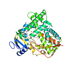
1PO5
 | | Structure of mammalian cytochrome P450 2B4 | | 分子名称: | Cytochrome P450 2B4, PROTOPORPHYRIN IX CONTAINING FE | | 著者 | Scott, E.E, He, Y.A, Wester, M.R, White, M.A, Chin, C.C, Halpert, J.R, Johnson, E.F, Stout, C.D. | | 登録日 | 2003-06-13 | | 公開日 | 2003-10-07 | | 最終更新日 | 2023-08-16 | | 実験手法 | X-RAY DIFFRACTION (1.6 Å) | | 主引用文献 | An open conformation of mammalian cytochrome P450 2B4 at 1.6 A resolution
Proc.Natl.Acad.Sci.USA, 100, 2003
|
|
1PQ2
 | | Crystal Structure of Human Drug Metabolizing Cytochrome P450 2C8 | | 分子名称: | Cytochrome P450 2C8, PALMITIC ACID, PHOSPHATE ION, ... | | 著者 | Schoch, G.A, Yano, J.K, Wester, M.R, Griffin, K.J, Stout, C.D, Johnson, E.F. | | 登録日 | 2003-06-17 | | 公開日 | 2004-01-13 | | 最終更新日 | 2023-08-16 | | 実験手法 | X-RAY DIFFRACTION (2.7 Å) | | 主引用文献 | Structure of human microsomal cytochrome P450 2C8. Evidence for a peripheral fatty acid binding site
J.Biol.Chem., 279, 2004
|
|
1N6B
 | | Microsomal Cytochrome P450 2C5/3LVdH Complex with a dimethyl derivative of sulfaphenazole | | 分子名称: | 4-METHYL-N-METHYL-N-(2-PHENYL-2H-PYRAZOL-3-YL)BENZENESULFONAMIDE, Cytochrome P450 2C5, PROTOPORPHYRIN IX CONTAINING FE, ... | | 著者 | Wester, M.R, Johnson, E.F, Marques-Soares, C, Dansette, P.M, Mansuy, D, Stout, C.D. | | 登録日 | 2002-11-09 | | 公開日 | 2003-06-03 | | 最終更新日 | 2024-02-14 | | 実験手法 | X-RAY DIFFRACTION (2.3 Å) | | 主引用文献 | Structure of a Substrate Complex of Mammalian Cytochrome P450 2C5 at 2.3 A Resolution: Evidence
for Multiple Substrate Binding Modes
Biochemistry, 42, 2003
|
|


