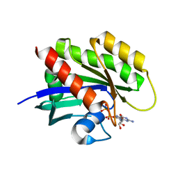
8RK1
 
 | |
4Z88
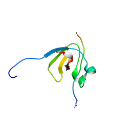
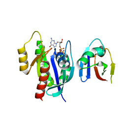
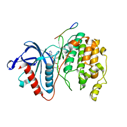
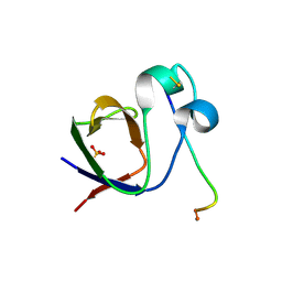 | | SH3-II of Drosophila Rim-binding protein with Aplip1 peptide | | 分子名称: | JNK-interacting protein 1, PHOSPHATE ION, RIM-binding protein, ... | | 著者 | Driller, J.H, Holton, N, Siebert, M, Boehme, M.A, Wahl, M.C, Sigrist, S.J, Loll, B. | | 登録日 | 2015-04-08 | | 公開日 | 2015-08-05 | | 最終更新日 | 2024-01-10 | | 実験手法 | X-RAY DIFFRACTION (2.09 Å) | | 主引用文献 | A high affinity RIM-binding protein/Aplip1 interaction prevents the formation of ectopic axonal active zones.
Elife, 4, 2015
|
|
1A8R
 | | GTP CYCLOHYDROLASE I (H112S MUTANT) IN COMPLEX WITH GTP | | 分子名称: | GTP CYCLOHYDROLASE I, GUANOSINE-5'-TRIPHOSPHATE | | 著者 | Auerbach, G, Nar, H, Bracher, A, Bacher, A, Huber, R. | | 登録日 | 1998-03-27 | | 公開日 | 1999-05-11 | | 最終更新日 | 2024-02-07 | | 実験手法 | X-RAY DIFFRACTION (2.1 Å) | | 主引用文献 | Biosynthesis of pteridines. Reaction mechanism of GTP cyclohydrolase I.
J.Mol.Biol., 326, 2003
|
|
8W0O
 | | GDH-105 crystal structure | | 分子名称: | CHLORIDE ION, Dehydrogenase, NICOTINAMIDE-ADENINE-DINUCLEOTIDE | | 著者 | Peat, T.S, Newman, J. | | 登録日 | 2024-02-13 | | 公開日 | 2024-07-24 | | 実験手法 | X-RAY DIFFRACTION (1.66 Å) | | 主引用文献 | Enhancing the Imine Reductase Activity of a Promiscuous Glucose Dehydrogenase for Scalable Manufacturing of a Chiral Neprilysin Inhibitor Precursor
Acs Catalysis, 14, 2024
|
|
8W0N
 | | IRED crystal structure | | 分子名称: | DI(HYDROXYETHYL)ETHER, DIMETHYL SULFOXIDE, IRED, ... | | 著者 | Peat, T.S, Newman, J. | | 登録日 | 2024-02-13 | | 公開日 | 2024-07-24 | | 実験手法 | X-RAY DIFFRACTION (1.37 Å) | | 主引用文献 | Enhancing the Imine Reductase Activity of a Promiscuous Glucose Dehydrogenase for Scalable Manufacturing of a Chiral Neprilysin Inhibitor Precursor
Acs Catalysis, 14, 2024
|
|
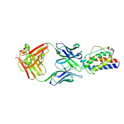
4Z8A
 | | SH3-III of Drosophila Rim-binding protein bound to a Cacophony derived peptide | | 分子名称: | RIM-binding protein, isoform F, Voltage-dependent calcium channel type A subunit alpha-1 | | 著者 | Driller, J.H, Holton, N, Siebert, M, Boehme, A.M, Wahl, M.C, Sigrist, S.J, Loll, B. | | 登録日 | 2015-04-08 | | 公開日 | 2015-08-05 | | 最終更新日 | 2024-01-10 | | 実験手法 | X-RAY DIFFRACTION (1.759 Å) | | 主引用文献 | A high affinity RIM-binding protein/Aplip1 interaction prevents the formation of ectopic axonal active zones.
Elife, 4, 2015
|
|
8OEI
 | | SFX structure of FutA after an accumulated dose of 350 kGy | | 分子名称: | FE (III) ION, Putative iron ABC transporter, substrate binding protein | | 著者 | Bolton, R, Tews, I. | | 登録日 | 2023-03-10 | | 公開日 | 2023-08-30 | | 最終更新日 | 2024-03-27 | | 実験手法 | X-RAY DIFFRACTION (1.65 Å) | | 主引用文献 | A redox switch allows binding of Fe(II) and Fe(III) ions in the cyanobacterial iron-binding protein FutA from Prochlorococcus.
Proc.Natl.Acad.Sci.USA, 121, 2024
|
|
8OEM
 | | Crystal structure of FutA bound to Fe(II) | | 分子名称: | FE (II) ION, Putative iron ABC transporter, substrate binding protein | | 著者 | Bolton, R, Tews, I. | | 登録日 | 2023-03-10 | | 公開日 | 2023-08-30 | | 最終更新日 | 2024-03-27 | | 実験手法 | X-RAY DIFFRACTION (1.7 Å) | | 主引用文献 | A redox switch allows binding of Fe(II) and Fe(III) ions in the cyanobacterial iron-binding protein FutA from Prochlorococcus.
Proc.Natl.Acad.Sci.USA, 121, 2024
|
|
8OGG
 | | Crystal structure of FutA after an accumulated dose of 5 kGy | | 分子名称: | FE (III) ION, Putative iron ABC transporter, substrate binding protein | | 著者 | Bolton, R, Tews, I. | | 登録日 | 2023-03-20 | | 公開日 | 2023-08-30 | | 最終更新日 | 2024-03-27 | | 実験手法 | X-RAY DIFFRACTION (1.76 Å) | | 主引用文献 | A redox switch allows binding of Fe(II) and Fe(III) ions in the cyanobacterial iron-binding protein FutA from Prochlorococcus.
Proc.Natl.Acad.Sci.USA, 121, 2024
|
|
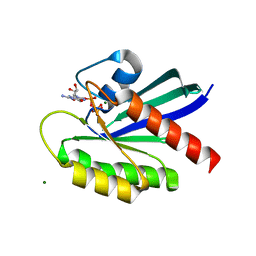
5LQB
 | |
8C4Y
 | | SFX structure of FutA bound to Fe(III) | | 分子名称: | FE (III) ION, Putative iron ABC transporter, substrate binding protein | | 著者 | Bolton, R, Tews, I. | | 登録日 | 2023-01-05 | | 公開日 | 2023-08-30 | | 最終更新日 | 2024-03-27 | | 実験手法 | X-RAY DIFFRACTION (1.6 Å) | | 主引用文献 | A redox switch allows binding of Fe(II) and Fe(III) ions in the cyanobacterial iron-binding protein FutA from Prochlorococcus.
Proc.Natl.Acad.Sci.USA, 121, 2024
|
|
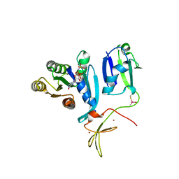
8EBZ
 | |
8ETR
 | | CryoEM Structure of NLRP3 NACHT domain in complex with G2394 | | 分子名称: | (6S,8R)-N-[(1,2,3,5,6,7-hexahydro-s-indacen-4-yl)carbamoyl]-6-(methylamino)-6,7-dihydro-5H-pyrazolo[5,1-b][1,3]oxazine-3-sulfonamide, ADENOSINE-5'-DIPHOSPHATE, MAGNESIUM ION, ... | | 著者 | Murray, J.M, Johnson, M.C. | | 登録日 | 2022-10-17 | | 公開日 | 2022-11-02 | | 最終更新日 | 2024-06-19 | | 実験手法 | ELECTRON MICROSCOPY (3.5 Å) | | 主引用文献 | Overcoming Preclinical Safety Obstacles to Discover ( S )- N -((1,2,3,5,6,7-Hexahydro- s -indacen-4-yl)carbamoyl)-6-(methylamino)-6,7-dihydro-5 H -pyrazolo[5,1- b ][1,3]oxazine-3-sulfonamide (GDC-2394): A Potent and Selective NLRP3 Inhibitor.
J.Med.Chem., 65, 2022
|
|
8EPW
 | | Crystal Structure of KRAS4b-G13D (GMPPNP-bound) in complex with RAS-binding domain (RBD) of RAF1/CRAF | | 分子名称: | GTPase KRas, MAGNESIUM ION, PHOSPHOAMINOPHOSPHONIC ACID-GUANYLATE ESTER, ... | | 著者 | Tran, T.H, Chan, A.H, Dharmaiah, S, Simanshu, D.K. | | 登録日 | 2022-10-06 | | 公開日 | 2023-06-07 | | 最終更新日 | 2023-10-25 | | 実験手法 | X-RAY DIFFRACTION (2 Å) | | 主引用文献 | Reduced dynamic complexity allows structure elucidation of an excited state of KRAS G13D .
Commun Biol, 6, 2023
|
|
4WNX
 | | Netrin 4 lacking the C-terminal Domain | | 分子名称: | 2-acetamido-2-deoxy-beta-D-glucopyranose, 2-acetamido-2-deoxy-beta-D-glucopyranose-(1-4)-2-acetamido-2-deoxy-beta-D-glucopyranose, CALCIUM ION, ... | | 著者 | McDougall, M, Patel, T, Reuten, R, Meier, M, Koch, M, Stetefeld, J. | | 登録日 | 2014-10-14 | | 公開日 | 2016-02-10 | | 最終更新日 | 2023-09-27 | | 実験手法 | X-RAY DIFFRACTION (2.723 Å) | | 主引用文献 | Structural decoding of netrin-4 reveals a regulatory function towards mature basement membranes.
Nat Commun, 7, 2016
|
|
6Q9M
 | | Central Fibronectin-III array of RIM-binding protein | | 分子名称: | PHOSPHATE ION, RIM-binding protein, isoform F | | 著者 | Driller, J.D, Habibi, S, Wahl, M.C, Loll, B. | | 登録日 | 2018-12-18 | | 公開日 | 2020-01-15 | | 最終更新日 | 2024-05-15 | | 実験手法 | X-RAY DIFFRACTION (2.453 Å) | | 主引用文献 | RIM-binding protein couples synaptic vesicle recruitment to release sites.
J.Cell Biol., 219, 2020
|
|
8T71
 | | Crystal Structure of WT KRAS4a with bound GDP and Mg ion | | 分子名称: | GTPase KRas, GUANOSINE-5'-DIPHOSPHATE, MAGNESIUM ION | | 著者 | Tran, T.H, Whitley, M.J, Dharmaiah, S, Simanshu, D.K. | | 登録日 | 2023-06-19 | | 公開日 | 2024-02-14 | | 最終更新日 | 2024-02-28 | | 実験手法 | X-RAY DIFFRACTION (1.8 Å) | | 主引用文献 | Comparative analysis of KRAS4a and KRAS4b splice variants reveals distinctive structural and functional properties.
Sci Adv, 10, 2024
|
|
8T73
 | | Crystal structure of KRAS4a-R151G with bound GDP and Mg ion | | 分子名称: | GTPase KRas, GUANOSINE-5'-DIPHOSPHATE, MAGNESIUM ION | | 著者 | Tran, T.H, Whitley, M.J, Dharmaiah, S, Simanshu, D.K. | | 登録日 | 2023-06-19 | | 公開日 | 2024-02-14 | | 最終更新日 | 2024-02-28 | | 実験手法 | X-RAY DIFFRACTION (1.5 Å) | | 主引用文献 | Comparative analysis of KRAS4a and KRAS4b splice variants reveals distinctive structural and functional properties.
Sci Adv, 10, 2024
|
|
8T72
 | | Crystal structure of WT KRAS4a with bound GMPPNP and Mg ion | | 分子名称: | GTPase KRas, MAGNESIUM ION, PHOSPHOAMINOPHOSPHONIC ACID-GUANYLATE ESTER | | 著者 | Tran, T.H, Whitley, M.J, Dharmaiah, S, Simanshu, D.K. | | 登録日 | 2023-06-19 | | 公開日 | 2024-02-14 | | 最終更新日 | 2024-02-28 | | 実験手法 | X-RAY DIFFRACTION (1.6 Å) | | 主引用文献 | Comparative analysis of KRAS4a and KRAS4b splice variants reveals distinctive structural and functional properties.
Sci Adv, 10, 2024
|
|
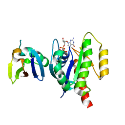
8T75
 | |
8T74
 | |
7W5C
 | | Crystal structure of Mitogen Activated Protein Kinase 4 (MPK4) from Arabidopsis thaliana | | 分子名称: | MAGNESIUM ION, Mitogen-activated protein kinase 4, Mitogen-activated protein kinase kinase 1, ... | | 著者 | Arold, S.T, Hameed, U.F.S. | | 登録日 | 2021-11-30 | | 公開日 | 2022-12-07 | | 最終更新日 | 2023-11-29 | | 実験手法 | X-RAY DIFFRACTION (2.201 Å) | | 主引用文献 | Essential role of the CD docking motif of MPK4 in plant immunity, growth, and development.
New Phytol., 239, 2023
|
|
6RU5
 | | human complement C3 in complex with the hC3Nb1 nanobody | | 分子名称: | 2-acetamido-2-deoxy-beta-D-glucopyranose, Complement C3, alpha-D-mannopyranose-(1-3)-[alpha-D-mannopyranose-(1-6)]beta-D-mannopyranose-(1-4)-2-acetamido-2-deoxy-beta-D-glucopyranose-(1-4)-2-acetamido-2-deoxy-beta-D-glucopyranose, ... | | 著者 | Jensen, R.K, Andersen, G.R. | | 登録日 | 2019-05-27 | | 公開日 | 2019-08-21 | | 最終更新日 | 2024-01-24 | | 実験手法 | X-RAY DIFFRACTION (3.9 Å) | | 主引用文献 | Structural Basis for Properdin Oligomerization and Convertase Stimulation in the Human Complement System.
Front Immunol, 10, 2019
|
|
6RU3
 | |
6RV6
 | | Structure of properdin lacking TSR3 based on anomalous data | | 分子名称: | 2-acetamido-2-deoxy-beta-D-glucopyranose-(1-4)-[alpha-L-fucopyranose-(1-6)]2-acetamido-2-deoxy-beta-D-glucopyranose, Properdin, alpha-D-mannopyranose, ... | | 著者 | Pedersen, D.V, Andersen, G.R. | | 登録日 | 2019-05-31 | | 公開日 | 2019-08-21 | | 最終更新日 | 2024-05-01 | | 実験手法 | X-RAY DIFFRACTION (3.507 Å) | | 主引用文献 | Structural Basis for Properdin Oligomerization and Convertase Stimulation in the Human Complement System.
Front Immunol, 10, 2019
|
|


