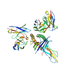
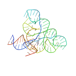
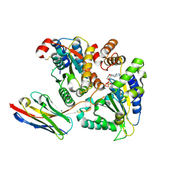
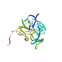
8EEY
 
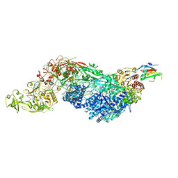 | | Cas7-11 in complex with DR-mismatched target RNA, Csx29 and Csx30 | | 分子名称: | Cas7-11, Csx29, Csx30, ... | | 著者 | Demircioglu, F.E, Wilkinson, M.E, Strecker, J, Li, D, Faure, G, Macrae, R.K, Zhang, F. | | 登録日 | 2022-09-07 | | 公開日 | 2022-11-16 | | 最終更新日 | 2024-06-19 | | 実験手法 | ELECTRON MICROSCOPY (2.53 Å) | | 主引用文献 | RNA-activated protein cleavage with a CRISPR-associated endopeptidase.
Science, 378, 2022
|
|
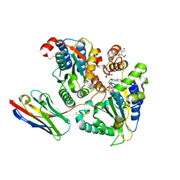
7AA4
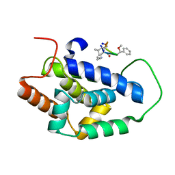 | | Structure of ClpC1-NTD bound to a CymA analogue | | 分子名称: | Negative regulator of genetic competence ClpC/mecB, polymer Cyclomarin A analogue | | 著者 | Meinhart, A, Morreale, F.E, Kaiser, M, Clausen, T. | | 登録日 | 2020-09-03 | | 公開日 | 2021-08-11 | | 最終更新日 | 2024-01-31 | | 実験手法 | X-RAY DIFFRACTION (1.68 Å) | | 主引用文献 | BacPROTACs mediate targeted protein degradation in bacteria.
Cell, 185, 2022
|
|
7ABR
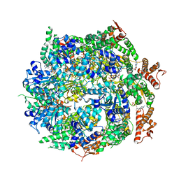 | | Cryo-EM structure of B. subtilis ClpC (DWB mutant) hexamer bound to a substrate polypeptide | | 分子名称: | ADENOSINE-5'-DIPHOSPHATE, ADENOSINE-5'-TRIPHOSPHATE, Negative regulator of genetic competence ClpC/MecB, ... | | 著者 | Morreale, F.E, Meinhart, A, Haselbach, D, Clausen, T. | | 登録日 | 2020-09-08 | | 公開日 | 2021-10-06 | | 最終更新日 | 2022-07-06 | | 実験手法 | ELECTRON MICROSCOPY (3.7 Å) | | 主引用文献 | BacPROTACs mediate targeted protein degradation in bacteria.
Cell, 185, 2022
|
|
8TAH
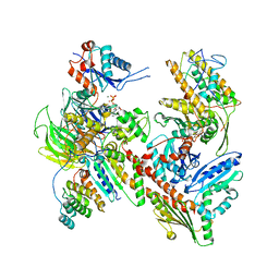 | | Cryo-EM structure of Cortactin-bound to Arp2/3 complex | | 分子名称: | ADENOSINE-5'-TRIPHOSPHATE, Actin-related protein 2, Actin-related protein 2/3 complex subunit 1A, ... | | 著者 | Fregoso, F.E, van Eeuwen, T, Dominguez, R. | | 登録日 | 2023-06-27 | | 公開日 | 2023-09-27 | | 最終更新日 | 2024-01-31 | | 実験手法 | ELECTRON MICROSCOPY (2.89 Å) | | 主引用文献 | Mechanism of synergistic activation of Arp2/3 complex by cortactin and WASP-family proteins.
Nat Commun, 14, 2023
|
|
8T6M
 | | Human leukocyte antigen bound by two alloreactive antibody Fabs | | 分子名称: | Beta-2-microglobulin, HLA_A0101_peptide, JTK191b_L02_Fab, ... | | 著者 | Kizziah, J.L, Killian, J.T, Diaz-Avalos, R, Qiu, S, Yang, G, Green, T, Lund, F.E. | | 登録日 | 2023-06-16 | | 公開日 | 2024-12-25 | | 最終更新日 | 2025-07-09 | | 実験手法 | ELECTRON MICROSCOPY (3.14 Å) | | 主引用文献 | Topography of the HLA-A protein enforces shared and convergent immunodominant B cell and antibody alloresponses in transplant recipients
To Be Published
|
|
8T7R
 | | Crystal structure of human leukocyte antigen A*0101 in complex with the Fab of alloreactive antibody E07 | | 分子名称: | Beta-2-microglobulin, Fab heavy chain from antibody JTK191b E07, Light chain from antibody JTK191b E07, ... | | 著者 | Green, T.J, Killian Jr, J.T, Qiu, S, Macon, K.J, Yang, G, King, R.G, Lund, F.E. | | 登録日 | 2023-06-21 | | 公開日 | 2024-12-25 | | 実験手法 | X-RAY DIFFRACTION (3.84 Å) | | 主引用文献 | B cell and antibody targeting of transplant alloantigen epitopes is supported by both self and allo-recognition
To Be Published
|
|
6CSE
 | | Crystal structure of sodium/alanine symporter AgcS with L-alanine bound | | 分子名称: | ALANINE, Monoclonal antibody FAB heavy chain, Monoclonal antibody FAB light chain, ... | | 著者 | Ma, J, Reyes, F.E, Gonen, T. | | 登録日 | 2018-03-20 | | 公開日 | 2019-01-30 | | 最終更新日 | 2024-11-13 | | 実験手法 | X-RAY DIFFRACTION (3.24 Å) | | 主引用文献 | Structural basis for substrate binding and specificity of a sodium-alanine symporter AgcS.
Proc. Natl. Acad. Sci. U.S.A., 116, 2019
|
|
6DN2
 | | CRYSTAL STRUCTURE OF THE FMN RIBOSWITCH BOUND TO BRX1354 SPLIT RNA | | 分子名称: | 4-{benzyl[2-(7,8-dimethyl-2,4-dioxo-3,4-dihydrobenzo[g]pteridin-10(2H)-yl)ethyl]amino}butanoic acid, MAGNESIUM ION, POTASSIUM ION, ... | | 著者 | Vicens, Q, Mondragon, E, Reyes, F.E, Berman, J, Kaur, H, Kells, K, Wickens, P, Wilson, J, Gadwood, R, Schostarez, H, Suto, R.K, Coish, P, Blount, K.F, Batey, R.T. | | 登録日 | 2018-06-05 | | 公開日 | 2018-09-05 | | 最終更新日 | 2023-10-11 | | 実験手法 | X-RAY DIFFRACTION (2.88 Å) | | 主引用文献 | Structure-Activity Relationship of Flavin Analogues That Target the Flavin Mononucleotide Riboswitch.
ACS Chem. Biol., 13, 2018
|
|
6DN3
 | | CRYSTAL STRUCTURE OF THE FMN RIBOSWITCH BOUND TO BRX1555 SPLIT RNA | | 分子名称: | 7,8-dimethyl-2,4-dioxo-10-(3-phenylpropyl)-1,2,3,4-tetrahydrobenzo[g]pteridin-10-ium, CHLORIDE ION, MAGNESIUM ION, ... | | 著者 | Vicens, Q, Mondragon, E, Reyes, F.E, Berman, J, Kaur, H, Kells, K, Wickens, P, Wilson, J, Gadwood, R, Schostarez, H, Suto, R.K, Coish, P, Blount, K.F, Batey, R.T. | | 登録日 | 2018-06-05 | | 公開日 | 2018-09-05 | | 最終更新日 | 2023-10-11 | | 実験手法 | X-RAY DIFFRACTION (2.8 Å) | | 主引用文献 | Structure-Activity Relationship of Flavin Analogues That Target the Flavin Mononucleotide Riboswitch.
ACS Chem. Biol., 13, 2018
|
|
6DN1
 | | CRYSTAL STRUCTURE OF THE FMN RIBOSWITCH BOUND TO BRX1151 SPLIT RNA | | 分子名称: | 10-(6-carboxyhexyl)-8-(cyclopentylamino)-2,4-dihydroxy-7-methylbenzo[g]pteridin-10-ium, MAGNESIUM ION, POTASSIUM ION, ... | | 著者 | Vicens, Q, Mondragon, E, Reyes, F.E, Berman, J, Kaur, H, Kells, K, Wickens, P, Wilson, J, Gadwood, R, Schostarez, H, Suto, R.K, Coish, P, Blount, K.F, Batey, R.T. | | 登録日 | 2018-06-05 | | 公開日 | 2018-09-05 | | 最終更新日 | 2023-10-11 | | 実験手法 | X-RAY DIFFRACTION (3.03 Å) | | 主引用文献 | Structure-Activity Relationship of Flavin Analogues That Target the Flavin Mononucleotide Riboswitch.
ACS Chem. Biol., 13, 2018
|
|
6CSF
 | | Crystal structure of sodium/alanine symporter AgcS with D-alanine bound | | 分子名称: | D-ALANINE, Monoclonal antibody FAB heavy chain, Monoclonal antibody FAB light chain, ... | | 著者 | Ma, J, Reyes, F.E, Gonen, T. | | 登録日 | 2018-03-20 | | 公開日 | 2019-01-30 | | 最終更新日 | 2024-10-16 | | 実験手法 | X-RAY DIFFRACTION (3.3 Å) | | 主引用文献 | Structural basis for substrate binding and specificity of a sodium-alanine symporter AgcS.
Proc. Natl. Acad. Sci. U.S.A., 116, 2019
|
|
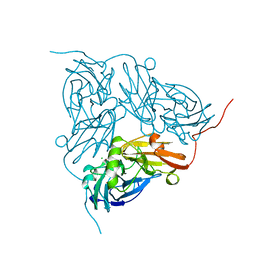
5I9S
 | | MicroED structure of proteinase K at 1.75 A resolution | | 分子名称: | Proteinase K, SULFATE ION | | 著者 | Hattne, J, Shi, D, de la Cruz, M.J, Reyes, F.E, Gonen, T. | | 登録日 | 2016-02-20 | | 公開日 | 2016-06-08 | | 最終更新日 | 2024-11-13 | | 実験手法 | ELECTRON CRYSTALLOGRAPHY (1.75 Å) | | 主引用文献 | Modeling truncated pixel values of faint reflections in MicroED images.
J.Appl.Crystallogr., 49, 2016
|
|
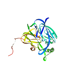
5HCL
 | | Crystal Structure of the first bromodomain of BRD4 in complex with DMA | | 分子名称: | 1,2-ETHANEDIOL, Bromodomain-containing protein 4, ~{N},~{N}-dimethylethanamide | | 著者 | Dong, J, Weber, F.E, Caflisch, A. | | 登録日 | 2016-01-04 | | 公開日 | 2017-01-25 | | 最終更新日 | 2024-01-10 | | 実験手法 | X-RAY DIFFRACTION (1.5 Å) | | 主引用文献 | N,N Dimethylacetamide a drug excipient that acts as bromodomain ligand for osteoporosis treatment.
Sci Rep, 7, 2017
|
|
5J1S
 | | TorsinA-LULL1 complex, H. sapiens, bound to VHH-BS2 | | 分子名称: | 2-(N-MORPHOLINO)-ETHANESULFONIC ACID, ADENOSINE-5'-TRIPHOSPHATE, CHLORIDE ION, ... | | 著者 | Demircioglu, F.E, Schwartz, T.U. | | 登録日 | 2016-03-29 | | 公開日 | 2016-08-17 | | 最終更新日 | 2024-10-16 | | 実験手法 | X-RAY DIFFRACTION (1.399 Å) | | 主引用文献 | Structures of TorsinA and its disease-mutant complexed with an activator reveal the molecular basis for primary dystonia.
Elife, 5, 2016
|
|
5J1T
 | | TorsinAdeltaE-LULL1 complex, H. sapiens, bound to VHH-BS2 | | 分子名称: | 2-[BIS-(2-HYDROXY-ETHYL)-AMINO]-2-HYDROXYMETHYL-PROPANE-1,3-DIOL, ADENOSINE-5'-TRIPHOSPHATE, CHLORIDE ION, ... | | 著者 | Demircioglu, F.E, Schwartz, T.U. | | 登録日 | 2016-03-29 | | 公開日 | 2016-08-17 | | 最終更新日 | 2024-10-09 | | 実験手法 | X-RAY DIFFRACTION (1.402 Å) | | 主引用文献 | Structures of TorsinA and its disease-mutant complexed with an activator reveal the molecular basis for primary dystonia.
Elife, 5, 2016
|
|
5TY4
 | | MicroED structure of a complex between monomeric TGF-b and its receptor, TbRII, at 2.9 A resolution | | 分子名称: | TGF-beta receptor type-2, mmTGF-b2-7m | | 著者 | Weiss, S.C, de la Cruz, M.J, Hattne, J, Shi, D, Reyes, F.E, Callero, G, Gonen, T. | | 登録日 | 2016-11-18 | | 公開日 | 2017-04-26 | | 最終更新日 | 2024-11-06 | | 実験手法 | ELECTRON CRYSTALLOGRAPHY (2.9 Å) | | 主引用文献 | Atomic-resolution structures from fragmented protein crystals with the cryoEM method MicroED.
Nat. Methods, 14, 2017
|
|
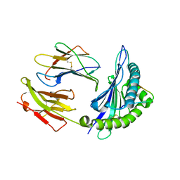
2PK8
 | | Crystal structure of an uncharacterized protein PF0899 from Pyrococcus furiosus | | 分子名称: | GOLD (I) CYANIDE ION, Uncharacterized protein PF0899 | | 著者 | Liu, Z.J, Tempel, W, Chen, L, Shah, A, Lee, D, Clancy-Kelley, L.L, Dillard, B.D, Rose, J.P, Sugar, F.J, Jenny Jr, F.E, Lee, H.S, Izumi, M, Shah, C, Poole III, F.L, Adams, M.W.W, Richardson, J.S, Richardson, D.C, Wang, B.-C, Southeast Collaboratory for Structural Genomics (SECSG) | | 登録日 | 2007-04-17 | | 公開日 | 2007-05-22 | | 最終更新日 | 2024-02-21 | | 実験手法 | X-RAY DIFFRACTION (1.85 Å) | | 主引用文献 | Structure of the hypothetical protein PF0899 from Pyrococcus furiosus at 1.85 A resolution.
Acta Crystallogr.,Sect.F, 63, 2007
|
|
2SEM
 | | SEM5 SH3 DOMAIN COMPLEXED WITH PEPTOID INHIBITOR | | 分子名称: | PROTEIN (SEX MUSCLE ABNORMAL PROTEIN 5), PROTEIN (SH3 PEPTOID INHIBITOR) | | 著者 | Nguyen, J.T, Turck, C.W, Cohen, F.E, Zuckermann, R.N, Lim, W.A. | | 登録日 | 1998-11-02 | | 公開日 | 1999-01-06 | | 最終更新日 | 2024-11-20 | | 実験手法 | X-RAY DIFFRACTION (2.2 Å) | | 主引用文献 | Exploiting the basis of proline recognition by SH3 and WW domains: design of N-substituted inhibitors.
Science, 282, 1998
|
|
1ZHK
 | | Crystal structure of HLA-B*3501 presenting 13-mer EBV antigen LPEPLPQGQLTAY | | 分子名称: | Beta-2-microglobulin, EBV-peptide LPEPLPQGQLTAY, HLA class I histocompatibility antigen, ... | | 著者 | Tynan, F.E, Borg, N.A, Miles, J.J, Beddoe, T, El-Hassen, D, Silins, S.L, van Zuylen, W.J, Purcell, A.W, Kjer-Nielsen, L, McCluskey, J, Burrows, S.R, Rossjohn, J. | | 登録日 | 2005-04-26 | | 公開日 | 2005-05-17 | | 最終更新日 | 2024-10-16 | | 実験手法 | X-RAY DIFFRACTION (1.6 Å) | | 主引用文献 | The high resolution structures of highly bulged viral epitopes bound to the major histocompatability class I: Implications for T-cell receptor engagement and T-cell immunodominance
J.Biol.Chem., 280, 2005
|
|
2AXG
 | | The Immunogenicity of a Viral Cytotoxic T Cell Epitope is controlled by its MHC-bound Conformation | | 分子名称: | 10-mer peptide from BZLF1 trans-activator protein, ACETIC ACID, Beta-2-microglobulin, ... | | 著者 | Tynan, F.E, Elhassen, D, Purcell, A.W, Burrows, J.M, Borg, N.A, Miles, J.J, Williamson, N.A, Green, K.J, Tellam, J, Kjer-Nielsen, L, McCluskey, J, Rossjohn, J, Burrows, S.R. | | 登録日 | 2005-09-05 | | 公開日 | 2005-11-29 | | 最終更新日 | 2024-10-30 | | 実験手法 | X-RAY DIFFRACTION (2 Å) | | 主引用文献 | The immunogenicity of a viral cytotoxic T cell epitope is controlled by its MHC-bound conformation
J.Exp.Med., 202, 2005
|
|
1NDT
 | | NITRITE REDUCTASE FROM ALCALIGENES XYLOSOXIDANS | | 分子名称: | CHLORIDE ION, COPPER (II) ION, PROTEIN (NITRITE REDUCTASE) | | 著者 | Dodd, F.E, Vanbeeumen, J, Eady, R.R, Hasnain, S.S. | | 登録日 | 1998-10-28 | | 公開日 | 1998-11-04 | | 最終更新日 | 2023-08-16 | | 実験手法 | X-RAY DIFFRACTION (2.1 Å) | | 主引用文献 | X-ray structure of a blue-copper nitrite reductase in two crystal forms. The nature of the copper sites, mode of substrate binding and recognition by redox partner.
J.Mol.Biol., 282, 1998
|
|
1NDR
 | | CRYSTALLOGRAPHIC STRUCTURE OF A BLUE COPPER NITRITE REDUCTASE FROM ALCALIGENES XYLOSOXIDANS | | 分子名称: | COPPER (II) ION, NITRITE REDUCTASE | | 著者 | Dodd, F.E, Hasnain, S.S, Abraham, Z.H.L, Eady, R.R, Smith, B.E. | | 登録日 | 1997-01-23 | | 公開日 | 1997-07-07 | | 最終更新日 | 2024-05-22 | | 実験手法 | X-RAY DIFFRACTION (3 Å) | | 主引用文献 | Structures of a blue-copper nitrite reductase and its substrate-bound complex.
Acta Crystallogr.,Sect.D, 53, 1997
|
|
1NDS
 | | CRYSTALLOGRAPHIC STRUCTURE OF A SUBSTRATE BOUND BLUE COPPER NITRITE REDUCTASE FROM ALCALIGENES XYLOSOXIDANS | | 分子名称: | COPPER (II) ION, NITRITE ION, NITRITE REDUCTASE | | 著者 | Dodd, F.E, Hasnain, S.S, Abraham, Z.H.L, Eady, R.R, Smith, B.E. | | 登録日 | 1997-01-23 | | 公開日 | 1997-07-07 | | 最終更新日 | 2024-05-22 | | 実験手法 | X-RAY DIFFRACTION (2.8 Å) | | 主引用文献 | Structures of a blue-copper nitrite reductase and its substrate-bound complex.
Acta Crystallogr.,Sect.D, 53, 1997
|
|
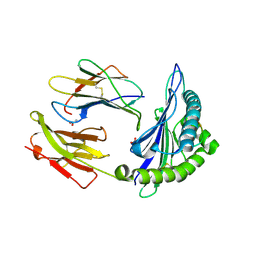
1ZTD
 | | Hypothetical Protein Pfu-631545-001 From Pyrococcus furiosus | | 分子名称: | Hypothetical Protein Pfu-631545-001 | | 著者 | Fu, Z.-Q, Horanyi, P, Florence, Q, Liu, Z.-J, Chen, L, Lee, D, Habel, J, Xu, H, Nguyen, D, Chang, S.-H, Zhou, W, Zhang, H, Jenney Jr, F.E, Sha, B, Adams, M.W.W, Rose, J.P, Wang, B.-C, Southeast Collaboratory for Structural Genomics (SECSG) | | 登録日 | 2005-05-26 | | 公開日 | 2005-06-07 | | 最終更新日 | 2024-02-14 | | 実験手法 | X-RAY DIFFRACTION (2 Å) | | 主引用文献 | Hypothetical Protein Pfu-631545-001 From Pyrococcus furiosus
To be Published
|
|
1ZSD
 | | Crystal Structure Of HLA-B*3501 Presenting an 11-Mer EBV Antigen EPLPQGQLTAY | | 分子名称: | BZLF1 trans-activator protein, Beta-2-microglobulin, HLA class I histocompatibility antigen, ... | | 著者 | Miles, J.J, Elhassen, D, Borg, N.A, Silins, S.L, Tynan, F.E, Burrows, J.M, Purcell, A.W, Kjer-Nielsen, L, Rossjohn, J, Burrows, S.R, McCluskey, J. | | 登録日 | 2005-05-24 | | 公開日 | 2005-06-07 | | 最終更新日 | 2024-10-09 | | 実験手法 | X-RAY DIFFRACTION (1.7 Å) | | 主引用文献 | CTL Recognition of a Bulged Viral Peptide Involves Biased TCR Selection.
J.Immunol., 175, 2005
|
|


