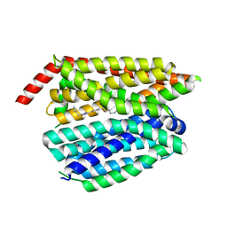
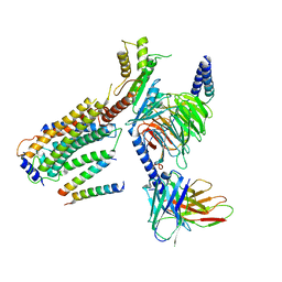
2PZP
 
 | |
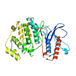
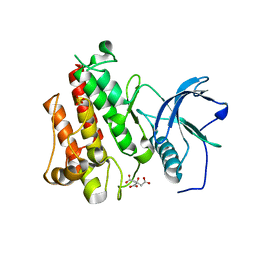
4IQ2
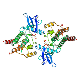
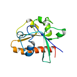
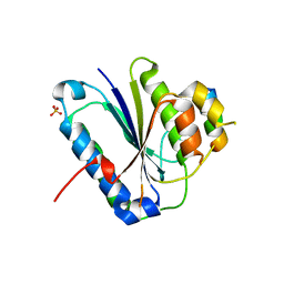
 | | P21 crystal form of FKBP12.6 | | 分子名称: | MALONIC ACID, Peptidyl-prolyl cis-trans isomerase FKBP1B | | 著者 | Chen, H, Mustafi, S.M, Li, H.M, LeMaster, D.M, Hernandez, G. | | 登録日 | 2013-01-10 | | 公開日 | 2014-01-15 | | 最終更新日 | 2023-09-20 | | 実験手法 | X-RAY DIFFRACTION (1.7 Å) | | 主引用文献 | Crystal structure and conformational flexibility of the unligated FK506-binding protein FKBP12.6.
Acta Crystallogr.,Sect.D, 70, 2014
|
|
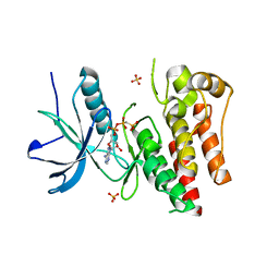
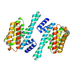
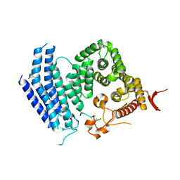
4IQC
 | | P3121 crystal form of FKBP12.6 | | 分子名称: | Peptidyl-prolyl cis-trans isomerase FKBP1B | | 著者 | Chen, H, Mustafi, S.M, Li, H.M, LeMaster, D.M, Hernandez, G. | | 登録日 | 2013-01-11 | | 公開日 | 2014-01-15 | | 最終更新日 | 2023-09-20 | | 実験手法 | X-RAY DIFFRACTION (1.903 Å) | | 主引用文献 | Crystal structure and conformational flexibility of the unligated FK506-binding protein FKBP12.6.
Acta Crystallogr.,Sect.D, 70, 2014
|
|
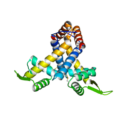
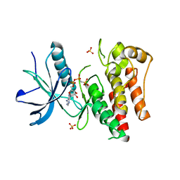
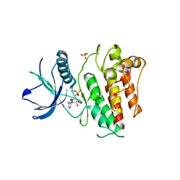
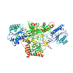
3CLY
 | |
3E17
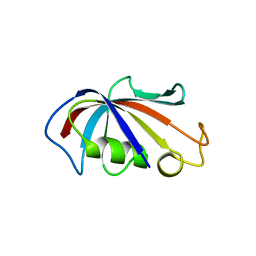
 | | Crystal structure of the second PDZ domain from human Zona Occludens-2 | | 分子名称: | Tight junction protein ZO-2 | | 著者 | Chen, H, Tong, S.L, Teng, M.K, Niu, L.W. | | 登録日 | 2008-08-01 | | 公開日 | 2009-07-21 | | 最終更新日 | 2023-11-01 | | 実験手法 | X-RAY DIFFRACTION (1.75 Å) | | 主引用文献 | Structure of the second PDZ domain from human zonula occludens 2
Acta Crystallogr.,Sect.F, 65, 2009
|
|
4DM4
 | | The conserved domain of yeast Cdc73 | | 分子名称: | Cell division control protein 73 | | 著者 | Chen, H, Shi, N, Gao, Y, Li, X, Niu, L, Teng, M. | | 登録日 | 2012-02-06 | | 公開日 | 2012-08-22 | | 最終更新日 | 2024-03-20 | | 実験手法 | X-RAY DIFFRACTION (2.19 Å) | | 主引用文献 | Crystallographic analysis of the conserved C-terminal domain of transcription factor Cdc73 from Saccharomyces cerevisiae reveals a GTPase-like fold.
Acta Crystallogr.,Sect.D, 68, 2012
|
|
3MEX
 | | Crystal structure of MexR in oxidized state | | 分子名称: | Multidrug resistance operon repressor | | 著者 | Chen, H, Yi, C, Zhang, J, Zhang, W, Yang, C.-G, He, C. | | 登録日 | 2010-04-01 | | 公開日 | 2010-07-28 | | 最終更新日 | 2023-11-01 | | 実験手法 | X-RAY DIFFRACTION (2.1 Å) | | 主引用文献 | Structural insight into the oxidation-sensing mechanism of the antibiotic resistance of regulator MexR
Embo Rep., 11, 2010
|
|
8G92
 | | Structure of inhibitor 16d-bound SPNS2 | | 分子名称: | 3-[3-(4-decylphenyl)-1,2,4-oxadiazol-5-yl]propan-1-amine, Sphingosine-1-phosphate transporter SPNS2 | | 著者 | Chen, H, Li, X. | | 登録日 | 2023-02-21 | | 公開日 | 2023-05-24 | | 最終更新日 | 2023-12-13 | | 実験手法 | ELECTRON MICROSCOPY (3.6 Å) | | 主引用文献 | Structural and functional insights into Spns2-mediated transport of sphingosine-1-phosphate.
Cell, 186, 2023
|
|
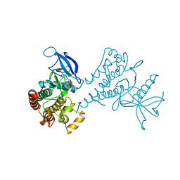
6AGK
 | | The structure of CH-II-77-tubulin complex | | 分子名称: | 2-(N-MORPHOLINO)-ETHANESULFONIC ACID, CALCIUM ION, GUANOSINE-5'-DIPHOSPHATE, ... | | 著者 | Chen, H, Arnst, K, Wang, Y, Miller, D, Li, W. | | 登録日 | 2018-08-13 | | 公開日 | 2019-08-21 | | 最終更新日 | 2023-11-22 | | 実験手法 | X-RAY DIFFRACTION (2.8 Å) | | 主引用文献 | Structure-Activity Relationship Study of Novel 6-Aryl-2-benzoyl-pyridines as Tubulin Polymerization Inhibitors with Potent Antiproliferative Properties.
J.Med.Chem., 63, 2020
|
|
5BYZ
 | | ERK5 in complex with small molecule | | 分子名称: | 4-({5-fluoro-4-[2-methyl-1-(propan-2-yl)-1H-imidazol-5-yl]pyrimidin-2-yl}amino)-N-[2-(piperidin-1-yl)ethyl]benzamide, GLYCEROL, Mitogen-activated protein kinase 7 | | 著者 | Chen, H, Tucker, J, Wang, X, Gavine, P.R, Philips, C, Augustin, M.A, Schreiner, P, Steinbacher, S, Preston, M, Ogg, D. | | 登録日 | 2015-06-11 | | 公開日 | 2016-05-04 | | 最終更新日 | 2024-05-08 | | 実験手法 | X-RAY DIFFRACTION (1.65 Å) | | 主引用文献 | Discovery of a novel allosteric inhibitor-binding site in ERK5: comparison with the canonical kinase hinge ATP-binding site.
Acta Crystallogr D Struct Biol, 72, 2016
|
|
5BYY
 | | ERK5 IN COMPLEX WITH SMALL MOLECULE | | 分子名称: | 2-{[2-ethoxy-4-(4-hydroxypiperidin-1-yl)phenyl]amino}-5,11-dimethyl-5,11-dihydro-6H-pyrimido[4,5-b][1,4]benzodiazepin-6-one, Mitogen-activated protein kinase 7 | | 著者 | Chen, H, Tucker, J, Wang, X, Gavine, P.R, Philips, C, Augustin, M.A, Schreiner, P, Steinbacher, S, Preston, M, Ogg, D. | | 登録日 | 2015-06-11 | | 公開日 | 2016-05-04 | | 最終更新日 | 2024-05-08 | | 実験手法 | X-RAY DIFFRACTION (2.79 Å) | | 主引用文献 | Discovery of a novel allosteric inhibitor-binding site in ERK5: comparison with the canonical kinase hinge ATP-binding site.
Acta Crystallogr D Struct Biol, 72, 2016
|
|
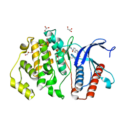
4J99
 | |
4J95
 | |
4J97
 | |
4J96
 | |
7D9V
 | |
7D8P
 | |
7D8H
 | |
8G94
 | | Structure of CD69-bound S1PR1 coupled to heterotrimeric Gi | | 分子名称: | Early activation antigen CD69, Guanine nucleotide-binding protein G(I)/G(S)/G(O) subunit gamma-2, Guanine nucleotide-binding protein G(I)/G(S)/G(T) subunit beta-1, ... | | 著者 | Chen, H, Li, X. | | 登録日 | 2023-02-21 | | 公開日 | 2023-04-19 | | 最終更新日 | 2023-04-26 | | 実験手法 | ELECTRON MICROSCOPY (3.15 Å) | | 主引用文献 | Transmembrane protein CD69 acts as an S1PR1 agonist.
Elife, 12, 2023
|
|
5Y59
 | | Crystal structure of Ku80 and Sir4 | | 分子名称: | ATP-dependent DNA helicase II subunit 2, SULFATE ION, Sir4p | | 著者 | Chen, H, Xue, J, Wu, J, Lei, M. | | 登録日 | 2017-08-08 | | 公開日 | 2017-12-20 | | 最終更新日 | 2024-03-27 | | 実験手法 | X-RAY DIFFRACTION (2.402 Å) | | 主引用文献 | Structural Insights into Yeast Telomerase Recruitment to Telomeres
Cell, 172, 2018
|
|
5Y5A
 | | Crystal structure of Est1 and Cdc13 | | 分子名称: | KLLA0F20702p, KLLA0F20922p | | 著者 | Chen, H, Xue, J, Wu, J, Lei, M. | | 登録日 | 2017-08-08 | | 公開日 | 2017-12-20 | | 最終更新日 | 2024-03-27 | | 実験手法 | X-RAY DIFFRACTION (2.206 Å) | | 主引用文献 | Structural Insights into Yeast Telomerase Recruitment to Telomeres
Cell, 172, 2018
|
|
5Y58
 | | Crystal structure of Ku70/80 and TLC1 | | 分子名称: | ATP-dependent DNA helicase II subunit 1, ATP-dependent DNA helicase II subunit 2, TLC1 | | 著者 | Chen, H, Xue, J, Wu, J, Lei, M. | | 登録日 | 2017-08-08 | | 公開日 | 2017-12-20 | | 最終更新日 | 2024-03-27 | | 実験手法 | X-RAY DIFFRACTION (2.8 Å) | | 主引用文献 | Structural Insights into Yeast Telomerase Recruitment to Telomeres
Cell, 172, 2018
|
|
7TUZ
 | | Cryo-EM structure of 7alpha,25-dihydroxycholesterol-bound EBI2/GPR183 in complex with Gi protein | | 分子名称: | (2S,4aS,4bS,7R,8S,8aS,9R,10aR)-7-[(2R,3R)-7-hydroxy-3,7-dimethyloctan-2-yl]-4a,7,8-trimethyltetradecahydrophenanthrene-2,9-diol, G-protein coupled receptor 183, Guanine nucleotide-binding protein G(I)/G(S)/G(O) subunit gamma-2, ... | | 著者 | Chen, H, Hung, W, Li, X. | | 登録日 | 2022-02-03 | | 公開日 | 2022-04-13 | | 最終更新日 | 2022-07-20 | | 実験手法 | ELECTRON MICROSCOPY (3.12 Å) | | 主引用文献 | Structures of oxysterol sensor EBI2/GPR183, a key regulator of the immune response.
Structure, 30, 2022
|
|
7TUY
 | | Cryo-EM structure of GSK682753A-bound EBI2/GPR183 | | 分子名称: | 8-[(2E)-3-(4-chlorophenyl)prop-2-enoyl]-3-[(3,4-dichlorophenyl)methyl]-1-oxa-3,8-diazaspiro[4.5]decan-2-one, G-protein coupled receptor 183,Soluble cytochrome b562 fusion, anti-BRIL Fab Heavy chain, ... | | 著者 | Chen, H, Huang, W, Li, X. | | 登録日 | 2022-02-03 | | 公開日 | 2022-04-13 | | 最終更新日 | 2022-07-20 | | 実験手法 | ELECTRON MICROSCOPY (2.98 Å) | | 主引用文献 | Structures of oxysterol sensor EBI2/GPR183, a key regulator of the immune response.
Structure, 30, 2022
|
|
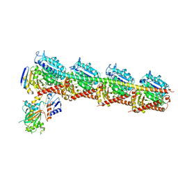
7D7X
 | |


