6N1I
 
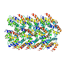
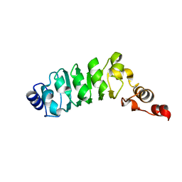
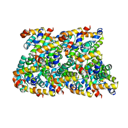 | | Cryo-EM structure of NLRC4-CARD filament | | 分子名称: | NLR family CARD domain-containing protein 4 | | 著者 | Li, Y, Fu, T, Wu, H. | | 登録日 | 2018-11-08 | | 公開日 | 2018-12-05 | | 最終更新日 | 2024-03-13 | | 実験手法 | ELECTRON MICROSCOPY (3.58 Å) | | 主引用文献 | Cryo-EM structures of ASC and NLRC4 CARD filaments reveal a unified mechanism of nucleation and activation of caspase-1.
Proc. Natl. Acad. Sci. U.S.A., 115, 2018
|
|
6MK7
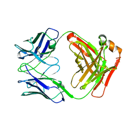
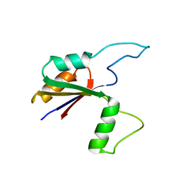 | | Solution structure of the large extracellular loop of FtsX in Streptococcus pneumoniae | | 分子名称: | Cell division protein FtsX | | 著者 | Edmonds, K.A, Fu, Y, Wu, H, Rued, B.E, Bruce, K.E, Winkler, M.E, Giedroc, D.P. | | 登録日 | 2018-09-25 | | 公開日 | 2019-02-13 | | 最終更新日 | 2024-05-15 | | 実験手法 | SOLUTION NMR | | 主引用文献 | Structure of the Large Extracellular Loop of FtsX and Its Interaction with the Essential Peptidoglycan Hydrolase PcsB in Streptococcus pneumoniae.
MBio, 10, 2019
|
|
6NDJ
 | |
6MIZ
 | |
6NCV
 | | Cryo-EM structure of NLRP6 PYD filament | | 分子名称: | NACHT, LRR and PYD domains-containing protein 6 | | 著者 | Shen, C, Fu, T.M, Wu, H. | | 登録日 | 2018-12-12 | | 公開日 | 2019-01-23 | | 最終更新日 | 2024-03-20 | | 実験手法 | ELECTRON MICROSCOPY (3.7 Å) | | 主引用文献 | Molecular mechanism for NLRP6 inflammasome assembly and activation.
Proc. Natl. Acad. Sci. U.S.A., 116, 2019
|
|
6MB2
 | |
6MIX
 | |
7DWO
 | |
7DWN
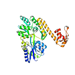
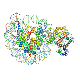
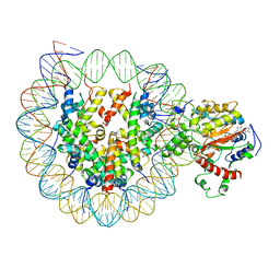
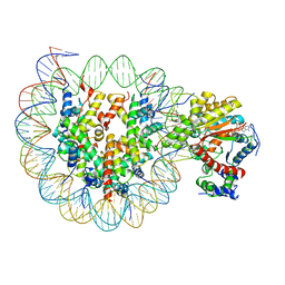
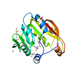
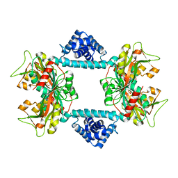 | | Crystal structure of Vibrio fischeri DarR in complex with DNA reveals the transcriptional activation mechanism of LTTR family members | | 分子名称: | 2-(N-MORPHOLINO)-ETHANESULFONIC ACID, Predicted DNA-binding transcriptional regulator | | 著者 | Wang, W.W, Wu, H, He, J.H, Yu, F. | | 登録日 | 2021-01-17 | | 公開日 | 2021-07-07 | | 最終更新日 | 2023-11-29 | | 実験手法 | X-RAY DIFFRACTION (2.32 Å) | | 主引用文献 | Crystal structure details of Vibrio fischeri DarR and mutant DarR-M202I from LTTR family reveals their activation mechanism.
Int.J.Biol.Macromol., 183, 2021
|
|
6N1H
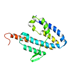
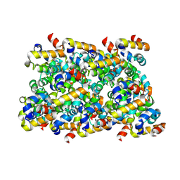 | | Cryo-EM structure of ASC-CARD filament | | 分子名称: | Apoptosis-associated speck-like protein containing a CARD | | 著者 | Li, Y, Fu, T, Wu, H. | | 登録日 | 2018-11-08 | | 公開日 | 2018-12-05 | | 最終更新日 | 2024-03-13 | | 実験手法 | ELECTRON MICROSCOPY (3.17 Å) | | 主引用文献 | Cryo-EM structures of ASC and NLRC4 CARD filaments reveal a unified mechanism of nucleation and activation of caspase-1.
Proc. Natl. Acad. Sci. U.S.A., 115, 2018
|
|
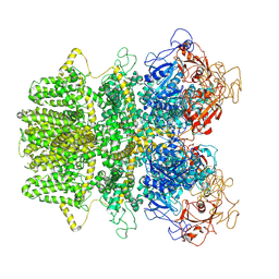
6IRO
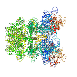
 | | the crosslinked complex of ISWI-nucleosome in the ADP-bound state | | 分子名称: | ADENOSINE-5'-DIPHOSPHATE, DNA (167-MER), Histone H2A, ... | | 著者 | Yan, L.J, Wu, H, Li, X.M, Gao, N, Chen, Z.C. | | 登録日 | 2018-11-13 | | 公開日 | 2019-04-03 | | 最終更新日 | 2024-03-27 | | 実験手法 | ELECTRON MICROSCOPY (3.4 Å) | | 主引用文献 | Structures of the ISWI-nucleosome complex reveal a conserved mechanism of chromatin remodeling.
Nat. Struct. Mol. Biol., 26, 2019
|
|
6K1P
 | | The complex of ISWI-nucleosome in the ADP.BeF-bound state | | 分子名称: | ADENOSINE-5'-DIPHOSPHATE, BERYLLIUM TRIFLUORIDE ION, DNA (167-MER), ... | | 著者 | Yan, L.J, Wu, H, Li, X.M, Gao, N, Chen, Z.C. | | 登録日 | 2019-05-10 | | 公開日 | 2019-05-29 | | 最終更新日 | 2024-03-27 | | 実験手法 | ELECTRON MICROSCOPY (3.87 Å) | | 主引用文献 | Structures of the ISWI-nucleosome complex reveal a conserved mechanism of chromatin remodeling.
Nat.Struct.Mol.Biol., 26, 2019
|
|
6JYL
 | | The crosslinked complex of ISWI-nucleosome in the ADP.BeF-bound state | | 分子名称: | ADENOSINE-5'-DIPHOSPHATE, BERYLLIUM TRIFLUORIDE ION, DNA (167-MER), ... | | 著者 | Yan, L.J, Wu, H, Li, X.M, Gao, N, Chen, Z.C. | | 登録日 | 2019-04-26 | | 公開日 | 2019-05-29 | | 最終更新日 | 2024-03-27 | | 実験手法 | ELECTRON MICROSCOPY (3.37 Å) | | 主引用文献 | Structures of the ISWI-nucleosome complex reveal a conserved mechanism of chromatin remodeling.
Nat.Struct.Mol.Biol., 26, 2019
|
|
5XF8
 | | Cryo-EM structure of the Cdt1-MCM2-7 complex in AMPPNP state | | 分子名称: | Cell division cycle protein CDT1, DNA replication licensing factor MCM2, DNA replication licensing factor MCM3, ... | | 著者 | Zhai, Y, Cheng, E, Wu, H, Li, N, Yung, P.Y, Gao, N, Tye, B.K. | | 登録日 | 2017-04-09 | | 公開日 | 2017-05-03 | | 実験手法 | ELECTRON MICROSCOPY (7.1 Å) | | 主引用文献 | Open-ringed structure of the Cdt1-Mcm2-7 complex as a precursor of the MCM double hexamer
Nat. Struct. Mol. Biol., 24, 2017
|
|
5ZCK
 | | Structure of the RIP3 core region | | 分子名称: | SODIUM ION, peptide from Receptor-interacting serine/threonine-protein kinase 3 | | 著者 | Li, J, Wu, H. | | 登録日 | 2018-02-18 | | 公開日 | 2018-04-18 | | 最終更新日 | 2023-11-22 | | 実験手法 | X-RAY DIFFRACTION (1.271 Å) | | 主引用文献 | The Structure of the Necrosome RIPK1-RIPK3 Core, a Human Hetero-Amyloid Signaling Complex.
Cell, 173, 2018
|
|
4ZSE
 | |
4WD5
 | |
1DS9
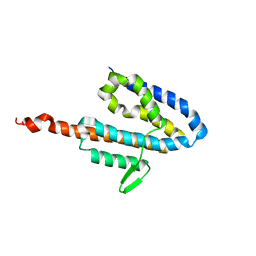
 | | SOLUTION STRUCTURE OF CHLAMYDOMONAS OUTER ARM DYNEIN LIGHT CHAIN 1 | | 分子名称: | OUTER ARM DYNEIN | | 著者 | Wu, H.W, Maciejewski, M.W, Marintchev, A, Benashski, S.E, Mullen, G.P, King, S.M. | | 登録日 | 2000-01-07 | | 公開日 | 2000-07-26 | | 最終更新日 | 2024-05-22 | | 実験手法 | SOLUTION NMR | | 主引用文献 | Solution structure of a dynein motor domain associated light chain.
Nat.Struct.Biol., 7, 2000
|
|
1M9L
 | | Relaxation-based Refined Structure Of Chlamydomonas Outer Arm Dynein Light Chain 1 | | 分子名称: | Outer Arm Dynein Light Chain 1 | | 著者 | Wu, H.W, Maciejewski, M.W, Marintchev, A, Benashski, S.E, Mullen, G.P, King, S.M. | | 登録日 | 2002-07-29 | | 公開日 | 2003-03-04 | | 最終更新日 | 2024-05-22 | | 実験手法 | SOLUTION NMR | | 主引用文献 | Relaxation-based structure refinement and backbone molecular dynamics of the
Dynein motor domain-associated light chain
Biochemistry, 42, 2003
|
|
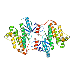
2H11
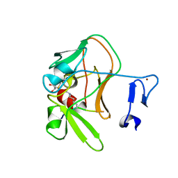
 | | Amino-terminal Truncated Thiopurine S-Methyltransferase Complexed with S-Adenosyl-L-Homocysteine | | 分子名称: | 2-[3-(2-HYDROXY-1,1-DIHYDROXYMETHYL-ETHYLAMINO)-PROPYLAMINO]-2-HYDROXYMETHYL-PROPANE-1,3-DIOL, S-ADENOSYL-L-HOMOCYSTEINE, THIOCYANATE ION, ... | | 著者 | Horton, J.R, Cheng, X. | | 登録日 | 2006-05-15 | | 公開日 | 2006-11-21 | | 最終更新日 | 2023-08-30 | | 実験手法 | X-RAY DIFFRACTION (1.89 Å) | | 主引用文献 | Structural basis of allele variation of human thiopurine-S-methyltransferase.
Proteins, 67, 2007
|
|
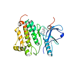
4C1Q
 | | Crystal structure of the PRDM9 SET domain in complex with H3K4me2 and AdoHcy. | | 分子名称: | GLYCEROL, HISTONE H3.1, HISTONE-LYSINE N-METHYLTRANSFERASE PRDM9, ... | | 著者 | Mathioudakis, N, Cusack, S, Kadlec, J. | | 登録日 | 2013-08-13 | | 公開日 | 2013-10-16 | | 最終更新日 | 2023-12-20 | | 実験手法 | X-RAY DIFFRACTION (2.3 Å) | | 主引用文献 | Molecular Basis for the Regulation of the H3K4 Methyltransferase Activity of Prdm9.
Cell Rep., 5, 2013
|
|
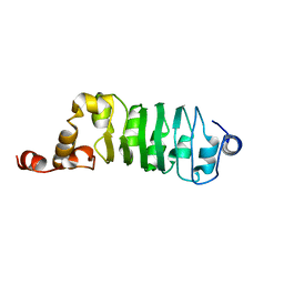
2JOL
 | | Average NMR structure of the catalytic domain of guanine nucleotide exchange factor BopE from Burkholderia pseudomallei | | 分子名称: | Putative G-nucleotide exchange factor | | 著者 | Wu, H, Upadhyay, A, Williams, C, Galyov, E.E, van den Elsen, J.M.H, Bagby, S. | | 登録日 | 2007-03-14 | | 公開日 | 2007-03-27 | | 最終更新日 | 2023-12-20 | | 実験手法 | SOLUTION NMR | | 主引用文献 | The guanine-nucleotide-exchange factor BopE from Burkholderia pseudomallei adopts a compact version of the Salmonella SopE/SopE2 fold and undergoes a closed-to-open conformational change upon interaction with Cdc42
Biochem.J., 411, 2008
|
|
2JOK
 | | NMR structure of the catalytic domain of guanine nucleotide exchange factor BopE from Burkholderia pseudomallei | | 分子名称: | Putative G-nucleotide exchange factor | | 著者 | Wu, H, Upadhyay, A, Williams, C, Galyov, E.E, van den Elsen, J.M.H, Bagby, S. | | 登録日 | 2007-03-14 | | 公開日 | 2007-09-18 | | 最終更新日 | 2023-12-20 | | 実験手法 | SOLUTION NMR | | 主引用文献 | The guanine-nucleotide-exchange factor BopE from Burkholderia pseudomallei adopts a compact version of the Salmonella SopE/SopE2 fold and undergoes a closed-to-open conformational change upon interaction with Cdc42
Biochem.J., 411, 2008
|
|
4UB0
 | |
5KSI
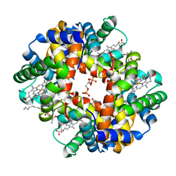 | | Crystal structure of deoxygenated hemoglobin in complex with sphingosine phosphate and 2,3-Bisphosphoglycerate | | 分子名称: | (2R)-2,3-diphosphoglyceric acid, (2S,3R,4E)-2-amino-3-hydroxyoctadec-4-en-1-yl dihydrogen phosphate, Hemoglobin subunit alpha, ... | | 著者 | Ahmed, M.H, Safo, M.K, Xia, Y. | | 登録日 | 2016-07-08 | | 公開日 | 2017-07-26 | | 最終更新日 | 2023-10-04 | | 実験手法 | X-RAY DIFFRACTION (1.8 Å) | | 主引用文献 | Structural and Functional Insight of Sphingosine 1-Phosphate-Mediated Pathogenic Metabolic Reprogramming in Sickle Cell Disease.
Sci Rep, 7, 2017
|
|


