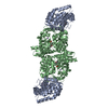
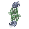
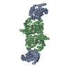
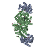
- PDB-6o1h: Tryptophan synthase Q114A mutant in complex with N-(4'-trifluorom... -
+
Open data
ID or keywords:
Loading...
-
Basic information
Entry
Database: PDB / ID: 6o1h
Title
Tryptophan synthase Q114A mutant in complex with N-(4'-trifluoromethoxybenzenesulfonyl)-2-amino-1-ethylphosphate (F9F) at the enzyme alpha-site, cesium ion at the metal coordination site, and 2-aminophenol quinonoid at the enzyme beta site
National Institutes of Health/National Human Genome Research Institute (NIH/NHGRI)
2R01GM097569-06A1
United States
Citation
Journal: To be Published Title: Crystal structure of Salmonella typhimurium Tryptophan Synthase mutant beta-Q114A with 2-({[4-(trifluoromethoxy)phenyl]sulfonyl}amino)ethyl dihydrogen phosphate (F9F) at the alpha-site, Cesium ...Title: Crystal structure of Salmonella typhimurium Tryptophan Synthase mutant beta-Q114A with 2-({[4-(trifluoromethoxy)phenyl]sulfonyl}amino)ethyl dihydrogen phosphate (F9F) at the alpha-site, Cesium ion at the metal coordination site, and [3-hydroxy-2-methyl-5-phosphonooxymethyl-pyridin-4-ylmethyl]-serine (PLS) at the beta-site. Authors: Hilario, E. / Dunn, M.F. / Mueller, L.J. / Fan, L.
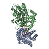
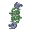
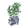
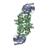
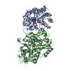
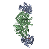
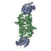
Evidence: gel filtration, To verify the oligomeric size of Tryptophan synthase (TS) mutant beta-Q114A in buffered solution, we have used a HiPrep 16/60 Sephacryl S-100 High Resolution column (GE ...Evidence: gel filtration, To verify the oligomeric size of Tryptophan synthase (TS) mutant beta-Q114A in buffered solution, we have used a HiPrep 16/60 Sephacryl S-100 High Resolution column (GE Healthcare) in 50mM bicine:CsCl, pH 7.80, containing 50mM CsCl and 0.01mM PLP at 10 degrees Celsius. 1mg of pure TS mutant beta-Q114A was loaded at a flowrate of 30mL/h and 2mL fraction size. Marker proteins were used for column calibration: ferritin (440 kDa), aldolase (158 kDa), conalbumin (75 kDa), and lactalbumin (14 kDa). The effluent was monitored by absorbance at 280nm and the protein sample from the peak fraction was loaded in a 15% SDS-PAGE gel. Tryptophan synthase is a tetrameter composed of two alpha-chains and two beta-chains (Alpha2-Beta2).
In the structure databanks used in Yorodumi, some data are registered as the other names, "COVID-19 virus" and "2019-nCoV". Here are the details of the virus and the list of structure data.
Jan 31, 2019. EMDB accession codes are about to change! (news from PDBe EMDB page)
EMDB accession codes are about to change! (news from PDBe EMDB page)
The allocation of 4 digits for EMDB accession codes will soon come to an end. Whilst these codes will remain in use, new EMDB accession codes will include an additional digit and will expand incrementally as the available range of codes is exhausted. The current 4-digit format prefixed with “EMD-” (i.e. EMD-XXXX) will advance to a 5-digit format (i.e. EMD-XXXXX), and so on. It is currently estimated that the 4-digit codes will be depleted around Spring 2019, at which point the 5-digit format will come into force.
The EM Navigator/Yorodumi systems omit the EMD- prefix.
Related info.:Q: What is EMD? / ID/Accession-code notation in Yorodumi/EM Navigator
Yorodumi is a browser for structure data from EMDB, PDB, SASBDB, etc.
This page is also the successor to EM Navigator detail page, and also detail information page/front-end page for Omokage search.
The word "yorodu" (or yorozu) is an old Japanese word meaning "ten thousand". "mi" (miru) is to see.
Related info.:EMDB / PDB / SASBDB / Comparison of 3 databanks / Yorodumi Search / Aug 31, 2016. New EM Navigator & Yorodumi / Yorodumi Papers / Jmol/JSmol / Function and homology information / Changes in new EM Navigator and Yorodumi
 Movie
Movie Controller

 Yorodumi
 Open data
 Basic information
 Components
 Keywords
 Function and homology information
 Salmonella typhimurium (bacteria)
 X-RAY DIFFRACTION /
  Authors
 United States, 1items
  Citation
 Structure visualization
 Downloads & links
 Other downloads


 PDBj
PDBj
 Assembly
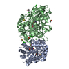


 Mass: 78.133 Da / Num. of mol.: 8 / Source method: obtained synthetically / Formula: C2H6OS / Comment: DMSO, precipitant*YM
 Mass: 365.220 Da / Num. of mol.: 1 / Source method: obtained synthetically / Formula: C9H11F3NO7PS
 Mass: 132.905 Da / Num. of mol.: 3 / Source method: obtained synthetically / Formula: Cs
 Mass: 35.453 Da / Num. of mol.: 16 / Source method: obtained synthetically / Formula: Cl
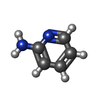
 Mass: 94.115 Da / Num. of mol.: 1 / Source method: obtained synthetically / Formula: C5H6N2
 Mass: 425.330 Da / Num. of mol.: 1 / Source method: obtained synthetically / Formula: C17H20N3O8P
 Sample preparation
 Processing