 Movie
Movie Controller

[English] 日本語
 Yorodumi
- PDB-4hn4: Tryptophan synthase in complex with alpha aminoacrylate E(A-A) fo... -
+ Open data

- Basic information
| Entry | Database: PDB / ID: 4hn4 | ||||||
|---|---|---|---|---|---|---|---|
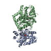
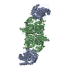
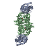
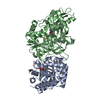
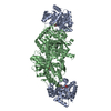
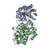
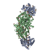
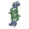
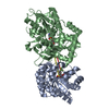
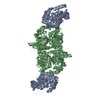
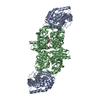
| Title | Tryptophan synthase in complex with alpha aminoacrylate E(A-A) form and the F9 inhibitor in the alpha site | ||||||
 Components Components | (Tryptophan synthase ...) x 2 | ||||||
 Keywords Keywords | LYASE/LYASE INHIBITOR / Lyase / carbon-oxygen lyase / tryptophan biosynthesis / Salmonella / F9F / Allosteric enzyme / Amino-acid biosynthesis / Aromatic amino acid biosynthesis / Pyridoxal phosphate / alpha amino acrylate / LYASE-LYASE INHIBITOR complex | ||||||
| Function / homology |  Function and homology information Function and homology informationtryptophan synthase / tryptophan synthase activity / L-tryptophan biosynthetic process / identical protein binding / cytoplasm / cytosol Similarity search - Function | ||||||
| Biological species |  Salmonella enterica subsp. enterica serovar Typhimurium (bacteria) Salmonella enterica subsp. enterica serovar Typhimurium (bacteria) | ||||||
| Method |  X-RAY DIFFRACTION / MOLECULAR REPLACEMENT / molecular replacement / Resolution: 1.64 Å X-RAY DIFFRACTION / MOLECULAR REPLACEMENT / molecular replacement / Resolution: 1.64 Å | ||||||
 Authors Authors | Hilario, E. / Niks, D. / Dunn, M.F. / Mueller, L.J. / Fan, L. | ||||||
 Citation Citation | Journal: Biochemistry / Year: 2013 Title: Allostery and substrate channeling in the tryptophan synthase bienzyme complex: evidence for two subunit conformations and four quaternary states. Authors: Niks, D. / Hilario, E. / Dierkers, A. / Ngo, H. / Borchardt, D. / Neubauer, T.J. / Fan, L. / Mueller, L.J. / Dunn, M.F. | ||||||
| History |
|
- Structure visualization
| Structure viewer | Molecule: MolmilJmol/JSmol |
|---|
- Downloads & links
-Download
| PDBx/mmCIF format | 4hn4.cif.gz | 304.3 KB | Display | PDBx/mmCIF format |
|---|---|---|---|---|
| PDB format | pdb4hn4.ent.gz | 244.3 KB | Display | PDB format |
| PDBx/mmJSON format | 4hn4.json.gz | Tree view | PDBx/mmJSON format | |
| Others |  Other downloads Other downloads |
-Validation report
| Arichive directory | https://data.pdbj.org/pub/pdb/validation_reports/hn/4hn4ftp://data.pdbj.org/pub/pdb/validation_reports/hn/4hn4 | HTTPS FTP |
|---|
-Related structure data
| Related structure data |  4hpjC  4hpxC  4ht3C  4kkxC 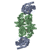 1tjpS C: citing same article ( S: Starting model for refinement |
|---|---|
| Similar structure data |


-Links
 PDBj
PDBj
- Assembly
| Deposited unit | 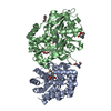
| |||||||||
|---|---|---|---|---|---|---|---|---|---|---|
| 1 | 
| |||||||||
| Unit cell |
| |||||||||
| Components on special symmetry positions |
|
-Components
-Tryptophan synthase ... , 2 types, 2 molecules AB
| #1: Protein | Mass: 28698.797 Da / Num. of mol.: 1 Source method: isolated from a genetically manipulated source Details: Yang et al (1996) Protein Expression and Purification, 8:126-136. Source: (gene. exp.) Salmonella enterica subsp. enterica serovar Typhimurium (bacteria)Gene: STM1727, trpA, trpA and trpB / Plasmid: pEBA-10 / Production host: |
|---|---|
| #2: Protein | Mass: 42918.879 Da / Num. of mol.: 1 / Source method: isolated from a natural source Source: (natural) Salmonella enterica subsp. enterica serovar Typhimurium (bacteria)References: UniProt: P0A2K1, tryptophan synthase |
-Non-polymers , 6 types, 686 molecules 
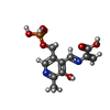


| #3: Chemical | ChemComp-F9F /  Mass: 365.220 Da / Num. of mol.: 1 / Source method: obtained synthetically / Formula: C9H11F3NO7PS Mass: 365.220 Da / Num. of mol.: 1 / Source method: obtained synthetically / Formula: C9H11F3NO7PS | ||||||
|---|---|---|---|---|---|---|---|
| #4: Chemical | ChemComp-0JO /  Mass: 316.204 Da / Num. of mol.: 1 / Source method: obtained synthetically / Formula: C11H13N2O7P Mass: 316.204 Da / Num. of mol.: 1 / Source method: obtained synthetically / Formula: C11H13N2O7P | ||||||
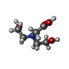
| #5: Chemical |  Mass: 106.120 Da / Num. of mol.: 3 / Source method: obtained synthetically / Formula: C4H10O3 Mass: 106.120 Da / Num. of mol.: 3 / Source method: obtained synthetically / Formula: C4H10O3#6: Chemical |  Mass: 163.172 Da / Num. of mol.: 3 / Source method: obtained synthetically / Formula: C6H13NO4 / Comment: pH buffer*YM Mass: 163.172 Da / Num. of mol.: 3 / Source method: obtained synthetically / Formula: C6H13NO4 / Comment: pH buffer*YM#7: Chemical |  Mass: 132.905 Da / Num. of mol.: 2 / Source method: obtained synthetically / Formula: Cs Mass: 132.905 Da / Num. of mol.: 2 / Source method: obtained synthetically / Formula: Cs#8: Water | ChemComp-HOH / | Mass: 18.015 Da / Num. of mol.: 676 / Source method: isolated from a natural source / Formula: H2O |
-Experimental details
-Experiment
| Experiment | Method: X-RAY DIFFRACTION / Number of used crystals: 1 |
|---|
- Sample preparation
| Crystal | Density Matthews: 2.58 Å3/Da / Density % sol: 52.26 % |
|---|---|
| Crystal grow | Temperature: 298 K / Method: vapor diffusion, hanging drop / pH: 7.8 Details: 50 mM Bicine-CsOH, 10% PEG 8000, 2 mM Spermine, pH 7.8, VAPOR DIFFUSION, HANGING DROP, temperature 298K |
-Data collection
| Diffraction | Mean temperature: 100 K | |||||||||||||||||||||||||||||||||||||||||||||||||||||||||||||||||||||||||||||
|---|---|---|---|---|---|---|---|---|---|---|---|---|---|---|---|---|---|---|---|---|---|---|---|---|---|---|---|---|---|---|---|---|---|---|---|---|---|---|---|---|---|---|---|---|---|---|---|---|---|---|---|---|---|---|---|---|---|---|---|---|---|---|---|---|---|---|---|---|---|---|---|---|---|---|---|---|---|---|
| Diffraction source | Source: ROTATING ANODE / Type: RIGAKU MICROMAX-007 HF / Wavelength: 1.5418 Å | |||||||||||||||||||||||||||||||||||||||||||||||||||||||||||||||||||||||||||||
| Detector | Type: RIGAKU RAXIS IV++ / Detector: IMAGE PLATE / Date: Jan 1, 2012 / Details: VariMax HF | |||||||||||||||||||||||||||||||||||||||||||||||||||||||||||||||||||||||||||||
| Radiation | Protocol: SINGLE WAVELENGTH / Monochromatic (M) / Laue (L): M / Scattering type: x-ray | |||||||||||||||||||||||||||||||||||||||||||||||||||||||||||||||||||||||||||||
| Radiation wavelength | Wavelength: 1.5418 Å / Relative weight: 1 | |||||||||||||||||||||||||||||||||||||||||||||||||||||||||||||||||||||||||||||
| Reflection | Redundancy: 3.3 % / Av σ(I) over netI: 9.6 / Number: 283827 / Rsym value: 0.049 / D res high: 1.635 Å / D res low: 91.677 Å / Num. obs: 86376 / % possible obs: 95.7 | |||||||||||||||||||||||||||||||||||||||||||||||||||||||||||||||||||||||||||||
| Diffraction reflection shell |
| |||||||||||||||||||||||||||||||||||||||||||||||||||||||||||||||||||||||||||||
| Reflection | Resolution: 1.63→30.74 Å / Num. all: 90177 / Num. obs: 86376 / % possible obs: 95.7 % / Observed criterion σ(F): 0 / Observed criterion σ(I): -3 / Redundancy: 3.3 % / Rmerge(I) obs: 0.049 / Net I/σ(I): 12.6 |
-Phasing
| Phasing | Method: molecular replacement | |||||||||
|---|---|---|---|---|---|---|---|---|---|---|
| Phasing MR | Model details: Phaser MODE: MR_AUTO
|
- Processing
| Software |
| |||||||||||||||||||||||||||||||||||||||||||||||||||||||||||||||||||||||||||||||||||||||||||||||||||||||||||||||||||||||||||||||||||||||||||||||||||||||||||||||||||||||||||||||||||||||||||||||||||||||||||||||||||||||||||||||||
|---|---|---|---|---|---|---|---|---|---|---|---|---|---|---|---|---|---|---|---|---|---|---|---|---|---|---|---|---|---|---|---|---|---|---|---|---|---|---|---|---|---|---|---|---|---|---|---|---|---|---|---|---|---|---|---|---|---|---|---|---|---|---|---|---|---|---|---|---|---|---|---|---|---|---|---|---|---|---|---|---|---|---|---|---|---|---|---|---|---|---|---|---|---|---|---|---|---|---|---|---|---|---|---|---|---|---|---|---|---|---|---|---|---|---|---|---|---|---|---|---|---|---|---|---|---|---|---|---|---|---|---|---|---|---|---|---|---|---|---|---|---|---|---|---|---|---|---|---|---|---|---|---|---|---|---|---|---|---|---|---|---|---|---|---|---|---|---|---|---|---|---|---|---|---|---|---|---|---|---|---|---|---|---|---|---|---|---|---|---|---|---|---|---|---|---|---|---|---|---|---|---|---|---|---|---|---|---|---|---|---|---|---|---|---|---|---|---|---|---|---|---|---|---|---|---|---|
| Refinement | Method to determine structure: MOLECULAR REPLACEMENT Starting model: 1TJP Resolution: 1.64→28.59 Å / Cor.coef. Fo:Fc: 0.972 / Cor.coef. Fo:Fc free: 0.955 / WRfactor Rfree: 0.2035 / WRfactor Rwork: 0.154 / Occupancy max: 1 / Occupancy min: 0.25 / FOM work R set: 0.856 / SU B: 3.755 / SU ML: 0.058 / SU R Cruickshank DPI: 0.1146 / SU Rfree: 0.0914 / Cross valid method: THROUGHOUT / σ(F): 0 / ESU R: 0.115 / ESU R Free: 0.091 / Stereochemistry target values: MAXIMUM LIKELIHOOD / Details: U VALUES: WITH TLS ADDED
| |||||||||||||||||||||||||||||||||||||||||||||||||||||||||||||||||||||||||||||||||||||||||||||||||||||||||||||||||||||||||||||||||||||||||||||||||||||||||||||||||||||||||||||||||||||||||||||||||||||||||||||||||||||||||||||||||
| Solvent computation | Ion probe radii: 0.8 Å / Shrinkage radii: 0.8 Å / VDW probe radii: 1.2 Å / Solvent model: MASK | |||||||||||||||||||||||||||||||||||||||||||||||||||||||||||||||||||||||||||||||||||||||||||||||||||||||||||||||||||||||||||||||||||||||||||||||||||||||||||||||||||||||||||||||||||||||||||||||||||||||||||||||||||||||||||||||||
| Displacement parameters | Biso max: 68.29 Å2 / Biso mean: 21.4911 Å2 / Biso min: 9.7 Å2
| |||||||||||||||||||||||||||||||||||||||||||||||||||||||||||||||||||||||||||||||||||||||||||||||||||||||||||||||||||||||||||||||||||||||||||||||||||||||||||||||||||||||||||||||||||||||||||||||||||||||||||||||||||||||||||||||||
| Refinement step | Cycle: LAST / Resolution: 1.64→28.59 Å
| |||||||||||||||||||||||||||||||||||||||||||||||||||||||||||||||||||||||||||||||||||||||||||||||||||||||||||||||||||||||||||||||||||||||||||||||||||||||||||||||||||||||||||||||||||||||||||||||||||||||||||||||||||||||||||||||||
| Refine LS restraints |
| |||||||||||||||||||||||||||||||||||||||||||||||||||||||||||||||||||||||||||||||||||||||||||||||||||||||||||||||||||||||||||||||||||||||||||||||||||||||||||||||||||||||||||||||||||||||||||||||||||||||||||||||||||||||||||||||||
| LS refinement shell | Resolution: 1.64→1.682 Å / Total num. of bins used: 20
| |||||||||||||||||||||||||||||||||||||||||||||||||||||||||||||||||||||||||||||||||||||||||||||||||||||||||||||||||||||||||||||||||||||||||||||||||||||||||||||||||||||||||||||||||||||||||||||||||||||||||||||||||||||||||||||||||
| Refinement TLS params. | Method: refined / Refine-ID: X-RAY DIFFRACTION
| |||||||||||||||||||||||||||||||||||||||||||||||||||||||||||||||||||||||||||||||||||||||||||||||||||||||||||||||||||||||||||||||||||||||||||||||||||||||||||||||||||||||||||||||||||||||||||||||||||||||||||||||||||||||||||||||||
| Refinement TLS group |
|
