 Movie
Movie Controller

[English] 日本語
 Yorodumi
- PDB-3zqh: Structure of Tetracycline repressor in complex with inducer pepti... -
+ Open data

- Basic information
| Entry | Database: PDB / ID: 3zqh | ||||||
|---|---|---|---|---|---|---|---|
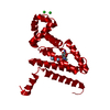
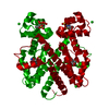
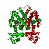
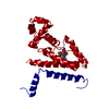
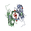
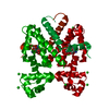
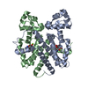
| Title | Structure of Tetracycline repressor in complex with inducer peptide- TIP3 | ||||||
 Components Components |
| ||||||
 Keywords Keywords | TRANSCRIPTION / TETR / PEPTIDIC EFFECTORS / ALLOSTERY | ||||||
| Function / homology |  Function and homology information Function and homology informationtranscription cis-regulatory region binding / DNA-binding transcription factor activity / response to antibiotic / negative regulation of DNA-templated transcription / metal ion binding Similarity search - Function | ||||||
| Biological species |  | ||||||
| Method |  X-RAY DIFFRACTION / SYNCHROTRON / MOLECULAR REPLACEMENT / Resolution: 1.6 Å X-RAY DIFFRACTION / SYNCHROTRON / MOLECULAR REPLACEMENT / Resolution: 1.6 Å | ||||||
 Authors Authors | Sevvana, M. / Goeke, D. / Stoeckle, C. / Kaspar, D. / Grubmueller, S. / Goetz, C. / Wimmer, C. / Berens, C. / Klotzsche, M. / Muller, Y.A. / Hillen, W. | ||||||
 Citation Citation | Journal: J.Mol.Biol. / Year: 2012 Title: An Exclusive Alpha/Beta Code Directs Allostery in Tetr-Peptide Complexes. Authors: Sevvana, M. / Goetz, C. / Goeke, D. / Wimmer, C. / Berens, C. / Hillen, W. / Muller, Y.A. | ||||||
| History |
|
- Structure visualization
| Structure viewer | Molecule: MolmilJmol/JSmol |
|---|
- Downloads & links
-Download
| PDBx/mmCIF format | 3zqh.cif.gz | 107.4 KB | Display | PDBx/mmCIF format |
|---|---|---|---|---|
| PDB format | pdb3zqh.ent.gz | 83 KB | Display | PDB format |
| PDBx/mmJSON format | 3zqh.json.gz | Tree view | PDBx/mmJSON format | |
| Others |  Other downloads Other downloads |
-Validation report
| Arichive directory | https://data.pdbj.org/pub/pdb/validation_reports/zq/3zqhftp://data.pdbj.org/pub/pdb/validation_reports/zq/3zqh | HTTPS FTP |
|---|
-Related structure data
| Related structure data |  3zqfC  3zqgC  3zqiC 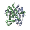 2ns8S C: citing same article ( S: Starting model for refinement |
|---|---|
| Similar structure data |


-Links
 PDBj
PDBj- Assembly
| Deposited unit | 
| ||||||||
|---|---|---|---|---|---|---|---|---|---|
| 1 | 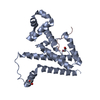
| ||||||||
| Unit cell |
| ||||||||
| Components on special symmetry positions |
|
-Components
| #1: Protein | Mass: 23531.662 Da / Num. of mol.: 1 / Fragment: RESIDUES 1-187,188-208 / Mutation: YES Source method: isolated from a genetically manipulated source Details: CHIMERIC PROTEIN, RESIDUES 1-187 ARE FROM VARIANT B, RESIDUES 188-208 ARE FROM VARIANT D Source: (gene. exp.) | ||||
|---|---|---|---|---|---|
| #2: Protein/peptide | Mass: 1849.248 Da / Num. of mol.: 1 / Source method: obtained synthetically / Source: (synth.) | ||||
| #3: Chemical |   Mass: 62.068 Da / Num. of mol.: 2 / Source method: obtained synthetically / Formula: C2H6O2 Mass: 62.068 Da / Num. of mol.: 2 / Source method: obtained synthetically / Formula: C2H6O2#4: Water | ChemComp-HOH / |  Mass: 18.015 Da / Num. of mol.: 182 / Source method: isolated from a natural source / Formula: H2O Mass: 18.015 Da / Num. of mol.: 182 / Source method: isolated from a natural source / Formula: H2OCompound details | ENGINEERED RESIDUE IN CHAIN A, CYS 68 TO SER ENGINEERED RESIDUE IN CHAIN A, CYS 88 TO ASN ...ENGINEERED | |
-Experimental details
-Experiment
| Experiment | Method: X-RAY DIFFRACTION / Number of used crystals: 1 |
|---|
- Sample preparation
| Crystal | Density Matthews: 2.27 Å3/Da / Density % sol: 45.9 % / Description: NONE |
|---|---|
| Crystal grow | pH: 9 / Details: 0.2 M NACL, 0.1 M TRIS PH 9.0, 25% PEG 3350. |
-Data collection
| Diffraction | Mean temperature: 100 K |
|---|---|
| Diffraction source | Source: SYNCHROTRON / Site: BESSY  / Beamline: 14.1 / Wavelength: 0.9184 / Beamline: 14.1 / Wavelength: 0.9184 |
| Detector | Type: MARRESEARCH MX-225 / Detector: CCD |
| Radiation | Protocol: SINGLE WAVELENGTH / Monochromatic (M) / Laue (L): M / Scattering type: x-ray |
| Radiation wavelength | Wavelength: 0.9184 Å / Relative weight: 1 |
| Reflection | Resolution: 1.6→20 Å / Num. obs: 28325 / % possible obs: 96.1 % / Observed criterion σ(I): 2 / Redundancy: 3.47 % / Biso Wilson estimate: 33.3 Å2 / Rmerge(I) obs: 0.05 / Net I/σ(I): 12.8 |
| Reflection shell | Resolution: 1.6→1.7 Å / Redundancy: 2.6 % / Rmerge(I) obs: 0.54 / Mean I/σ(I) obs: 1.71 / % possible all: 80.1 |
- Processing
| Software |
| ||||||||||||||||||||||||||||||||||||||||||||||||||||||||||||||||||||||||||||||||||||||||||||||||||||||||||||||||||||||||||||||||||||||||||||||||||||||||||||||||||||||||||||||||||||||
|---|---|---|---|---|---|---|---|---|---|---|---|---|---|---|---|---|---|---|---|---|---|---|---|---|---|---|---|---|---|---|---|---|---|---|---|---|---|---|---|---|---|---|---|---|---|---|---|---|---|---|---|---|---|---|---|---|---|---|---|---|---|---|---|---|---|---|---|---|---|---|---|---|---|---|---|---|---|---|---|---|---|---|---|---|---|---|---|---|---|---|---|---|---|---|---|---|---|---|---|---|---|---|---|---|---|---|---|---|---|---|---|---|---|---|---|---|---|---|---|---|---|---|---|---|---|---|---|---|---|---|---|---|---|---|---|---|---|---|---|---|---|---|---|---|---|---|---|---|---|---|---|---|---|---|---|---|---|---|---|---|---|---|---|---|---|---|---|---|---|---|---|---|---|---|---|---|---|---|---|---|---|---|---|
| Refinement | Method to determine structure: MOLECULAR REPLACEMENT Starting model: PDB ENTRY 2NS8 Resolution: 1.6→24.72 Å / Cor.coef. Fo:Fc: 0.967 / Cor.coef. Fo:Fc free: 0.95 / SU B: 4.222 / SU ML: 0.067 / Cross valid method: THROUGHOUT / ESU R: 0.095 / ESU R Free: 0.099 / Stereochemistry target values: MAXIMUM LIKELIHOOD / Details: HYDROGENS HAVE BEEN ADDED IN THE RIDING POSITIONS.
| ||||||||||||||||||||||||||||||||||||||||||||||||||||||||||||||||||||||||||||||||||||||||||||||||||||||||||||||||||||||||||||||||||||||||||||||||||||||||||||||||||||||||||||||||||||||
| Solvent computation | Ion probe radii: 0.8 Å / Shrinkage radii: 0.8 Å / VDW probe radii: 1.2 Å / Solvent model: MASK | ||||||||||||||||||||||||||||||||||||||||||||||||||||||||||||||||||||||||||||||||||||||||||||||||||||||||||||||||||||||||||||||||||||||||||||||||||||||||||||||||||||||||||||||||||||||
| Displacement parameters | Biso mean: 29.144 Å2
| ||||||||||||||||||||||||||||||||||||||||||||||||||||||||||||||||||||||||||||||||||||||||||||||||||||||||||||||||||||||||||||||||||||||||||||||||||||||||||||||||||||||||||||||||||||||
| Refinement step | Cycle: LAST / Resolution: 1.6→24.72 Å
| ||||||||||||||||||||||||||||||||||||||||||||||||||||||||||||||||||||||||||||||||||||||||||||||||||||||||||||||||||||||||||||||||||||||||||||||||||||||||||||||||||||||||||||||||||||||
| Refine LS restraints |
|