transcription cis-regulatory region binding / DNA-binding transcription factor activity / response to antibiotic / negative regulation of DNA-templated transcription / metal ion binding Similarity search - Function
#259 - Jul 2021 Designed Proteins and Citizen Science similarity (1)
-
Assembly
Deposited unit
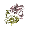
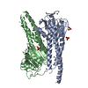
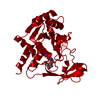
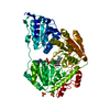
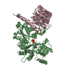
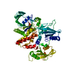
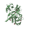
M: DNA (5'-D(*CP*CP*TP*AP*TP*CP*AP*AP*TP*GP*AP*TP*AP*GP*A)-3') N: DNA (5'-D(*TP*CP*TP*AP*TP*CP*AP*TP*TP*GP*AP*TP*AP*GP*G)-3') A: PROTEIN (TETRACYCLINE REPRESSOR) hetero molecules
M: DNA (5'-D(*CP*CP*TP*AP*TP*CP*AP*AP*TP*GP*AP*TP*AP*GP*A)-3') N: DNA (5'-D(*TP*CP*TP*AP*TP*CP*AP*TP*TP*GP*AP*TP*AP*GP*G)-3') A: PROTEIN (TETRACYCLINE REPRESSOR) hetero molecules
M: DNA (5'-D(*CP*CP*TP*AP*TP*CP*AP*AP*TP*GP*AP*TP*AP*GP*A)-3') N: DNA (5'-D(*TP*CP*TP*AP*TP*CP*AP*TP*TP*GP*AP*TP*AP*GP*G)-3') A: PROTEIN (TETRACYCLINE REPRESSOR) hetero molecules
Protocol: SINGLE WAVELENGTH / Monochromatic (M) / Laue (L): M / Scattering type: x-ray
Radiation wavelength
Wavelength: 1.5418 Å / Relative weight: 1
Reflection
Resolution: 2.45→34.5 Å / Num. obs: 12437 / % possible obs: 97.2 % / Observed criterion σ(I): 0 / Redundancy: 3.4 % / Biso Wilson estimate: 58 Å2 / Rmerge(I) obs: 0.09 / Net I/σ(I): 12.1
Reflection shell
Resolution: 2.45→2.55 Å / Redundancy: 3 % / Rmerge(I) obs: 0.287 / % possible all: 93.3
Reflection
*PLUS
Lowest resolution: 24.5 Å / Num. obs: 11520 / Num. measured all: 42624 / Biso Wilson estimate: 57.9 Å2
Reflection shell
*PLUS
% possible obs: 94.4 % / Mean I/σ(I) obs: 3.3
-
Processing
Software
Name
Version
Classification
AMoRE
phasing
X-PLOR
3.851
refinement
DENZO
datareduction
SCALEPACK
datascaling
Refinement
Resolution: 2.5→34.5 Å / Cross valid method: THROUGHOUT / σ(F): 2 Details: USED WEIGHTED FULL MATRIX LEAST SQUARE PROCEDURE. THE DNA BASE PAIRS 1, 8 AND 15 ARE NOT PALINDROMIC. IN REFINEMENT THE CENTRAL BASE M 8 WAS MODELED USING ADENINE AND THYMINE WITH 50 % ...Details: USED WEIGHTED FULL MATRIX LEAST SQUARE PROCEDURE. THE DNA BASE PAIRS 1, 8 AND 15 ARE NOT PALINDROMIC. IN REFINEMENT THE CENTRAL BASE M 8 WAS MODELED USING ADENINE AND THYMINE WITH 50 % OCCUPANCY EACH, RESPECTIVELY. BASE M 1 WAS EITHER CYTOSINE OR THYMINE AND BASE M 15 WAS EITHER GUANINE OR ADENINE. DUE TO CLOSE CRYSTAL PACKING CONTACTS THESE TERMINAL BASES WERE FOUND DISORDERED AND WERE MODELED AS CYTOSINE (BASE M 1) AND ADENINE (BASE M 15), RESPECTIVELY.
In the structure databanks used in Yorodumi, some data are registered as the other names, "COVID-19 virus" and "2019-nCoV". Here are the details of the virus and the list of structure data.
Jan 31, 2019. EMDB accession codes are about to change! (news from PDBe EMDB page)
EMDB accession codes are about to change! (news from PDBe EMDB page)
The allocation of 4 digits for EMDB accession codes will soon come to an end. Whilst these codes will remain in use, new EMDB accession codes will include an additional digit and will expand incrementally as the available range of codes is exhausted. The current 4-digit format prefixed with “EMD-” (i.e. EMD-XXXX) will advance to a 5-digit format (i.e. EMD-XXXXX), and so on. It is currently estimated that the 4-digit codes will be depleted around Spring 2019, at which point the 5-digit format will come into force.
The EM Navigator/Yorodumi systems omit the EMD- prefix.
Related info.:Q: What is EMD? / ID/Accession-code notation in Yorodumi/EM Navigator
Yorodumi is a browser for structure data from EMDB, PDB, SASBDB, etc.
This page is also the successor to EM Navigator detail page, and also detail information page/front-end page for Omokage search.
The word "yorodu" (or yorozu) is an old Japanese word meaning "ten thousand". "mi" (miru) is to see.
Related info.:EMDB / PDB / SASBDB / Comparison of 3 databanks / Yorodumi Search / Aug 31, 2016. New EM Navigator & Yorodumi / Yorodumi Papers / Jmol/JSmol / Function and homology information / Changes in new EM Navigator and Yorodumi
 Movie
Movie Controller


 Open data
 Basic information
 Components
 Keywords
 Function and homology information
 X-RAY DIFFRACTION / Resolution: 2.5 Å
  Authors
 Citation
 Structure visualization
 Downloads & links
 Other downloads


 PDBj
PDBj


 Assembly
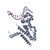


 Mass: 69.085 Da / Num. of mol.: 1 / Source method: obtained synthetically / Formula: C3H5N2
 Mass: 18.015 Da / Num. of mol.: 41 / Source method: isolated from a natural source / Formula: H2O
 Sample preparation
 Processing