 Movie
Movie Controller


+ Open data

- Basic information
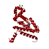
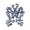
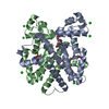
| Entry | Database: PDB / ID: 1a6i | ||||||
|---|---|---|---|---|---|---|---|
| Title | TET REPRESSOR, CLASS D VARIANT | ||||||
 Components Components | TETRACYCLINE REPRESSOR PROTEIN CLASS D | ||||||
 Keywords Keywords | TRANSCRIPTION REGULATION / REPRESSOR / DNA-BINDING | ||||||
| Function / homology |  Function and homology information Function and homology informationtranscription cis-regulatory region binding / DNA-binding transcription factor activity / response to antibiotic / negative regulation of DNA-templated transcription / metal ion binding Similarity search - Function | ||||||
| Biological species |  | ||||||
| Method |  X-RAY DIFFRACTION / SYNCHROTRON / MOLECULAR REPLACEMENT / Resolution: 2.4 Å X-RAY DIFFRACTION / SYNCHROTRON / MOLECULAR REPLACEMENT / Resolution: 2.4 Å | ||||||
 Authors Authors | Orth, P. / Cordes, F. / Schnappinger, D. / Hillen, W. / Saenger, W. / Hinrichs, W. | ||||||
 Citation Citation | Journal: J.Mol.Biol. / Year: 1998 Title: Conformational changes of the Tet repressor induced by tetracycline trapping. Authors: Orth, P. / Cordes, F. / Schnappinger, D. / Hillen, W. / Saenger, W. / Hinrichs, W. #1: Journal: J.Mol.Biol. / Year: 1995Title: The Complex Formed between Tet Repressor and Tetracycline-Mg2+ Reveals Mechanism of Antibiotic Resistance Authors: Kisker, C. / Hinrichs, W. / Tovar, K. / Hillen, W. / Saenger, W. #2: Journal: Science / Year: 1994Title: Structure of the Tet Repressor-Tetracycline Complex and Regulation of Antibiotic Resistance Authors: Hinrichs, W. / Kisker, C. / Duvel, M. / Muller, A. / Tovar, K. / Hillen, W. / Saenger, W. | ||||||
| History |
|
- Structure visualization
| Structure viewer | Molecule: MolmilJmol/JSmol |
|---|
- Downloads & links
-Download
| PDBx/mmCIF format | 1a6i.cif.gz | 50.4 KB | Display | PDBx/mmCIF format |
|---|---|---|---|---|
| PDB format | pdb1a6i.ent.gz | 36.6 KB | Display | PDB format |
| PDBx/mmJSON format | 1a6i.json.gz | Tree view | PDBx/mmJSON format | |
| Others |  Other downloads Other downloads |
-Validation report
| Arichive directory | https://data.pdbj.org/pub/pdb/validation_reports/a6/1a6iftp://data.pdbj.org/pub/pdb/validation_reports/a6/1a6i | HTTPS FTP |
|---|
-Related structure data
| Related structure data |  2tctS S: Starting model for refinement |
|---|---|
| Similar structure data |


-Links
 PDBj
PDBj- Assembly
| Deposited unit | 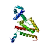
| ||||||||
|---|---|---|---|---|---|---|---|---|---|
| 1 | 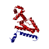
| ||||||||
| Unit cell |
|
-Components
| #1: Protein | Mass: 24319.691 Da / Num. of mol.: 1 Mutation: A2S, N5D, R6K, E7S, S8K, D11N, A12S, T20V, D23E, I36V Source method: isolated from a genetically manipulated source Source: (gene. exp.) |
|---|---|
| #2: Water | ChemComp-HOH /  Mass: 18.015 Da / Num. of mol.: 28 / Source method: isolated from a natural source / Formula: H2O Mass: 18.015 Da / Num. of mol.: 28 / Source method: isolated from a natural source / Formula: H2O |
-Experimental details
-Experiment
| Experiment | Method: X-RAY DIFFRACTION / Number of used crystals: 1 |
|---|
- Sample preparation
| Crystal | Density Matthews: 2.4 Å3/Da / Density % sol: 49 % Description: DATA WERE COLLECTED USING THE OSCILLATION METHOD | ||||||||||||||||||||||||||||||||||||
|---|---|---|---|---|---|---|---|---|---|---|---|---|---|---|---|---|---|---|---|---|---|---|---|---|---|---|---|---|---|---|---|---|---|---|---|---|---|
| Crystal grow | pH: 8 / Details: pH 8.0 | ||||||||||||||||||||||||||||||||||||
| Crystal grow | *PLUS Temperature: 18 ℃ / Method: vapor diffusion, hanging dropDetails: 0.01ml protein solution was mixed with 0.005ml reservoir | ||||||||||||||||||||||||||||||||||||
| Components of the solutions | *PLUS
|
-Data collection
| Diffraction | Mean temperature: 277 K |
|---|---|
| Diffraction source | Source: SYNCHROTRON / Site: LURE  / Beamline: DW32 / Wavelength: 0.92 / Beamline: DW32 / Wavelength: 0.92 |
| Detector | Type: MARRESEARCH / Detector: IMAGE PLATE / Date: Dec 1, 1995 |
| Radiation | Monochromatic (M) / Laue (L): M / Scattering type: x-ray |
| Radiation wavelength | Wavelength: 0.92 Å / Relative weight: 1 |
| Reflection | Resolution: 2.4→18.5 Å / Num. obs: 9076 / % possible obs: 97.8 % / Observed criterion σ(I): 0 / Redundancy: 4.8 % / Biso Wilson estimate: 42.4 Å2 / Rsym value: 0.073 / Net I/σ(I): 6.13 |
| Reflection shell | Resolution: 2.4→2.47 Å / Redundancy: 4.9 % / Mean I/σ(I) obs: 2.9 / Rsym value: 0.248 / % possible all: 98.6 |
| Reflection | *PLUS Rmerge(I) obs: 0.073 |
| Reflection shell | *PLUS % possible obs: 98.6 % / Rmerge(I) obs: 0.248 |
- Processing
| Software |
| ||||||||||||||||||||||||||||||||||||||||||||||||||||||||||||||||||||||||||||||||
|---|---|---|---|---|---|---|---|---|---|---|---|---|---|---|---|---|---|---|---|---|---|---|---|---|---|---|---|---|---|---|---|---|---|---|---|---|---|---|---|---|---|---|---|---|---|---|---|---|---|---|---|---|---|---|---|---|---|---|---|---|---|---|---|---|---|---|---|---|---|---|---|---|---|---|---|---|---|---|---|---|---|
| Refinement | Method to determine structure: MOLECULAR REPLACEMENT Starting model: 2TCT Resolution: 2.4→18.5 Å / Data cutoff high absF: 100000 / Data cutoff low absF: 0.1 / Isotropic thermal model: RESTRAINED / Cross valid method: THROUGHOUT / σ(F): 0
| ||||||||||||||||||||||||||||||||||||||||||||||||||||||||||||||||||||||||||||||||
| Displacement parameters | Biso mean: 47.6 Å2 | ||||||||||||||||||||||||||||||||||||||||||||||||||||||||||||||||||||||||||||||||
| Refine analyze | Luzzati coordinate error obs: 0.35 Å / Luzzati d res low obs: 10 Å | ||||||||||||||||||||||||||||||||||||||||||||||||||||||||||||||||||||||||||||||||
| Refinement step | Cycle: LAST / Resolution: 2.4→18.5 Å
| ||||||||||||||||||||||||||||||||||||||||||||||||||||||||||||||||||||||||||||||||
| Refine LS restraints |
| ||||||||||||||||||||||||||||||||||||||||||||||||||||||||||||||||||||||||||||||||
| Xplor file |
| ||||||||||||||||||||||||||||||||||||||||||||||||||||||||||||||||||||||||||||||||
| Software | *PLUS Name: X-PLOR / Version: 3.1 / Classification: refinement | ||||||||||||||||||||||||||||||||||||||||||||||||||||||||||||||||||||||||||||||||
| Refinement | *PLUS | ||||||||||||||||||||||||||||||||||||||||||||||||||||||||||||||||||||||||||||||||
| Solvent computation | *PLUS | ||||||||||||||||||||||||||||||||||||||||||||||||||||||||||||||||||||||||||||||||
| Displacement parameters | *PLUS | ||||||||||||||||||||||||||||||||||||||||||||||||||||||||||||||||||||||||||||||||
| Refine LS restraints | *PLUS
|
