 Movie
Movie Controller

[English] 日本語
 Yorodumi
- PDB-2c7o: HhaI DNA methyltransferase complex with 13mer oligonucleotide con... -
+ Open data

- Basic information
| Entry | Database: PDB / ID: 2c7o | ||||||
|---|---|---|---|---|---|---|---|
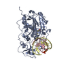
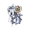
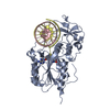
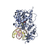
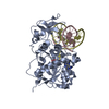
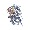
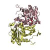
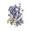
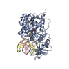
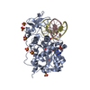
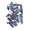
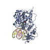
| Title | HhaI DNA methyltransferase complex with 13mer oligonucleotide containing 2-aminopurine adjacent to the target base (PCGC:GMGC) and SAH | ||||||
 Components Components |
| ||||||
 Keywords Keywords | TRANSFERASE/DNA / TRANSFERASE-DNA COMPLEX / BASE FLIPPING / TRANSFERASE RESTRICTION SYSTEM | ||||||
| Function / homology |  Function and homology information Function and homology informationDNA (cytosine-5-)-methyltransferase / DNA (cytosine-5-)-methyltransferase activity / DNA restriction-modification system / methylation / DNA binding Similarity search - Function | ||||||
| Biological species |  HAEMOPHILUS HAEMOLYTICUS (bacteria) HAEMOPHILUS HAEMOLYTICUS (bacteria)SYNTHETIC CONSTRUCT (others) | ||||||
| Method |  X-RAY DIFFRACTION / SYNCHROTRON / MOLECULAR REPLACEMENT / Resolution: 1.9 Å X-RAY DIFFRACTION / SYNCHROTRON / MOLECULAR REPLACEMENT / Resolution: 1.9 Å | ||||||
 Authors Authors | Daujotyte, D. / Grazulis, S. | ||||||
 Citation Citation | Journal: Nucleic Acids Res. / Year: 2005 Title: Time-Resolved Fluorescence of 2-Aminopurine as a Probe of Base Flipping in M.HhaI-DNA Complexes. Authors: Neely, R.K. / Daujotyte, D. / Grazulis, S. / Magennis, S.W. / Dryden, D.T.F. / Klimasauskas, S. / Jones, A.C. | ||||||
| History |
|
- Structure visualization
| Structure viewer | Molecule: MolmilJmol/JSmol |
|---|
- Downloads & links
-Download
| PDBx/mmCIF format | 2c7o.cif.gz | 105 KB | Display | PDBx/mmCIF format |
|---|---|---|---|---|
| PDB format | pdb2c7o.ent.gz | 77.5 KB | Display | PDB format |
| PDBx/mmJSON format | 2c7o.json.gz | Tree view | PDBx/mmJSON format | |
| Others |  Other downloads Other downloads |
-Validation report
| Arichive directory | https://data.pdbj.org/pub/pdb/validation_reports/c7/2c7oftp://data.pdbj.org/pub/pdb/validation_reports/c7/2c7o | HTTPS FTP |
|---|
-Related structure data
| Related structure data |  2c7pC  2c7qC 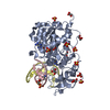 2c7rC 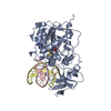 3mhtS S: Starting model for refinement C: citing same article ( |
|---|---|
| Similar structure data |


-Links
 PDBj
PDBj


- Assembly
| Deposited unit | 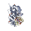
| ||||||||||||
|---|---|---|---|---|---|---|---|---|---|---|---|---|---|
| 1 |
| ||||||||||||
| Unit cell |
| ||||||||||||
| Components on special symmetry positions |
| ||||||||||||
| Details | THE OLIGOMERIC STATE OF HHAI METHYLTRANSFERASE IS MONOMERIC, BUT SINCE IN THIS ENTRY, THE PROTEIN IS IN COMPLEX WITH DNA, THE QUATERNARY STRUCTURE FOR THIS ENTRY IS MARKED AS TRIMERIC. |
-Components
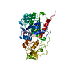
-Protein , 1 types, 1 molecules A
| #1: Protein | Mass: 37042.207 Da / Num. of mol.: 1 Source method: isolated from a genetically manipulated source Source: (gene. exp.) HAEMOPHILUS HAEMOLYTICUS (bacteria) / Plasmid: PHH553 / Production host: References: UniProt: P05102, DNA (cytosine-5-)-methyltransferase |
|---|
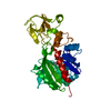
-DNA chain , 2 types, 2 molecules CD
| #2: DNA chain | Mass: 4021.636 Da / Num. of mol.: 1 / Source method: obtained synthetically / Source: (synth.) SYNTHETIC CONSTRUCT (others) |
|---|---|
| #3: DNA chain | Mass: 3911.562 Da / Num. of mol.: 1 / Source method: obtained synthetically / Source: (synth.) SYNTHETIC CONSTRUCT (others) |
-Non-polymers , 3 types, 346 molecules 


| #4: Chemical | ChemComp-SAH /  Mass: 384.411 Da / Num. of mol.: 1 / Source method: obtained synthetically / Formula: C14H20N6O5S Mass: 384.411 Da / Num. of mol.: 1 / Source method: obtained synthetically / Formula: C14H20N6O5S | ||
|---|---|---|---|
| #5: Chemical | ChemComp-SO4 /  Mass: 96.063 Da / Num. of mol.: 4 / Source method: obtained synthetically / Formula: SO4 Mass: 96.063 Da / Num. of mol.: 4 / Source method: obtained synthetically / Formula: SO4#6: Water | ChemComp-HOH / | Mass: 18.015 Da / Num. of mol.: 341 / Source method: isolated from a natural source / Formula: H2O |
-Details
| Compound details | RECOGNIZES THE DOUBLE-STRANDED SEQUENCE GCGC, CAUSES SPECIFIC METHYLATION AND PROTECTS THE DNA FROM ...RECOGNIZES |
|---|---|
| Has protein modification | N |
-Experimental details
-Experiment
| Experiment | Method: X-RAY DIFFRACTION / Number of used crystals: 1 |
|---|
- Sample preparation
| Crystal | Density Matthews: 2.74 Å3/Da / Density % sol: 54.79 % |
|---|---|
| Crystal grow | pH: 5.6 Details: 50 MM NA CITRATE PH 5.6, 1.6 M AMMONIUM SULFATE, 5 % GLUCOSE. |
-Data collection
| Diffraction | Mean temperature: 100 K |
|---|---|
| Diffraction source | Source: SYNCHROTRON / Site: EMBL/DESY, HAMBURG  / Beamline: X11 / Wavelength: 0.812 / Beamline: X11 / Wavelength: 0.812 |
| Detector | Type: MARRESEARCH / Detector: IMAGE PLATE / Date: Mar 18, 2004 / Details: MIRRORS |
| Radiation | Monochromator: SI(111) / Protocol: SINGLE WAVELENGTH / Monochromatic (M) / Laue (L): M / Scattering type: x-ray |
| Radiation wavelength | Wavelength: 0.812 Å / Relative weight: 1 |
| Reflection | Resolution: 1.9→23.2 Å / Num. obs: 43361 / % possible obs: 99.6 % / Observed criterion σ(I): 3.9 / Redundancy: 21.6 % / Biso Wilson estimate: 10.9 Å2 / Rmerge(I) obs: 0.05 / Net I/σ(I): 11.4 |
| Reflection shell | Resolution: 1.9→2 Å / Redundancy: 20.8 % / Rmerge(I) obs: 0.2 / Mean I/σ(I) obs: 3.9 / % possible all: 98.5 |
- Processing
| Software |
| ||||||||||||||||||||||||||||||||||||||||||||||||||||||||||||||||||||||||||||||||
|---|---|---|---|---|---|---|---|---|---|---|---|---|---|---|---|---|---|---|---|---|---|---|---|---|---|---|---|---|---|---|---|---|---|---|---|---|---|---|---|---|---|---|---|---|---|---|---|---|---|---|---|---|---|---|---|---|---|---|---|---|---|---|---|---|---|---|---|---|---|---|---|---|---|---|---|---|---|---|---|---|---|
| Refinement | Method to determine structure: MOLECULAR REPLACEMENT Starting model: PDB ENTRY 3MHT Resolution: 1.9→23.08 Å / Rfactor Rfree error: 0.003 / Data cutoff high absF: 34200167.59 / Isotropic thermal model: RESTRAINED / Cross valid method: THROUGHOUT / σ(F): 0 Details: RESOLUTION-DEPENDENT WEIGHTING SCHEME USED. BULK SOLVENT MODEL USED
| ||||||||||||||||||||||||||||||||||||||||||||||||||||||||||||||||||||||||||||||||
| Solvent computation | Solvent model: FLAT MODEL / Bsol: 51.1244 Å2 / ksol: 0.385184 e/Å3 | ||||||||||||||||||||||||||||||||||||||||||||||||||||||||||||||||||||||||||||||||
| Displacement parameters | Biso mean: 18.8 Å2
| ||||||||||||||||||||||||||||||||||||||||||||||||||||||||||||||||||||||||||||||||
| Refine analyze |
| ||||||||||||||||||||||||||||||||||||||||||||||||||||||||||||||||||||||||||||||||
| Refinement step | Cycle: LAST / Resolution: 1.9→23.08 Å
| ||||||||||||||||||||||||||||||||||||||||||||||||||||||||||||||||||||||||||||||||
| Refine LS restraints |
| ||||||||||||||||||||||||||||||||||||||||||||||||||||||||||||||||||||||||||||||||
| LS refinement shell | Resolution: 1.9→2.02 Å / Rfactor Rfree error: 0.008 / Total num. of bins used: 6
| ||||||||||||||||||||||||||||||||||||||||||||||||||||||||||||||||||||||||||||||||
| Xplor file |
|
