 Movie
Movie Controller


+ Open data

- Basic information
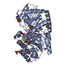
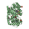
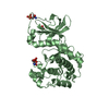
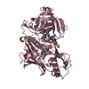
| Entry | Database: PDB / ID: 5its | ||||||
|---|---|---|---|---|---|---|---|
| Title | Crystal structure of LOG from Corynebacterium glutamicum | ||||||
 Components Components | Cytokinin riboside 5'-monophosphate phosphoribohydrolase | ||||||
 Keywords Keywords | HYDROLASE / Rossmann fold / nucleotide-binding domain / phosphoribohydrolase | ||||||
| Function / homology |  Function and homology information Function and homology informationcytokinin riboside 5'-monophosphate phosphoribohydrolase activity / : / cytokinin biosynthetic process / nucleotide binding / cytosol Similarity search - Function | ||||||
| Biological species |  Corynebacterium glutamicum (bacteria) Corynebacterium glutamicum (bacteria) | ||||||
| Method |  X-RAY DIFFRACTION / SYNCHROTRON / MOLECULAR REPLACEMENT / Resolution: 2.3 Å X-RAY DIFFRACTION / SYNCHROTRON / MOLECULAR REPLACEMENT / Resolution: 2.3 Å | ||||||
 Authors Authors | Seo, H.-G. / Kim, K.-J. | ||||||
 Citation Citation | Journal: Sci Rep / Year: 2016 Title: Structural basis for cytokinin production by LOG from Corynebacterium glutamicum Authors: Seo, H.-G. / Kim, S. / Sagong, H.Y. / Son, H.F. / Jin, K.S. / Kim, I.K. / Kim, K.J. | ||||||
| History |
|
- Structure visualization
| Structure viewer | Molecule: MolmilJmol/JSmol |
|---|
- Downloads & links
-Download
| PDBx/mmCIF format | 5its.cif.gz | 159.4 KB | Display | PDBx/mmCIF format |
|---|---|---|---|---|
| PDB format | pdb5its.ent.gz | 125.9 KB | Display | PDB format |
| PDBx/mmJSON format | 5its.json.gz | Tree view | PDBx/mmJSON format | |
| Others |  Other downloads Other downloads |
-Validation report
| Arichive directory | https://data.pdbj.org/pub/pdb/validation_reports/it/5itsftp://data.pdbj.org/pub/pdb/validation_reports/it/5its | HTTPS FTP |
|---|
-Related structure data
| Related structure data |  2a33S S: Starting model for refinement |
|---|---|
| Similar structure data |


-Links
 PDBj
PDBj- Assembly
| Deposited unit | 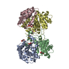
| ||||||||
|---|---|---|---|---|---|---|---|---|---|
| 1 | 
| ||||||||
| 2 | 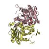
| ||||||||
| Unit cell |
|
-Components
| #1: Protein | Mass: 22581.900 Da / Num. of mol.: 4 Source method: isolated from a genetically manipulated source Source: (gene. exp.) Corynebacterium glutamicum (strain ATCC 13032 / DSM 20300 / JCM 1318 / LMG 3730 / NCIMB 10025) (bacteria)Strain: ATCC 13032 / DSM 20300 / JCM 1318 / LMG 3730 / NCIMB 10025 Gene: Cgl2379 / Plasmid: pET30a / Production host: References: UniProt: Q8NN34, Hydrolases; Glycosylases; Hydrolysing N-glycosyl compounds #2: Chemical |   Mass: 94.971 Da / Num. of mol.: 2 / Source method: obtained synthetically / Formula: PO4 Mass: 94.971 Da / Num. of mol.: 2 / Source method: obtained synthetically / Formula: PO4#3: Chemical | ChemComp-GOL /   Mass: 92.094 Da / Num. of mol.: 6 / Source method: obtained synthetically / Formula: C3H8O3 Mass: 92.094 Da / Num. of mol.: 6 / Source method: obtained synthetically / Formula: C3H8O3#4: Water | ChemComp-HOH / |  Mass: 18.015 Da / Num. of mol.: 204 / Source method: isolated from a natural source / Formula: H2O Mass: 18.015 Da / Num. of mol.: 204 / Source method: isolated from a natural source / Formula: H2O |
|---|
-Experimental details
-Experiment
| Experiment | Method: X-RAY DIFFRACTION / Number of used crystals: 1 |
|---|
- Sample preparation
| Crystal | Density Matthews: 3.06 Å3/Da / Density % sol: 59.76 % |
|---|---|
| Crystal grow | Temperature: 293 K / Method: vapor diffusion, hanging drop / pH: 7 / Details: PEG 3350, DL-malic acid |
-Data collection
| Diffraction | Mean temperature: 100 K |
|---|---|
| Diffraction source | Source: SYNCHROTRON / Site: PAL/PLS  / Beamline: 7A (6B, 6C1) / Wavelength: 0.97934 Å / Beamline: 7A (6B, 6C1) / Wavelength: 0.97934 Å |
| Detector | Type: ADSC QUANTUM 270 / Detector: CCD / Date: Oct 28, 2014 |
| Radiation | Monochromator: Double Crystal Monochromator / Protocol: SINGLE WAVELENGTH / Monochromatic (M) / Laue (L): M / Scattering type: x-ray |
| Radiation wavelength | Wavelength: 0.97934 Å / Relative weight: 1 |
| Reflection | Resolution: 2.3→95.62 Å / Num. obs: 44133 / % possible obs: 95.2 % / Redundancy: 6.8 % / Biso Wilson estimate: 23.3 Å2 / Rmerge(I) obs: 0.102 / Net I/σ(I): 17.5 |
| Reflection shell | Resolution: 2.3→2.34 Å / Redundancy: 3.2 % / Rmerge(I) obs: 0.32 / Mean I/σ(I) obs: 3.5 / % possible all: 87 |
- Processing
| Software |
| |||||||||||||||||||||||||||||||||||||||||||||||||||||||||||||||||||||||||||
|---|---|---|---|---|---|---|---|---|---|---|---|---|---|---|---|---|---|---|---|---|---|---|---|---|---|---|---|---|---|---|---|---|---|---|---|---|---|---|---|---|---|---|---|---|---|---|---|---|---|---|---|---|---|---|---|---|---|---|---|---|---|---|---|---|---|---|---|---|---|---|---|---|---|---|---|---|
| Refinement | Method to determine structure: MOLECULAR REPLACEMENT Starting model: 2A33 Resolution: 2.3→95.62 Å / Cor.coef. Fo:Fc: 0.952 / Cor.coef. Fo:Fc free: 0.921 / SU B: 7.513 / SU ML: 0.167 / Cross valid method: THROUGHOUT / σ(F): 0 / ESU R: 0.248 / ESU R Free: 0.208 / Stereochemistry target values: MAXIMUM LIKELIHOOD Details: HYDROGENS HAVE BEEN ADDED IN THE RIDING POSITIONS U VALUES : REFINED INDIVIDUALLY
| |||||||||||||||||||||||||||||||||||||||||||||||||||||||||||||||||||||||||||
| Solvent computation | Ion probe radii: 0.8 Å / Shrinkage radii: 0.8 Å / VDW probe radii: 1.2 Å / Solvent model: MASK | |||||||||||||||||||||||||||||||||||||||||||||||||||||||||||||||||||||||||||
| Displacement parameters | Biso max: 95.95 Å2 / Biso mean: 29.687 Å2 / Biso min: 12.12 Å2
| |||||||||||||||||||||||||||||||||||||||||||||||||||||||||||||||||||||||||||
| Refinement step | Cycle: final / Resolution: 2.3→95.62 Å
| |||||||||||||||||||||||||||||||||||||||||||||||||||||||||||||||||||||||||||
| Refine LS restraints |
| |||||||||||||||||||||||||||||||||||||||||||||||||||||||||||||||||||||||||||
| LS refinement shell | Resolution: 2.303→2.363 Å / Total num. of bins used: 20
|
