 Movie
Movie Controller

[English] 日本語
 Yorodumi
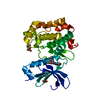
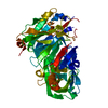
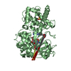
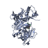
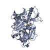
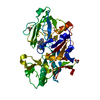
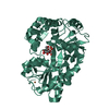
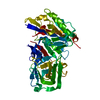
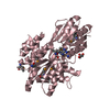
- PDB-2jed: The crystal structure of the kinase domain of the protein kinase ... -
+ Open data

- Basic information
| Entry | Database: PDB / ID: 2jed | ||||||
|---|---|---|---|---|---|---|---|
| Title | The crystal structure of the kinase domain of the protein kinase C theta in complex with NVP-XAA228 at 2.32A resolution. | ||||||
 Components Components | PROTEIN KINASE C THETA | ||||||
 Keywords Keywords | TRANSFERASE / PHOSPHORYLATION / NUCLEOTIDE-BINDING / SERINE/THREONINE-PROTEIN KINASE / ATP-BINDING / POLYMORPHISM / METAL-BINDING / ZINC / KINASE / PKC THETA / MAGNESIUM / ZINC-FINGER / ALTERNATIVE SPLICING / PHORBOL-ESTER BINDING | ||||||
| Function / homology |  Function and homology information Function and homology informationpositive regulation of T-helper 2 cell activation / Netrin-1 signaling / protein kinase C / diacylglycerol-dependent serine/threonine kinase activity / regulation of platelet aggregation / Effects of PIP2 hydrolysis / CD4-positive, alpha-beta T cell proliferation / negative regulation of T cell apoptotic process / positive regulation of CD4-positive, alpha-beta T cell proliferation / positive regulation of T-helper 17 type immune response ...positive regulation of T-helper 2 cell activation / Netrin-1 signaling / protein kinase C / diacylglycerol-dependent serine/threonine kinase activity / regulation of platelet aggregation / Effects of PIP2 hydrolysis / CD4-positive, alpha-beta T cell proliferation / negative regulation of T cell apoptotic process / positive regulation of CD4-positive, alpha-beta T cell proliferation / positive regulation of T-helper 17 type immune response / aggresome / Apoptotic cleavage of cellular proteins / positive regulation of interleukin-4 production / Fc-epsilon receptor signaling pathway / positive regulation of interleukin-17 production / membrane protein ectodomain proteolysis / positive regulation of telomere maintenance / immunological synapse / positive regulation of interleukin-2 production / negative regulation of insulin receptor signaling pathway / axon guidance / : / cell chemotaxis / regulation of cell growth / RUNX1 regulates genes involved in megakaryocyte differentiation and platelet function / positive regulation of T cell activation / FCERI mediated NF-kB activation / centriolar satellite / RAS processing / G alpha (z) signalling events / Downstream TCR signaling / Inactivation, recovery and regulation of the phototransduction cascade / protein kinase activity / intracellular signal transduction / inflammatory response / protein serine kinase activity / protein serine/threonine kinase activity / regulation of DNA-templated transcription / zinc ion binding / ATP binding / plasma membrane / cytosol Similarity search - Function | ||||||
| Biological species |  HOMO SAPIENS (human) HOMO SAPIENS (human) | ||||||
| Method |  X-RAY DIFFRACTION / SYNCHROTRON / MOLECULAR REPLACEMENT / Resolution: 2.32 Å X-RAY DIFFRACTION / SYNCHROTRON / MOLECULAR REPLACEMENT / Resolution: 2.32 Å | ||||||
 Authors Authors | Stark, W. / Bitsch, F. / Berner, A. / Buelens, F. / Graff, P. / Depersin, H. / Geiser, M. / Knecht, R. / Rahuel, J. / Rummel, G. ...Stark, W. / Bitsch, F. / Berner, A. / Buelens, F. / Graff, P. / Depersin, H. / Geiser, M. / Knecht, R. / Rahuel, J. / Rummel, G. / Schlaeppi, J.M. / Schmitz, R. / Strauss, A. / Wagner, J. | ||||||
 Citation Citation | Journal: To be Published Title: The Crystal Structure of the Kinase Domain of the Protein Kinase C Theta in Complex with Nvp-Xaa228 Authors: Stark, W. / Bitsch, F. / Berner, A. / Buelens, F. / Graff, P. / Depersin, H. / Fendrich, G. / Geiser, M. / Knecht, R. / Rahuel, J. / Rummel, G. / Schlaeppi, J.M. / Schmitz, R. / Strauss, A. / Wagner, J. #1: Journal: Protein Expression Purif. / Year: 2007 Title: Improved Expression of Kinases in Baculovirus-Infected Insect Cells Upon Addition of Specific Kinase Inhibitors to the Culture Helpful for Structural Studies. Authors: Strauss, A. / Fendrich, G. / Horisberger, M.A. / Liebetanz, J. / Meyhack, B. / Schlaeppi, J.-M. / Schmitz, R. | ||||||
| History |
|
- Structure visualization
| Structure viewer | Molecule: MolmilJmol/JSmol |
|---|
- Downloads & links
-Download
| PDBx/mmCIF format | 2jed.cif.gz | 156.1 KB | Display | PDBx/mmCIF format |
|---|---|---|---|---|
| PDB format | pdb2jed.ent.gz | 123.1 KB | Display | PDB format |
| PDBx/mmJSON format | 2jed.json.gz | Tree view | PDBx/mmJSON format | |
| Others |  Other downloads Other downloads |
-Validation report
| Arichive directory | https://data.pdbj.org/pub/pdb/validation_reports/je/2jedftp://data.pdbj.org/pub/pdb/validation_reports/je/2jed | HTTPS FTP |
|---|
-Related structure data
| Related structure data | 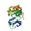 1fotS S: Starting model for refinement |
|---|---|
| Similar structure data |


-Links
 PDBj
PDBj


- Assembly
| Deposited unit | 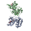
| ||||||||
|---|---|---|---|---|---|---|---|---|---|
| 1 | 
| ||||||||
| 2 | 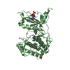
| ||||||||
| Unit cell |
|
-Components
| #1: Protein | Mass: 41823.965 Da / Num. of mol.: 2 / Fragment: KINASE DOMAIN 361-706 / Mutation: YES / Source method: isolated from a natural source Details: PHOSPHOSERINE 676, PHOSPHOSERINE 695, C540 MODIFIED Source: (natural) HOMO SAPIENS (human) / References: UniProt: Q04759, protein kinase C#2: Chemical | 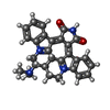  Mass: 452.548 Da / Num. of mol.: 2 / Source method: obtained synthetically / Formula: C28H28N4O2 Mass: 452.548 Da / Num. of mol.: 2 / Source method: obtained synthetically / Formula: C28H28N4O2#3: Chemical | ChemComp-MPD / ( |   Mass: 118.174 Da / Num. of mol.: 1 / Source method: obtained synthetically / Formula: C6H14O2 / Comment: precipitant*YM Mass: 118.174 Da / Num. of mol.: 1 / Source method: obtained synthetically / Formula: C6H14O2 / Comment: precipitant*YM#4: Water | ChemComp-HOH / |  Mass: 18.015 Da / Num. of mol.: 412 / Source method: isolated from a natural source / Formula: H2O Mass: 18.015 Da / Num. of mol.: 412 / Source method: isolated from a natural source / Formula: H2OCompound details | ENGINEERED RESIDUE IN CHAIN A, ILE 381 TO GLU ENGINEERED RESIDUE IN CHAIN A, THR 538 TO GLU ...ENGINEERED | Has protein modification | Y | Sequence details | MUTATIONS I381E, T538E | |
|---|
-Experimental details
-Experiment
| Experiment | Method: X-RAY DIFFRACTION / Number of used crystals: 1 |
|---|
- Sample preparation
| Crystal | Density Matthews: 3.14 Å3/Da / Density % sol: 56 % / Description: NONE |
|---|---|
| Crystal grow | Temperature: 277 K / Method: vapor diffusion / pH: 6.5 Details: VAPOR DIFFUSION AT 4C PROTEIN SOLUTION: 10MG/ML PROTEIN IN 0.2M NACL, 0.05M IMIDAZOLE, 0.001M NAF, 0.05M TCEP, PH = 8.0 RESERVOIR SOLUTION: 0.1 M NA-CACODYLATE PH = 6.5 24% MPD (V/V) 4% ...Details: VAPOR DIFFUSION AT 4C PROTEIN SOLUTION: 10MG/ML PROTEIN IN 0.2M NACL, 0.05M IMIDAZOLE, 0.001M NAF, 0.05M TCEP, PH = 8.0 RESERVOIR SOLUTION: 0.1 M NA-CACODYLATE PH = 6.5 24% MPD (V/V) 4% PEG8000 (W/V) DROP: 1:1 SMALL ORGANIC MOLECULES LIKE 2,5-HEXANEDIOL AND SULFOBETAINE-195 HAVE A POSITIVE EFFECT ON THE CRYSTAL GROWTH |
-Data collection
| Diffraction | Mean temperature: 100 K |
|---|---|
| Diffraction source | Source: SYNCHROTRON / Site: SLS  / Beamline: X06SA / Wavelength: 0.918396 / Beamline: X06SA / Wavelength: 0.918396 |
| Detector | Type: MARRESEARCH / Detector: CCD / Date: Sep 6, 2002 |
| Radiation | Protocol: SINGLE WAVELENGTH / Monochromatic (M) / Laue (L): M / Scattering type: x-ray |
| Radiation wavelength | Wavelength: 0.918396 Å / Relative weight: 1 |
| Reflection | Resolution: 2.32→50 Å / Num. obs: 42831 / % possible obs: 99.7 % / Redundancy: 4.8 % / Rmerge(I) obs: 0.06 / Net I/σ(I): 10.4 |
| Reflection shell | Resolution: 2.32→2.36 Å / Rmerge(I) obs: 0.48 / % possible all: 100 |
- Processing
| Software |
| ||||||||||||||||||||||||||||||||||||||||||||||||||||||||||||||||||||||||||||||||||||||||||||||||||||||||||||||||||||||||||||||||||||||||||||||||||||||||||||||||||||||||||||||||||||||
|---|---|---|---|---|---|---|---|---|---|---|---|---|---|---|---|---|---|---|---|---|---|---|---|---|---|---|---|---|---|---|---|---|---|---|---|---|---|---|---|---|---|---|---|---|---|---|---|---|---|---|---|---|---|---|---|---|---|---|---|---|---|---|---|---|---|---|---|---|---|---|---|---|---|---|---|---|---|---|---|---|---|---|---|---|---|---|---|---|---|---|---|---|---|---|---|---|---|---|---|---|---|---|---|---|---|---|---|---|---|---|---|---|---|---|---|---|---|---|---|---|---|---|---|---|---|---|---|---|---|---|---|---|---|---|---|---|---|---|---|---|---|---|---|---|---|---|---|---|---|---|---|---|---|---|---|---|---|---|---|---|---|---|---|---|---|---|---|---|---|---|---|---|---|---|---|---|---|---|---|---|---|---|---|
| Refinement | Method to determine structure: MOLECULAR REPLACEMENT Starting model: PDB ENTRY 1FOT Resolution: 2.32→19.88 Å / Cor.coef. Fo:Fc: 0.955 / Cor.coef. Fo:Fc free: 0.941 / SU B: 10.404 / SU ML: 0.134 / Cross valid method: THROUGHOUT / ESU R: 0.257 / ESU R Free: 0.198 / Stereochemistry target values: MAXIMUM LIKELIHOOD / Details: HYDROGENS HAVE BEEN ADDED IN THE RIDING POSITIONS
| ||||||||||||||||||||||||||||||||||||||||||||||||||||||||||||||||||||||||||||||||||||||||||||||||||||||||||||||||||||||||||||||||||||||||||||||||||||||||||||||||||||||||||||||||||||||
| Solvent computation | Ion probe radii: 0.8 Å / Shrinkage radii: 0.8 Å / VDW probe radii: 1.4 Å / Solvent model: MASK | ||||||||||||||||||||||||||||||||||||||||||||||||||||||||||||||||||||||||||||||||||||||||||||||||||||||||||||||||||||||||||||||||||||||||||||||||||||||||||||||||||||||||||||||||||||||
| Displacement parameters | Biso mean: 49.2 Å2
| ||||||||||||||||||||||||||||||||||||||||||||||||||||||||||||||||||||||||||||||||||||||||||||||||||||||||||||||||||||||||||||||||||||||||||||||||||||||||||||||||||||||||||||||||||||||
| Refinement step | Cycle: LAST / Resolution: 2.32→19.88 Å
| ||||||||||||||||||||||||||||||||||||||||||||||||||||||||||||||||||||||||||||||||||||||||||||||||||||||||||||||||||||||||||||||||||||||||||||||||||||||||||||||||||||||||||||||||||||||
| Refine LS restraints |
|