[English] 日本語
Yorodumi
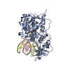
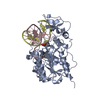
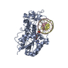
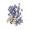
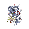
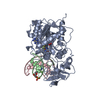
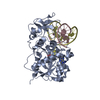
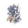
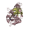
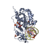
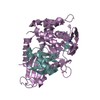
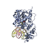
- PDB-2zcj: Ternary structure of the Glu119Gln M.HhaI, C5-Cytosine DNA methyl... -

+
Open data


ID or keywords:

Loading...

-
Basic information

Entry
Database: PDB / ID: 2zcj
Title
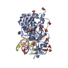
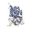
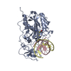
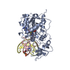
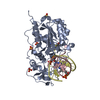
Ternary structure of the Glu119Gln M.HhaI, C5-Cytosine DNA methyltransferase, with unmodified DNA and AdoHcy
Components
  • DNA (5'-D(*DGP*DAP*DTP*DAP*DGP*DCP*DGP*DCP*DTP*DAP*DTP*DC)-3')
  • DNA (5'-D(*DTP*DGP*DAP*DTP*DAP*DGP*DCP*DGP*DCP*DTP*DAP*DTP*DC)-3')
  • Modification methylase HhaI
KeywordsTRANSFERASE/DNA / Alpha and beta / 3-layer Sandwich / Methyltransferase / Restriction system / S-adenosyl-L-methionine / TRANSFERASE-DNA COMPLEX
Function / homology
Function and homology information


DNA (cytosine-5-)-methyltransferase / DNA (cytosine-5-)-methyltransferase activity / DNA restriction-modification system / methylation / DNA binding
Similarity search - Function
DNA Methylase, subunit A, domain 2 / DNA Methylase; Chain A, domain 2 / DNA methylase, C-5 cytosine-specific, conserved site / C-5 cytosine-specific DNA methylases C-terminal signature. / DNA methylase, C-5 cytosine-specific, active site / C-5 cytosine-specific DNA methylases active site. / C-5 cytosine-specific DNA methylase (Dnmt) domain profile. / C-5 cytosine methyltransferase / C-5 cytosine-specific DNA methylase / Vaccinia Virus protein VP39 ...DNA Methylase, subunit A, domain 2 / DNA Methylase; Chain A, domain 2 / DNA methylase, C-5 cytosine-specific, conserved site / C-5 cytosine-specific DNA methylases C-terminal signature. / DNA methylase, C-5 cytosine-specific, active site / C-5 cytosine-specific DNA methylases active site. / C-5 cytosine-specific DNA methylase (Dnmt) domain profile. / C-5 cytosine methyltransferase / C-5 cytosine-specific DNA methylase / Vaccinia Virus protein VP39 / S-adenosyl-L-methionine-dependent methyltransferase superfamily / Alpha-Beta Complex / Rossmann fold / 3-Layer(aba) Sandwich / Alpha Beta
Similarity search - Domain/homology
S-ADENOSYL-L-HOMOCYSTEINE / DNA / DNA (> 10) / Type II methyltransferase M.HhaI
Similarity search - Component
Biological speciesHaemophilus parahaemolyticus (bacteria)
MethodX-RAY DIFFRACTION / simulation annealing / Resolution: 2.75 Å
AuthorsShieh, F.K. / Reich, N.O.
CitationJournal: J.Mol.Biol. / Year: 2007
Title: AdoMet-dependent Methyl-transfer: Glu(119) Is Essential for DNA C5-Cytosine Methyltransferase M.HhaI
Authors: Shieh, F.K. / Reich, N.O.
History
DepositionNov 9, 2007Deposition site: PDBJ / Processing site: PDBJ
Revision 1.0Dec 4, 2007Provider: repository / Type: Initial release
Revision 1.1Jul 13, 2011Group: Version format compliance
Revision 1.2Nov 10, 2021Group: Database references / Derived calculations / Category: database_2 / struct_ref_seq_dif / struct_site
Item: _database_2.pdbx_DOI / _database_2.pdbx_database_accession ..._database_2.pdbx_DOI / _database_2.pdbx_database_accession / _struct_ref_seq_dif.details / _struct_site.pdbx_auth_asym_id / _struct_site.pdbx_auth_comp_id / _struct_site.pdbx_auth_seq_id
Revision 1.3May 29, 2024Group: Data collection / Category: chem_comp_atom / chem_comp_bond

-
Structure visualization

Structure viewerMolecule:
MolmilJmol/JSmol

Downloads & links

-
Assembly

Deposited unit
C: DNA (5'-D(*DGP*DAP*DTP*DAP*DGP*DCP*DGP*DCP*DTP*DAP*DTP*DC)-3')
D: DNA (5'-D(*DTP*DGP*DAP*DTP*DAP*DGP*DCP*DGP*DCP*DTP*DAP*DTP*DC)-3')
A: Modification methylase HhaI
hetero molecules


Theoretical massNumber of molelcules
Total (without water)45,0554
Polymers44,6703
Non-polymers3841
Water1,29772
1


  • Idetical with deposited unit
  • defined by author&software
TypeNameSymmetry operationNumber
identity operation1_555x,y,z1
Buried area4410 Å2
MethodPISA
Unit cell
Length a, b, c (Å)98.470, 98.470, 321.615
Angle α, β, γ (deg.)90.00, 90.00, 120.00
Int Tables number155
Space group name H-MH32

-
Components

#1: DNA chain DNA (5'-D(*DGP*DAP*DTP*DAP*DGP*DCP*DGP*DCP*DTP*DAP*DTP*DC)-3')


Mass: 3662.404 Da / Num. of mol.: 1 / Source method: obtained synthetically / Details: Synthetic Construction
#2: DNA chain DNA (5'-D(*DTP*DGP*DAP*DTP*DAP*DGP*DCP*DGP*DCP*DTP*DAP*DTP*DC)-3')


Mass: 3966.597 Da / Num. of mol.: 1 / Source method: obtained synthetically / Details: Synthetic Construction
#3: Protein Modification methylase HhaI / Cytosine-specific methyltransferase HhaI / M.HhaI


Mass: 37041.223 Da / Num. of mol.: 1 / Mutation: E119Q
Source method: isolated from a genetically manipulated source
Source: (gene. exp.) Haemophilus parahaemolyticus (bacteria)
Gene: hhaIM / Plasmid: pHSHW-5 / Production host: Escherichia coli (E. coli)
References: UniProt: P05102, DNA (cytosine-5-)-methyltransferase
#4: Chemical ChemComp-SAH / S-ADENOSYL-L-HOMOCYSTEINE


Type: L-peptide linking / Mass: 384.411 Da / Num. of mol.: 1 / Source method: obtained synthetically / Formula: C14H20N6O5S
#5: Water ChemComp-HOH / water


Mass: 18.015 Da / Num. of mol.: 72 / Source method: isolated from a natural source / Formula: H2O

-
Experimental details

-
Experiment

ExperimentMethod: X-RAY DIFFRACTION / Number of used crystals: 1

-
Sample preparation

CrystalDensity Matthews: 3.36 Å3/Da / Density % sol: 63.38 %
Crystal growTemperature: 290 K / Method: vapor diffusion, hanging drop / pH: 6
Details: 100 mM Sodium Citrate, 1.6-2.1 Ammonium Sulfate, pH 6.0, VAPOR DIFFUSION, HANGING DROP, temperature 290K
Components of the solutions
IDNameCrystal-IDSol-ID
1sodium citrate11
2ammonium sulfate11
3H2O11
4sodium citrate12
5ammonium sulfate12
6H2O12

-
Data collection

DiffractionMean temperature: 100 K
Diffraction sourceSource: ROTATING ANODE / Type: RIGAKU RU200
DetectorType: MAR scanner 180 mm plate / Detector: IMAGE PLATE / Date: Aug 1, 2006 / Details: mirrors
RadiationProtocol: SINGLE WAVELENGTH / Monochromatic (M) / Laue (L): M / Scattering type: x-ray
Radiation wavelengthRelative weight: 1
ReflectionResolution: 2.5→10 Å / Num. obs: 18897 / Observed criterion σ(F): 2 / Observed criterion σ(I): 2 / Rmerge(I) obs: 0.106 / Rsym value: 0.071
Reflection shellResolution: 2.6→2.75 Å / Rmerge(I) obs: 0.365 / Num. unique all: 2691 / Rsym value: 0.509

-
Processing

Software
NameClassification
MAR345dtbdata collection
CNSrefinement
MOSFLMdata reduction
SCALAdata scaling
CNSphasing
RefinementMethod to determine structure: simulation annealing / Resolution: 2.75→8.64 Å / σ(F): 2 / σ(I): 2 /
RfactorNum. reflection
Rfree0.266 -
Rwork0.218 -
obs-16061
Refinement stepCycle: LAST / Resolution: 2.75→8.64 Å
ProteinNucleic acidLigandSolventTotal
Num. atoms2606 506 26 72 3210
LS refinement shellResolution: 2.75→2.78 Å /
RfactorNum. reflection
Rfree0.34 -
Rwork0.293 -
obs-53

+
About Yorodumi

-
News

-
Feb 9, 2022. New format data for meta-information of EMDB entries

New format data for meta-information of EMDB entries

  • Version 3 of the EMDB header file is now the official format.
  • The previous official version 1.9 will be removed from the archive.

Related info.:EMDB header

External links:wwPDB to switch to version 3 of the EMDB data model

-
Aug 12, 2020. Covid-19 info

Covid-19 info

URL: https://pdbjlvh1.pdbj.org/emnavi/covid19.php

New page: Covid-19 featured information page in EM Navigator.

Related info.:Covid-19 info / Mar 5, 2020. Novel coronavirus structure data

+
Mar 5, 2020. Novel coronavirus structure data

Novel coronavirus structure data

Related info.:Yorodumi Speices / Aug 12, 2020. Covid-19 info

External links:COVID-19 featured content - PDBj / Molecule of the Month (242):Coronavirus Proteases

+
Jan 31, 2019. EMDB accession codes are about to change! (news from PDBe EMDB page)

EMDB accession codes are about to change! (news from PDBe EMDB page)

  • The allocation of 4 digits for EMDB accession codes will soon come to an end. Whilst these codes will remain in use, new EMDB accession codes will include an additional digit and will expand incrementally as the available range of codes is exhausted. The current 4-digit format prefixed with “EMD-” (i.e. EMD-XXXX) will advance to a 5-digit format (i.e. EMD-XXXXX), and so on. It is currently estimated that the 4-digit codes will be depleted around Spring 2019, at which point the 5-digit format will come into force.
  • The EM Navigator/Yorodumi systems omit the EMD- prefix.

Related info.:Q: What is EMD? / ID/Accession-code notation in Yorodumi/EM Navigator

External links:EMDB Accession Codes are Changing Soon! / Contact to PDBj

+
Jul 12, 2017. Major update of PDB

Major update of PDB

  • wwPDB released updated PDB data conforming to the new PDBx/mmCIF dictionary.
  • This is a major update changing the version number from 4 to 5, and with Remediation, in which all the entries are updated.
  • In this update, many items about electron microscopy experimental information are reorganized (e.g. em_software).
  • Now, EM Navigator and Yorodumi are based on the updated data.

External links:wwPDB Remediation / Enriched Model Files Conforming to OneDep Data Standards Now Available in the PDB FTP Archive

-
Yorodumi

Thousand views of thousand structures

  • Yorodumi is a browser for structure data from EMDB, PDB, SASBDB, etc.
  • This page is also the successor to EM Navigator detail page, and also detail information page/front-end page for Omokage search.
  • The word "yorodu" (or yorozu) is an old Japanese word meaning "ten thousand". "mi" (miru) is to see.

Related info.:EMDB / PDB / SASBDB / Comparison of 3 databanks / Yorodumi Search / Aug 31, 2016. New EM Navigator & Yorodumi / Yorodumi Papers / Jmol/JSmol / Function and homology information / Changes in new EM Navigator and Yorodumi

Read more