 Movie
Movie Controller

[English] 日本語
 Yorodumi
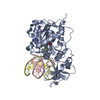
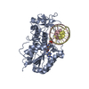
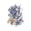
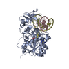
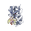
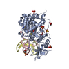
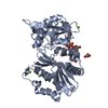
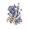
- PDB-1mht: COVALENT TERNARY STRUCTURE OF HHAI METHYLTRANSFERASE, DNA AND S-A... -
+ Open data

- Basic information
| Entry | Database: PDB / ID: 1mht | ||||||
|---|---|---|---|---|---|---|---|
| Title | COVALENT TERNARY STRUCTURE OF HHAI METHYLTRANSFERASE, DNA AND S-ADENOSYL-L-HOMOCYSTEINE | ||||||
 Components Components |
| ||||||
 Keywords Keywords | TRANSFERASE/DNA / PROTEIN-DNA COMPLEX / DOUBLE HELIX / OVERHANGING BASE / FLIPPED-OUT BASE / MODIFIED / TRANSFERASE-DNA COMPLEX | ||||||
| Function / homology |  Function and homology information Function and homology informationDNA (cytosine-5-)-methyltransferase / DNA (cytosine-5-)-methyltransferase activity / DNA restriction-modification system / methylation / DNA binding Similarity search - Function | ||||||
| Biological species |  Haemophilus haemolyticus (bacteria) Haemophilus haemolyticus (bacteria) | ||||||
| Method |  X-RAY DIFFRACTION / SYNCHROTRON / Resolution: 2.6 Å X-RAY DIFFRACTION / SYNCHROTRON / Resolution: 2.6 Å | ||||||
 Authors Authors | Cheng, X. | ||||||
 Citation Citation | Journal: Cell(Cambridge,Mass.) / Year: 1994 Title: HhaI methyltransferase flips its target base out of the DNA helix. Authors: Klimasauskas, S. / Kumar, S. / Roberts, R.J. / Cheng, X. #1: Journal: Cell(Cambridge,Mass.) / Year: 1993Title: Crystal Structure of the HhaI DNA Methyltransferase Complexed with S-Adenosyl-L-Methionine Authors: Cheng, X. / Kumar, S. / Posfai, J. / Pflugrath, J.W. / Roberts, R.J. | ||||||
| History |
|
- Structure visualization
| Structure viewer | Molecule: MolmilJmol/JSmol |
|---|
- Downloads & links
-Download
| PDBx/mmCIF format | 1mht.cif.gz | 95.8 KB | Display | PDBx/mmCIF format |
|---|---|---|---|---|
| PDB format | pdb1mht.ent.gz | 69.8 KB | Display | PDB format |
| PDBx/mmJSON format | 1mht.json.gz | Tree view | PDBx/mmJSON format | |
| Others |  Other downloads Other downloads |
-Validation report
| Arichive directory | https://data.pdbj.org/pub/pdb/validation_reports/mh/1mhtftp://data.pdbj.org/pub/pdb/validation_reports/mh/1mht | HTTPS FTP |
|---|
-Related structure data
| Similar structure data |
|---|


-Links
 PDBj
PDBj


- Assembly
| Deposited unit | 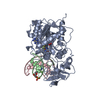
| ||||||||||
|---|---|---|---|---|---|---|---|---|---|---|---|
| 1 |
| ||||||||||
| Unit cell |
|
-Components
| #1: DNA chain | Mass: 3712.436 Da / Num. of mol.: 1 / Source method: obtained synthetically |
|---|---|
| #2: DNA chain | Mass: 4016.629 Da / Num. of mol.: 1 / Source method: obtained synthetically |
| #3: Protein | Mass: 37042.207 Da / Num. of mol.: 1 Source method: isolated from a genetically manipulated source Source: (gene. exp.) Haemophilus haemolyticus (bacteria) / References: UniProt: P05102, EC: 2.1.1.73 |
| #4: Chemical | ChemComp-SAH /   Mass: 384.411 Da / Num. of mol.: 1 / Source method: obtained synthetically / Formula: C14H20N6O5S Mass: 384.411 Da / Num. of mol.: 1 / Source method: obtained synthetically / Formula: C14H20N6O5S |
| Has protein modification | Y |
-Experimental details
-Experiment
| Experiment | Method: X-RAY DIFFRACTION / Number of used crystals: 1 |
|---|
- Sample preparation
| Crystal | Density Matthews: 3.51 Å3/Da / Density % sol: 65 % | ||||||||||||||||||||||||||||||||||||||||||||||||
|---|---|---|---|---|---|---|---|---|---|---|---|---|---|---|---|---|---|---|---|---|---|---|---|---|---|---|---|---|---|---|---|---|---|---|---|---|---|---|---|---|---|---|---|---|---|---|---|---|---|
| Crystal grow | Temperature: 289 K / Method: vapor diffusion, hanging drop / pH: 7.25 Details: BIS-TRIS-PROPANE_HCL, NACL, EDTA, pH 7.25, VAPOR DIFFUSION, HANGING DROP, temperature 289.00K | ||||||||||||||||||||||||||||||||||||||||||||||||
| Components of the solutions |
| ||||||||||||||||||||||||||||||||||||||||||||||||
| Crystal grow | *PLUS Temperature: 16 ℃ | ||||||||||||||||||||||||||||||||||||||||||||||||
| Components of the solutions | *PLUS
|
-Data collection
| Diffraction source | Source: SYNCHROTRON / Site: NSLS  / Type: NSLS / Wavelength: 1 / Type: NSLS / Wavelength: 1 |
|---|---|
| Detector | Type: ENRAF-NONIUS FAST / Detector: DIFFRACTOMETER |
| Radiation | Protocol: SINGLE WAVELENGTH / Monochromatic (M) / Laue (L): M / Scattering type: x-ray |
| Radiation wavelength | Wavelength: 1 Å / Relative weight: 1 |
| Reflection | Highest resolution: 2.8 Å / Num. obs: 17839 / Rmerge(I) obs: 0.0765 |
| Reflection | *PLUS Highest resolution: 2.8 Å |
- Processing
| Software |
| ||||||||||||||||||||||||||||||||||||||||||||||||||||||||||||
|---|---|---|---|---|---|---|---|---|---|---|---|---|---|---|---|---|---|---|---|---|---|---|---|---|---|---|---|---|---|---|---|---|---|---|---|---|---|---|---|---|---|---|---|---|---|---|---|---|---|---|---|---|---|---|---|---|---|---|---|---|---|
| Refinement | Resolution: 2.6→10 Å /
| ||||||||||||||||||||||||||||||||||||||||||||||||||||||||||||
| Refinement step | Cycle: LAST / Resolution: 2.6→10 Å
| ||||||||||||||||||||||||||||||||||||||||||||||||||||||||||||
| Refine LS restraints |
| ||||||||||||||||||||||||||||||||||||||||||||||||||||||||||||
| Software | *PLUS Name: X-PLOR / Classification: refinement | ||||||||||||||||||||||||||||||||||||||||||||||||||||||||||||
| Refinement | *PLUS Highest resolution: 2.8 Å | ||||||||||||||||||||||||||||||||||||||||||||||||||||||||||||
| Solvent computation | *PLUS | ||||||||||||||||||||||||||||||||||||||||||||||||||||||||||||
| Displacement parameters | *PLUS | ||||||||||||||||||||||||||||||||||||||||||||||||||||||||||||
| Refine LS restraints | *PLUS Type: x_angle_deg / Dev ideal: 1.6 |
