 Movie
Movie Controller

[English] 日本語
 Yorodumi
- PDB-2z6a: S-Adenosyl-L-methionine-Dependent Methyl Transfer: Observable Pre... -
+ Open data

- Basic information
| Entry | Database: PDB / ID: 2z6a | ||||||
|---|---|---|---|---|---|---|---|
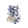
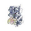
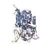
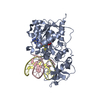
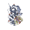
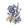
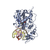
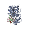
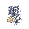
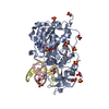
| Title | S-Adenosyl-L-methionine-Dependent Methyl Transfer: Observable Precatalytic Intermediates during DNA Cytosine Methylation | ||||||
 Components Components |
| ||||||
 Keywords Keywords | TRANSFERASE/DNA / Protein-DNA complex / Methyltransferase / Restriction system / S-adenosyl-L-methionine / Transferase / TRANSFERASE-DNA COMPLEX | ||||||
| Function / homology |  Function and homology information Function and homology informationDNA (cytosine-5-)-methyltransferase / DNA (cytosine-5-)-methyltransferase activity / DNA restriction-modification system / methylation / DNA binding Similarity search - Function | ||||||
| Biological species |  Haemophilus parahaemolyticus (bacteria) Haemophilus parahaemolyticus (bacteria)synthetic construct (others) | ||||||
| Method |  X-RAY DIFFRACTION / Simulation annealing / Resolution: 2.88 Å X-RAY DIFFRACTION / Simulation annealing / Resolution: 2.88 Å | ||||||
 Authors Authors | Shieh, F.K. | ||||||
 Citation Citation | Journal: Biochemistry / Year: 2007 Title: S-Adenosyl-l-methionine-Dependent Methyl Transfer: Observable Precatalytic Intermediates during DNA Cytosine Methylation Authors: Youngblood, B. / Shieh, F.K. / Buller, F. / Bullock, T. / Reich, N.O. | ||||||
| History |
|
- Structure visualization
| Structure viewer | Molecule: MolmilJmol/JSmol |
|---|
- Downloads & links
-Download
| PDBx/mmCIF format | 2z6a.cif.gz | 95.5 KB | Display | PDBx/mmCIF format |
|---|---|---|---|---|
| PDB format | pdb2z6a.ent.gz | 69.3 KB | Display | PDB format |
| PDBx/mmJSON format | 2z6a.json.gz | Tree view | PDBx/mmJSON format | |
| Others |  Other downloads Other downloads |
-Validation report
| Arichive directory | https://data.pdbj.org/pub/pdb/validation_reports/z6/2z6aftp://data.pdbj.org/pub/pdb/validation_reports/z6/2z6a | HTTPS FTP |
|---|
-Related structure data
| Related structure data |  2hr1S S: Starting model for refinement |
|---|---|
| Similar structure data |

-Links
 PDBj
PDBj


- Assembly
| Deposited unit | 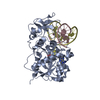
| ||||||||
|---|---|---|---|---|---|---|---|---|---|
| 1 |
| ||||||||
| Unit cell |
|
-Components
| #1: DNA chain | Mass: 3662.404 Da / Num. of mol.: 1 / Source method: obtained synthetically / Source: (synth.) synthetic construct (others) |
|---|---|
| #2: DNA chain | Mass: 3966.597 Da / Num. of mol.: 1 / Source method: obtained synthetically / Source: (synth.) synthetic construct (others) |
| #3: Protein | Mass: 37010.145 Da / Num. of mol.: 1 / Mutation: C81A Source method: isolated from a genetically manipulated source Source: (gene. exp.) Haemophilus parahaemolyticus (bacteria)Gene: hhaIM / Plasmid: pHSHW-5 / Production host: References: UniProt: P05102, DNA (cytosine-5-)-methyltransferase |
| #4: Chemical | ChemComp-SAH /   Type: L-peptide linking / Mass: 384.411 Da / Num. of mol.: 1 / Source method: obtained synthetically / Formula: C14H20N6O5S Type: L-peptide linking / Mass: 384.411 Da / Num. of mol.: 1 / Source method: obtained synthetically / Formula: C14H20N6O5S |
| #5: Water | ChemComp-HOH /  Mass: 18.015 Da / Num. of mol.: 55 / Source method: isolated from a natural source / Formula: H2O Mass: 18.015 Da / Num. of mol.: 55 / Source method: isolated from a natural source / Formula: H2O |
-Experimental details
-Experiment
| Experiment | Method: X-RAY DIFFRACTION / Number of used crystals: 1 |
|---|
- Sample preparation
| Crystal | Density Matthews: 3.38 Å3/Da / Density % sol: 63.65 % | ||||||||||||||||||||||||||||
|---|---|---|---|---|---|---|---|---|---|---|---|---|---|---|---|---|---|---|---|---|---|---|---|---|---|---|---|---|---|
| Crystal grow | Temperature: 290 K / Method: vapor diffusion, hanging drop / pH: 6.5 Details: 2.0M Ammonium Sulfate, 50mM Sodium Citrate, pH 6.5, VAPOR DIFFUSION, HANGING DROP, temperature 290K | ||||||||||||||||||||||||||||
| Components of the solutions |
|
-Data collection
| Diffraction | Mean temperature: 100 K |
|---|---|
| Diffraction source | Source: ROTATING ANODE / Type: RIGAKU RU300 / Wavelength: 1.5418 Å |
| Detector | Type: MAR scanner 180 mm plate / Detector: IMAGE PLATE / Date: Dec 1, 2005 / Details: mirrors |
| Radiation | Monochromator: YALE MIRRORS / Protocol: SINGLE WAVELENGTH / Monochromatic (M) / Laue (L): M / Scattering type: x-ray |
| Radiation wavelength | Wavelength: 1.5418 Å / Relative weight: 1 |
| Reflection | Resolution: 2.88→20 Å / Num. obs: 14120 / % possible obs: 99.9 % / Observed criterion σ(F): 2 / Observed criterion σ(I): 2 / Redundancy: 3.5 % / Biso Wilson estimate: 65.6 Å2 / Rmerge(I) obs: 0.16 / Rsym value: 0.09 / Net I/σ(I): 6.3 |
| Reflection shell | Resolution: 2.88→2.98 Å / Redundancy: 3.6 % / Rmerge(I) obs: 0.4 / Mean I/σ(I) obs: 2 / Num. unique all: 2034 / Rsym value: 0.354 / % possible all: 99.9 |
- Processing
| Software |
| ||||||||||||||||||||
|---|---|---|---|---|---|---|---|---|---|---|---|---|---|---|---|---|---|---|---|---|---|
| Refinement | Method to determine structure: Simulation annealing Starting model: PDB Entry 2HR1 Resolution: 2.88→8.85 Å / σ(F): 2 / σ(I): 2
| ||||||||||||||||||||
| Refinement step | Cycle: LAST / Resolution: 2.88→8.85 Å
| ||||||||||||||||||||
| Refine LS restraints |
| ||||||||||||||||||||
| LS refinement shell | Resolution: 2.88→2.92 Å / Rfactor Rfree error: 0.03
|
