 Movie
Movie Controller

[English] 日本語
 Yorodumi
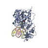
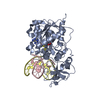
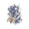
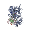
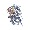
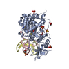
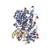
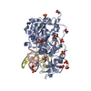
- PDB-1m0e: ZEBULARINE: A NOVEL DNA METHYLATION INHIBITOR THAT FORMS A COVALE... -
+ Open data

- Basic information
| Entry | Database: PDB / ID: 1m0e | |||||||||
|---|---|---|---|---|---|---|---|---|---|---|
| Title | ZEBULARINE: A NOVEL DNA METHYLATION INHIBITOR THAT FORMS A COVALENT COMPLEX WITH DNA METHYLTRANSFERASE | |||||||||
 Components Components |
| |||||||||
 Keywords Keywords | TRANSFERASE/DNA / Protein-DNA covalent complex / Mechanism based DNA methylation inhibitors / Zebularine / TRANSFERASE-DNA COMPLEX | |||||||||
| Function / homology |  Function and homology information Function and homology informationDNA (cytosine-5-)-methyltransferase / DNA (cytosine-5-)-methyltransferase activity / DNA restriction-modification system / methylation / DNA binding Similarity search - Function | |||||||||
| Biological species |  Haemophilus haemolyticus (bacteria) Haemophilus haemolyticus (bacteria) | |||||||||
| Method |  X-RAY DIFFRACTION / SYNCHROTRON / MOLECULAR REPLACEMENT / Resolution: 2.5 Å X-RAY DIFFRACTION / SYNCHROTRON / MOLECULAR REPLACEMENT / Resolution: 2.5 Å | |||||||||
 Authors Authors | Zhou, L. / Cheng, X. / Connolly, B.A. / Dickman, M.J. / Hurd, P.J. / Hornby, D.P. | |||||||||
 Citation Citation | Journal: J.MOL.BIOL. / Year: 2002 Title: ZEBULARINE: A NOVEL DNA METHYLATION INHIBITOR THAT FORMS A COVALENT COMPLEX WITH DNA METHYLTRANSFERASES Authors: Zhou, L. / Cheng, X. / Connolly, B.A. / Dickman, M.J. / Hurd, P.J. / Hornby, D.P. | |||||||||
| History |
|
- Structure visualization
| Structure viewer | Molecule: MolmilJmol/JSmol |
|---|
- Downloads & links
-Download
| PDBx/mmCIF format | 1m0e.cif.gz | 101.1 KB | Display | PDBx/mmCIF format |
|---|---|---|---|---|
| PDB format | pdb1m0e.ent.gz | 73.9 KB | Display | PDB format |
| PDBx/mmJSON format | 1m0e.json.gz | Tree view | PDBx/mmJSON format | |
| Others |  Other downloads Other downloads |
-Validation report
| Arichive directory | https://data.pdbj.org/pub/pdb/validation_reports/m0/1m0eftp://data.pdbj.org/pub/pdb/validation_reports/m0/1m0e | HTTPS FTP |
|---|
-Related structure data
| Related structure data |  9mhtS S: Starting model for refinement |
|---|---|
| Similar structure data |


-Links
 PDBj
PDBj


- Assembly
| Deposited unit | 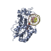
| ||||||||
|---|---|---|---|---|---|---|---|---|---|
| 1 |
| ||||||||
| Unit cell |
| ||||||||
| Components on special symmetry positions |
|
-Components
| #1: DNA chain | Mass: 3623.368 Da / Num. of mol.: 1 / Source method: obtained synthetically |
|---|---|
| #2: DNA chain | Mass: 3688.401 Da / Num. of mol.: 1 / Source method: obtained synthetically |
| #3: Protein | Mass: 37042.207 Da / Num. of mol.: 1 Source method: isolated from a genetically manipulated source Source: (gene. exp.) Haemophilus haemolyticus (bacteria) / Production host: |
| #4: Chemical | ChemComp-SAH /   Mass: 384.411 Da / Num. of mol.: 1 / Source method: obtained synthetically / Formula: C14H20N6O5S Mass: 384.411 Da / Num. of mol.: 1 / Source method: obtained synthetically / Formula: C14H20N6O5S |
| #5: Water | ChemComp-HOH /  Mass: 18.015 Da / Num. of mol.: 245 / Source method: isolated from a natural source / Formula: H2O Mass: 18.015 Da / Num. of mol.: 245 / Source method: isolated from a natural source / Formula: H2O |
| Has protein modification | Y |
-Experimental details
-Experiment
| Experiment | Method: X-RAY DIFFRACTION / Number of used crystals: 1 |
|---|
- Sample preparation
| Crystal | Density Matthews: 3.15 Å3/Da / Density % sol: 60.91 % | ||||||||||||||||||||||||||||||
|---|---|---|---|---|---|---|---|---|---|---|---|---|---|---|---|---|---|---|---|---|---|---|---|---|---|---|---|---|---|---|---|
| Crystal grow | Temperature: 289 K / Method: vapor diffusion, hanging drop / pH: 6.5 Details: PEG 8000, Ca acetate, sodium cacodylate, pH 6.5, VAPOR DIFFUSION, HANGING DROP, temperature 289K | ||||||||||||||||||||||||||||||
| Crystal grow | *PLUS Temperature: 16 ℃ / Method: vapor diffusion | ||||||||||||||||||||||||||||||
| Components of the solutions | *PLUS
|
-Data collection
| Diffraction | Mean temperature: 100 K |
|---|---|
| Diffraction source | Source: SYNCHROTRON / Site: NSLS  / Beamline: X8C / Wavelength: 0.979 Å / Beamline: X8C / Wavelength: 0.979 Å |
| Detector | Type: ADSC QUANTUM 4 / Detector: CCD / Date: Sep 16, 1999 Details: a parabolic collimating mirror placed upstream of monochromator |
| Radiation | Protocol: SINGLE WAVELENGTH / Monochromatic (M) / Laue (L): M / Scattering type: x-ray |
| Radiation wavelength | Wavelength: 0.979 Å / Relative weight: 1 |
| Reflection | Resolution: 2.5→39.7 Å / Num. obs: 19591 / % possible obs: 98.1 % / Observed criterion σ(F): -3 / Observed criterion σ(I): -3 |
| Reflection shell | Resolution: 2.5→2.61 Å / % possible all: 96.3 |
| Reflection | *PLUS Highest resolution: 2.5 Å |
- Processing
| Software |
| |||||||||||||||||||||||||
|---|---|---|---|---|---|---|---|---|---|---|---|---|---|---|---|---|---|---|---|---|---|---|---|---|---|---|
| Refinement | Method to determine structure: MOLECULAR REPLACEMENT Starting model: PDB entry 9MHT Resolution: 2.5→39.7 Å / Cross valid method: THROUGHOUT / σ(F): 2
| |||||||||||||||||||||||||
| Displacement parameters | Biso mean: 31.1 Å2
| |||||||||||||||||||||||||
| Refine analyze | Luzzati coordinate error obs: 0.24 Å / Luzzati d res low obs: 39.7 Å / Luzzati sigma a obs: 0.26 Å | |||||||||||||||||||||||||
| Refinement step | Cycle: LAST / Resolution: 2.5→39.7 Å
| |||||||||||||||||||||||||
| Refine LS restraints |
| |||||||||||||||||||||||||
| LS refinement shell | Resolution: 2.5→2.66 Å / Rfactor Rfree error: 0.019
| |||||||||||||||||||||||||
| Refinement | *PLUS % reflection Rfree: 10 % / Rfactor Rfree: 0.243 / Rfactor Rwork: 0.177 | |||||||||||||||||||||||||
| Solvent computation | *PLUS | |||||||||||||||||||||||||
| Displacement parameters | *PLUS | |||||||||||||||||||||||||
| Refine LS restraints | *PLUS
| |||||||||||||||||||||||||
| LS refinement shell | *PLUS Rfactor Rfree: 0.329 / Rfactor Rwork: 0.268 |
