[English] 日本語
Yorodumi
- PDB-2c7q: HhaI DNA methyltransferase complex with oligonucleotide containin... -

+
Open data


ID or keywords:

Loading...

-
Basic information

Entry
Database: PDB / ID: 2c7q
TitleHhaI DNA methyltransferase complex with oligonucleotide containing 2- aminopurine outside the recognition sequence (paired with G) and SAH
Components
  • 5'-D(*T*GP*GP*(2PR)*GP*GP*(5CM)*GP*CP*TP*GP* AP*C)-3'
  • 5'-D(*T*GP*TP*CP*AP*GP*CP*GP*CP*CP*GP*CP*C)-3'
  • MODIFICATION METHYLASE HHAI
Keywords
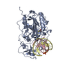
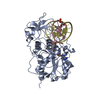
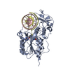
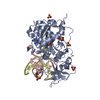
TRANSFERASE/DNA / BASE FLIPPING / RESTRICTION SYSTEM / TRANSFERASE-DNA COMPLEX
Function / homology
Function and homology information


DNA (cytosine-5-)-methyltransferase / DNA (cytosine-5-)-methyltransferase activity / DNA restriction-modification system / DNA binding
Similarity search - Function
DNA Methylase, subunit A, domain 2 / DNA Methylase; Chain A, domain 2 / DNA methylase, C-5 cytosine-specific, conserved site / C-5 cytosine-specific DNA methylases C-terminal signature. / DNA methylase, C-5 cytosine-specific, active site / C-5 cytosine-specific DNA methylases active site. / C-5 cytosine-specific DNA methylase (Dnmt) domain profile. / C-5 cytosine methyltransferase / C-5 cytosine-specific DNA methylase / Vaccinia Virus protein VP39 ...DNA Methylase, subunit A, domain 2 / DNA Methylase; Chain A, domain 2 / DNA methylase, C-5 cytosine-specific, conserved site / C-5 cytosine-specific DNA methylases C-terminal signature. / DNA methylase, C-5 cytosine-specific, active site / C-5 cytosine-specific DNA methylases active site. / C-5 cytosine-specific DNA methylase (Dnmt) domain profile. / C-5 cytosine methyltransferase / C-5 cytosine-specific DNA methylase / Vaccinia Virus protein VP39 / S-adenosyl-L-methionine-dependent methyltransferase superfamily / Alpha-Beta Complex / Rossmann fold / 3-Layer(aba) Sandwich / Alpha Beta
Similarity search - Domain/homology
CITRIC ACID / S-ADENOSYL-L-HOMOCYSTEINE / DNA / DNA (> 10) / Type II methyltransferase M.HhaI
Similarity search - Component
Biological speciesHAEMOPHILUS HAEMOLYTICUS (bacteria)
SYNTHETIC CONSTRUCT (others)
MethodX-RAY DIFFRACTION / SYNCHROTRON / MOLECULAR REPLACEMENT / Resolution: 1.85 Å
AuthorsDaujotyte, D. / Grazulis, S.
CitationJournal: Nucleic Acids Res. / Year: 2005
Title: Time-Resolved Fluorescence of 2-Aminopurine as a Probe of Base Flipping in M.HhaI-DNA Complexes
Authors: Neely, R.K. / Daujotyte, D. / Grazulis, S. / Magennis, S.W. / Dryden, D.T.F. / Klimasauskas, S. / Jones, A.C.
History
DepositionNov 27, 2005Deposition site: PDBE / Processing site: PDBE
Revision 1.0Dec 14, 2005Provider: repository / Type: Initial release
Revision 1.1Jan 29, 2014Group: Atomic model / Data collection ...Atomic model / Data collection / Derived calculations / Non-polymer description / Other / Refinement description / Source and taxonomy / Structure summary / Version format compliance
Revision 1.2Jul 24, 2019Group: Data collection / Derived calculations / Category: diffrn_source / struct_conn
Item: _diffrn_source.pdbx_synchrotron_site / _struct_conn.pdbx_leaving_atom_flag
Revision 1.3Dec 13, 2023Group: Data collection / Database references ...Data collection / Database references / Derived calculations / Other / Refinement description
Category: chem_comp_atom / chem_comp_bond ...chem_comp_atom / chem_comp_bond / database_2 / pdbx_database_status / pdbx_initial_refinement_model / struct_site
Item: _database_2.pdbx_DOI / _database_2.pdbx_database_accession ..._database_2.pdbx_DOI / _database_2.pdbx_database_accession / _pdbx_database_status.status_code_sf / _struct_site.pdbx_auth_asym_id / _struct_site.pdbx_auth_comp_id / _struct_site.pdbx_auth_seq_id

-
Structure visualization

Structure viewerMolecule:
MolmilJmol/JSmol

Downloads & links

-
Assembly

Deposited unit
A: MODIFICATION METHYLASE HHAI
C: 5'-D(*T*GP*GP*(2PR)*GP*GP*(5CM)*GP*CP*TP*GP* AP*C)-3'
D: 5'-D(*T*GP*TP*CP*AP*GP*CP*GP*CP*CP*GP*CP*C)-3'
hetero molecules


Theoretical massNumber of molelcules
Total (without water)45,8828
Polymers45,0173
Non-polymers8655
Water6,593366
1


  • Idetical with deposited unit
  • defined by author&software
TypeNameSymmetry operationNumber
identity operation1_555x,y,z1
Buried area6600 Å2
ΔGint-34.8 kcal/mol
Surface area15520 Å2
MethodPISA
Unit cell
Length a, b, c (Å)95.100, 95.100, 312.180
Angle α, β, γ (deg.)90.00, 90.00, 120.00
Int Tables number155
Space group name H-MH32
Components on special symmetry positions
IDModelComponents
11A-2139-

HOH

Details
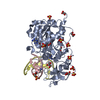
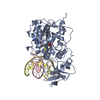
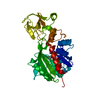
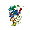
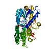
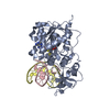
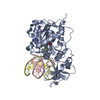
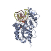
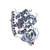
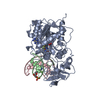
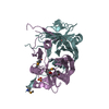
THE OLIGOMERIC STATE OF HHAI METHYLTRANSFERASE IS MONOMERIC, BUT SINCE IN THIS ENTRY, THE PROTEIN IS IN COMPLEX WITH DNA, THE QUATERNARY STRUCTURE FOR THIS ENTRY IS MARKED AS TRIMERIC.

-
Components

-
Protein , 1 types, 1 molecules A

#1: Protein MODIFICATION METHYLASE HHAI / DNA METHYLTRANSFERASE HHAI / CYTOSINE-SPECIFIC METHYLTRANSFERASE HHAI


Mass: 37042.207 Da / Num. of mol.: 1
Source method: isolated from a genetically manipulated source
Source: (gene. exp.) HAEMOPHILUS HAEMOLYTICUS (bacteria) / Plasmid: PHH553 / Production host: ESCHERICHIA COLI (E. coli) / Strain (production host): ER1727
References: UniProt: P05102, DNA (cytosine-5-)-methyltransferase

-
DNA chain , 2 types, 2 molecules CD

#2: DNA chain 5'-D(*T*GP*GP*(2PR)*GP*GP*(5CM)*GP*CP*TP*GP* AP*C)-3'


Mass: 4046.648 Da / Num. of mol.: 1 / Source method: obtained synthetically / Source: (synth.) SYNTHETIC CONSTRUCT (others)
#3: DNA chain 5'-D(*T*GP*TP*CP*AP*GP*CP*GP*CP*CP*GP*CP*C)-3'


Mass: 3928.549 Da / Num. of mol.: 1 / Source method: obtained synthetically / Source: (synth.) SYNTHETIC CONSTRUCT (others)

-
Non-polymers , 4 types, 371 molecules

#4: Chemical ChemComp-SAH / S-ADENOSYL-L-HOMOCYSTEINE / S-Adenosyl-L-homocysteine


Type: L-peptide linking / 
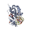
Mass: 384.411 Da / Num. of mol.: 1 / Source method: obtained synthetically / Formula: C14H20N6O5S
#5: Chemical ChemComp-SO4 / SULFATE ION / Sulfate


Mass: 96.063 Da / Num. of mol.: 3 / Source method: obtained synthetically / Formula: SO4
#6: Chemical ChemComp-CIT / CITRIC ACID / Citric acid


Mass: 192.124 Da / Num. of mol.: 1 / Source method: obtained synthetically / Formula: C6H8O7
#7: Water ChemComp-HOH / water / Water


Mass: 18.015 Da / Num. of mol.: 366 / Source method: isolated from a natural source / Formula: H2O

-
Details

Compound detailsRECOGNIZES THE DOUBLE-STRANDED SEQUENCE GCGC, CAUSES SPECIFIC METHYLATION AND PROTECTS THE DNA FROM ...RECOGNIZES THE DOUBLE-STRANDED SEQUENCE GCGC, CAUSES SPECIFIC METHYLATION AND PROTECTS THE DNA FROM CLEAVAGE BY THE HHAI ENDONUCLEASE.

-
Experimental details

-
Experiment

ExperimentMethod: X-RAY DIFFRACTION / Number of used crystals: 1

-
Sample preparation

CrystalDensity Matthews: 2.72 Å3/Da / Density % sol: 54.38 %
Crystal growpH: 5.6 / Details: 50 MM NA CITRATE PH 5.6, 1.6 M AMMONIUM SULFATE

-
Data collection

DiffractionMean temperature: 100 K
Diffraction sourceSource: SYNCHROTRON / Site: EMBL/DESY, HAMBURG / Beamline: X13 / Wavelength: 1.05
DetectorType: MARRESEARCH / Detector: IMAGE PLATE / Date: Jul 30, 2004 / Details: MIRRORS
RadiationMonochromator: SI(111) / Protocol: SINGLE WAVELENGTH / Monochromatic (M) / Laue (L): M / Scattering type: x-ray
Radiation wavelengthWavelength: 1.05 Å / Relative weight: 1
ReflectionResolution: 1.85→31 Å / Num. obs: 45796 / % possible obs: 98.7 % / Observed criterion σ(I): 3.7 / Redundancy: 8.7 % / Biso Wilson estimate: 10.6 Å2 / Rmerge(I) obs: 0.07 / Net I/σ(I): 8.5
Reflection shellResolution: 1.85→1.96 Å / Redundancy: 3.6 % / Rmerge(I) obs: 0.2 / Mean I/σ(I) obs: 3.7 / % possible all: 94.5

-
Processing

Software
NameVersionClassification
CNS1refinement
MOSFLMdata reduction
SCALAdata scaling
AMoREphasing
RefinementMethod to determine structure: MOLECULAR REPLACEMENT
Starting model: PDB ENTRY 3MHT
Resolution: 1.85→30.98 Å / Rfactor Rfree error: 0.003 / Data cutoff high absF: 50617366.94 / Isotropic thermal model: RESTRAINED / Cross valid method: THROUGHOUT / σ(F): 0
Details: RESOLUTION-DEPENDENT WEIGHTING SCHEME USED. BULK SOLVENT MODEL USED
RfactorNum. reflection% reflectionSelection details
Rfree0.21 4631 10.1 %RANDOM
Rwork0.187 ---
obs0.187 45794 98.3 %-
Solvent computationSolvent model: FLAT MODEL / Bsol: 56.7045 Å2 / ksol: 0.396895 e/Å3
Displacement parametersBiso mean: 15.9 Å2
Baniso -1Baniso -2Baniso -3
1-1.14 Å20.77 Å20 Å2
2--1.14 Å20 Å2
3----2.28 Å2
Refine analyze
FreeObs
Luzzati coordinate error0.21 Å0.19 Å
Luzzati d res low-5 Å
Luzzati sigma a0.14 Å0.14 Å
Refinement stepCycle: LAST / Resolution: 1.85→30.98 Å
ProteinNucleic acidLigandSolventTotal
Num. atoms2574 512 54 366 3506
Refine LS restraints
Refine-IDTypeDev idealDev ideal target
X-RAY DIFFRACTIONc_bond_d0.006
X-RAY DIFFRACTIONc_bond_d_na
X-RAY DIFFRACTIONc_bond_d_prot
X-RAY DIFFRACTIONc_angle_d
X-RAY DIFFRACTIONc_angle_d_na
X-RAY DIFFRACTIONc_angle_d_prot
X-RAY DIFFRACTIONc_angle_deg1.3
X-RAY DIFFRACTIONc_angle_deg_na
X-RAY DIFFRACTIONc_angle_deg_prot
X-RAY DIFFRACTIONc_dihedral_angle_d22.9
X-RAY DIFFRACTIONc_dihedral_angle_d_na
X-RAY DIFFRACTIONc_dihedral_angle_d_prot
X-RAY DIFFRACTIONc_improper_angle_d1.11
X-RAY DIFFRACTIONc_improper_angle_d_na
X-RAY DIFFRACTIONc_improper_angle_d_prot
X-RAY DIFFRACTIONc_mcbond_it0.471.5
X-RAY DIFFRACTIONc_mcangle_it0.792
X-RAY DIFFRACTIONc_scbond_it0.672
X-RAY DIFFRACTIONc_scangle_it1.152.5
LS refinement shellResolution: 1.85→1.97 Å / Rfactor Rfree error: 0.009 / Total num. of bins used: 6
RfactorNum. reflection% reflection
Rfree0.235 720 9.9 %
Rwork0.212 6523 -
obs--94.7 %
Xplor file
Refine-IDSerial noParam fileTopol file
X-RAY DIFFRACTION1PROTEIN_REP.PARAMPROTEIN.TOP
X-RAY DIFFRACTION2DNA-RNA_REP.PARAMDNA-RNA.TOP
X-RAY DIFFRACTION3WATER.PARAMWATER.TOP
X-RAY DIFFRACTION42AP.PAR2AP.TOP
X-RAY DIFFRACTION55MC.PAR5MC.TOP
X-RAY DIFFRACTION6CIT.PARCIT.TOP
X-RAY DIFFRACTION7SAH.PARSAH.TOP
X-RAY DIFFRACTION8SO4.PARSO4.TOP
X-RAY DIFFRACTION9EPE.PAREPE.TOP

+
About Yorodumi

-
News

-
Feb 9, 2022. New format data for meta-information of EMDB entries

New format data for meta-information of EMDB entries

  • Version 3 of the EMDB header file is now the official format.
  • The previous official version 1.9 will be removed from the archive.

Related info.:EMDB header

External links:wwPDB to switch to version 3 of the EMDB data model

-
Aug 12, 2020. Covid-19 info

Covid-19 info

URL: https://pdbj.org/emnavi/covid19.php

New page: Covid-19 featured information page in EM Navigator.

Related info.:Covid-19 info / Mar 5, 2020. Novel coronavirus structure data

+
Mar 5, 2020. Novel coronavirus structure data

Novel coronavirus structure data

Related info.:Yorodumi Speices / Aug 12, 2020. Covid-19 info

External links:COVID-19 featured content - PDBj / Molecule of the Month (242):Coronavirus Proteases

+
Jan 31, 2019. EMDB accession codes are about to change! (news from PDBe EMDB page)

EMDB accession codes are about to change! (news from PDBe EMDB page)

  • The allocation of 4 digits for EMDB accession codes will soon come to an end. Whilst these codes will remain in use, new EMDB accession codes will include an additional digit and will expand incrementally as the available range of codes is exhausted. The current 4-digit format prefixed with “EMD-” (i.e. EMD-XXXX) will advance to a 5-digit format (i.e. EMD-XXXXX), and so on. It is currently estimated that the 4-digit codes will be depleted around Spring 2019, at which point the 5-digit format will come into force.
  • The EM Navigator/Yorodumi systems omit the EMD- prefix.

Related info.:Q: What is EMD? / ID/Accession-code notation in Yorodumi/EM Navigator

External links:EMDB Accession Codes are Changing Soon! / Contact to PDBj

+
Jul 12, 2017. Major update of PDB

Major update of PDB

  • wwPDB released updated PDB data conforming to the new PDBx/mmCIF dictionary.
  • This is a major update changing the version number from 4 to 5, and with Remediation, in which all the entries are updated.
  • In this update, many items about electron microscopy experimental information are reorganized (e.g. em_software).
  • Now, EM Navigator and Yorodumi are based on the updated data.

External links:wwPDB Remediation / Enriched Model Files Conforming to OneDep Data Standards Now Available in the PDB FTP Archive

-
Yorodumi

Thousand views of thousand structures

  • Yorodumi is a browser for structure data from EMDB, PDB, SASBDB, etc.
  • This page is also the successor to EM Navigator detail page, and also detail information page/front-end page for Omokage search.
  • The word "yorodu" (or yorozu) is an old Japanese word meaning "ten thousand". "mi" (miru) is to see.

Related info.:EMDB / PDB / SASBDB / Comparison of 3 databanks / Yorodumi Search / Aug 31, 2016. New EM Navigator & Yorodumi / Yorodumi Papers / Jmol/JSmol / Function and homology information / Changes in new EM Navigator and Yorodumi

Read more