 Movie
Movie Controller

[English] 日本語
 Yorodumi
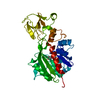
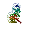
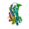
- PDB-2hmy: BINARY COMPLEX OF HHAI METHYLTRANSFERASE WITH ADOMET FORMED IN TH... -
+ Open data

- Basic information
| Entry | Database: PDB / ID: 2hmy | ||||||
|---|---|---|---|---|---|---|---|
| Title | BINARY COMPLEX OF HHAI METHYLTRANSFERASE WITH ADOMET FORMED IN THE PRESENCE OF A SHORT NONPSECIFIC DNA OLIGONUCLEOTIDE | ||||||
 Components Components | PROTEIN (CYTOSINE-SPECIFIC METHYLTRANSFERASE HHAI) | ||||||
 Keywords Keywords | TRANSFERASE (METHYLTRANSFERASE) | ||||||
| Function / homology |  Function and homology information Function and homology informationDNA (cytosine-5-)-methyltransferase / DNA (cytosine-5-)-methyltransferase activity / DNA restriction-modification system / methylation / DNA binding Similarity search - Function | ||||||
| Biological species |  Haemophilus haemolyticus (bacteria) Haemophilus haemolyticus (bacteria) | ||||||
| Method |  X-RAY DIFFRACTION / SYNCHROTRON / OTHER / Resolution: 2.61 Å X-RAY DIFFRACTION / SYNCHROTRON / OTHER / Resolution: 2.61 Å | ||||||
 Authors Authors | O'Gara, M. / Zhang, X. / Roberts, R.J. / Cheng, X. | ||||||
 Citation Citation | Journal: J.Mol.Biol. / Year: 1999 Title: Structure of a binary complex of HhaI methyltransferase with S-adenosyl-L-methionine formed in the presence of a short non-specific DNA oligonucleotide. Authors: O'Gara, M. / Zhang, X. / Roberts, R.J. / Cheng, X. #1: Journal: Cell(Cambridge,Mass.) / Year: 1993Title: Crystal structure of the HhaI DNA methyltransferase complexed with S-adenosyl-L-methionine. Authors: Cheng, X. / Kumar, S. / Posfai, J. / Pflugrath, J.W. / Roberts, R.J. #2: Journal: Biochemistry / Year: 1992 Title: Purification, crystallization, and preliminary X-ray diffraction analysis of an M.HhaI-AdoMet complex. Authors: Kumar, S. / Cheng, X. / Pflugrath, J.W. / Roberts, R.J. | ||||||
| History |
|
- Structure visualization
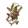
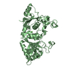
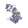
| Structure viewer | Molecule: MolmilJmol/JSmol |
|---|
- Downloads & links
-Download
| PDBx/mmCIF format | 2hmy.cif.gz | 80.2 KB | Display | PDBx/mmCIF format |
|---|---|---|---|---|
| PDB format | pdb2hmy.ent.gz | 59 KB | Display | PDB format |
| PDBx/mmJSON format | 2hmy.json.gz | Tree view | PDBx/mmJSON format | |
| Others |  Other downloads Other downloads |
-Validation report
| Arichive directory | https://data.pdbj.org/pub/pdb/validation_reports/hm/2hmyftp://data.pdbj.org/pub/pdb/validation_reports/hm/2hmy | HTTPS FTP |
|---|
-Related structure data
| Related structure data |  1hmyS S: Starting model for refinement |
|---|---|
| Similar structure data |


-Links
 PDBj
PDBj
- Assembly
| Deposited unit | 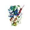
| ||||||||
|---|---|---|---|---|---|---|---|---|---|
| 1 |
| ||||||||
| Unit cell |
| ||||||||
| Noncrystallographic symmetry (NCS) | NCS oper: (Code: given Matrix: (-0.5025, -0.8406, -0.2021), Vector: |
-Components
| #1: Protein | Mass: 37042.207 Da / Num. of mol.: 1 Source method: isolated from a genetically manipulated source Source: (gene. exp.) Haemophilus haemolyticus (bacteria) / Production host: |
|---|---|
| #2: Chemical | ChemComp-SAM /   Mass: 398.437 Da / Num. of mol.: 1 / Source method: obtained synthetically / Formula: C15H22N6O5S Mass: 398.437 Da / Num. of mol.: 1 / Source method: obtained synthetically / Formula: C15H22N6O5S |
| #3: Water | ChemComp-HOH /  Mass: 18.015 Da / Num. of mol.: 63 / Source method: isolated from a natural source / Formula: H2O Mass: 18.015 Da / Num. of mol.: 63 / Source method: isolated from a natural source / Formula: H2O |
-Experimental details
-Experiment
| Experiment | Method: X-RAY DIFFRACTION / Number of used crystals: 1 |
|---|
- Sample preparation
| Crystal | Density Matthews: 4.94 Å3/Da / Density % sol: 75.1 % | ||||||||||||||||||||
|---|---|---|---|---|---|---|---|---|---|---|---|---|---|---|---|---|---|---|---|---|---|
| Crystal grow | pH: 6 / Details: 2.2M AMMONIUM SULFATE 50 MMSODIUM CITRATE PH 6.0 | ||||||||||||||||||||
| Crystal grow | *PLUS Method: vapor diffusion | ||||||||||||||||||||
| Components of the solutions | *PLUS
|
-Data collection
| Diffraction | Mean temperature: 95 K |
|---|---|
| Diffraction source | Source: SYNCHROTRON / Site: NSLS  / Beamline: X25 / Wavelength: 1 / Beamline: X25 / Wavelength: 1 |
| Detector | Type: MAR scanner 300 mm plate / Detector: IMAGE PLATE |
| Radiation | Protocol: SINGLE WAVELENGTH / Monochromatic (M) / Laue (L): M / Scattering type: x-ray |
| Radiation wavelength | Wavelength: 1 Å / Relative weight: 1 |
| Reflection | Resolution: 2.61→34.5 Å / Num. obs: 20104 / % possible obs: 91.3 % / Redundancy: 2 % |
- Processing
| Software |
| ||||||||||||||||||||||||||||||||||||||||||||||||||||||||||||
|---|---|---|---|---|---|---|---|---|---|---|---|---|---|---|---|---|---|---|---|---|---|---|---|---|---|---|---|---|---|---|---|---|---|---|---|---|---|---|---|---|---|---|---|---|---|---|---|---|---|---|---|---|---|---|---|---|---|---|---|---|---|
| Refinement | Method to determine structure: OTHER Starting model: PDB ENTRY 1HMY Resolution: 2.61→34.5 Å / σ(F): 0
| ||||||||||||||||||||||||||||||||||||||||||||||||||||||||||||
| Refinement step | Cycle: LAST / Resolution: 2.61→34.5 Å
| ||||||||||||||||||||||||||||||||||||||||||||||||||||||||||||
| Refine LS restraints |
| ||||||||||||||||||||||||||||||||||||||||||||||||||||||||||||
| LS refinement shell | Resolution: 2.61→2.75 Å / Total num. of bins used: 7
| ||||||||||||||||||||||||||||||||||||||||||||||||||||||||||||
| Software | *PLUS Name: X-PLOR / Classification: refinement | ||||||||||||||||||||||||||||||||||||||||||||||||||||||||||||
| Refinement | *PLUS Rfactor obs: 0.198 | ||||||||||||||||||||||||||||||||||||||||||||||||||||||||||||
| Solvent computation | *PLUS | ||||||||||||||||||||||||||||||||||||||||||||||||||||||||||||
| Displacement parameters | *PLUS | ||||||||||||||||||||||||||||||||||||||||||||||||||||||||||||
| Refine LS restraints | *PLUS
| ||||||||||||||||||||||||||||||||||||||||||||||||||||||||||||
| LS refinement shell | *PLUS Rfactor obs: 0.2945 |
